ROLE OF ESTROGEN ON THE MAINTENANCE AND
HOMING CAPACITY OF BONE MARROW DERIVED RAT
MESENCHYMAL STEM CELLS
A THESIS SUBMITTED TO
THE DEPARTMENT OF MOLECULAR BIOLOGY AND GENETICS
AND
THE GRADUATE SCHOOL OF ENGINEERING AND SCIENCE OF
BILKENT UNIVERSITY
IN PARTIAL FULFILLMENT OF THE REQUIREMENTS FOR
THE DEGREE OF DOCTOR OF PHILOSOPHY
BY
FATMA AYALOĞLU BÜTÜN
AUGUST 2011
I certify that I have read this thesis and that in my opinion it is fully adequate, in scope and in quality, as a thesis for the degree of Doctor of Philosophy
Assist. Prof. Dr. K. Can Akçalı I certify that I have read this thesis and that in my opinion it is fully adequate, in scope and in quality, as a thesis for the degree of Doctor of Philosophy
Prof. Dr. Mehmet Uğur I certify that I have read this thesis and that in my opinion it is fully adequate, in scope and in quality, as a thesis for the degree of Doctor of Philosophy
Assoc. Prof. Dr. İhsan Gürsel I certify that I have read this thesis and that in my opinion it is fully adequate, in scope and in quality, as a thesis for the degree of Doctor of Philosophy
Assoc. Prof. Dr. Sibel Yıldırım I certify that I have read this thesis and that in my opinion it is fully adequate, in scope and in quality, as a thesis for the degree of Doctor of Philosophy
Assist. Prof. Dr. H. Uygar Tazebay
Approved for the Graduate School of Engineering and Science
Director of Graduate School of Engineering and Science Prof. Dr. Levent Onural
i
ABSTRACT
ROLE OF ESTROGEN ON THE MAINTENANCE AND
HOMING CAPACITY OF BONE MARROW DERIVED RAT
MESENCHYMAL STEM CELLS
Fatma AYALOĞLU BÜTÜN Ph.D. in Molecular Biology and Genetics Supervisor: Assist. Prof. Dr. K. Can Akçalı
August 2011, 130 Pages
Mesenchymal Stem Cells (MSCs) can self renew and differentiate into different cell types like, adipocytes, osteoblasts, chondrocytes, neurons, hepatocytes and endothelial cells. Their ability to differentiate into wide variety of cell types, non-immunogenic characteristics, along with homing capacity to injured tissue and the absence of any ethical issue related to their uses, make MSCs important in regenerative medicine and tissue engineering. Their ability to migrate to the site of injury raises the opportunity of these stem cells to be considered also as in vivo delivery agents. However, one major obstacle in using MSCs in cell based therapies is their limited numbers. Estrogen is known to have role in growth, proliferation and apoptosis. Therefore our aim was to study the possible role and mechanism of estrogen in their maintenance and their homing capacity. We used MSCs derived from female and ovariectomized female rat bone marrow in our research. Our
ii
results revealed that estrogen treatment increased the number of colonies and the number of cells constituting a colony. Estrogen decreased the differentiation capacity of MSCs to the adipogenic lineages as shown by reduced of Oil Red O staining Estrogen also affected MSCs’ homing capacity. Estrogen treatment resulted in the migration of increased number of DiI labeled MSCs to the site of injury after partial hepatectomy (PH) compared to that of untreated MSCs. Furthermore, estrogen treatment decreased the rate of apoptosis. Our data showed that estrogen regulates apoptosis through Bcl-2 family of genes in MSCs. This regulation was at the protein level but not at the transcript level. Estrogen addition increased the expression of anti-apoptotic members of the Bcl-2 family of proteins, Bcl-2 and Bcl-xL. The decrease in the apoptosis was not observed when Bcl-xL and
Bcl-2 genes were knocked down. The silencing histone code H3K27me3 was also decreased in estrogen treated MSCs, suggesting an epigenetic regulation of MSCs upon estrogen treatment. Altogether our results show that estrogen increased the number of functional MSCs, decreased spontaneous apoptosis in these cells, and improved the homing capacity of rat bone marrow derived MSCs. Therefore, estrogen treatment of MSCs may offer new opportunities for the therapeutic actions of these cells.
iii
ÖZET
ÖSTROJEN HORMONUNUN SIÇAN KEMİK İLİĞİNDEN
ALINMIŞ MEZENKİMAL KÖK HÜCRELERİNİN
MUHAFAZASINDA VE HASARLI DOKUYA
ULAŞMASINDAKİ ROLÜ
Fatma AYALOĞLU BÜTÜN Moleküler Biyoloji ve Genetik Doktorası
Tez Yöneticisi: Doç. Dr. K. Can Akçalı Ağustos 2011, 130 Sayfa
Mezenkimal Kök Hücreler (MKH) kendilerini yenileyebilen ve adiposit, osteosit, kondrosit, sinir hücresi, hepatosit ve endotel hücresi gibi farklı hücrelere farklılaşabilen hücrelerdir. MKHlerin yüksek farklılaşma kapasitesi, immün reaksiyon oluşturmama özelliği, hasarlı dokuya gitme özelliği ve kullanımı ile ilgili etik sorunların olmaması, bu hücrelerin rejeneratif tıp ve doku mühendisliğinde önemli bir yerde olmalarının sebebidir. MKHlerin hasarlı dokuya ulaşabilmeleri aynı zamanda bu hücrelerin in vivo taşıyıcı ajan olarak kullanılması olasılığını da ortaya çıkarmıştır. Fakat MKHleri terapide kullanmanın önündeki en büyük zorluk onların sayıca az olmalarıdır. Östrojen hormonu büyüme, proliferasyon ve apoptozda rol aldığı bilinmektedir. Bu yüzden amacımız, östrojenin MKHlerin hasarlı dokuya yönelme ve yerleşmesindeki ve bu hücrelerin devamlılığındakı
iv
rolünü calışmaktı. Çalışmalarımızda dişi sıçanın kemik iliğinden alınan MKHler ve overleri çıkarılmış dişi sıçanın kemik iliğinden alınmış MKHler kullanıldı. Östrojen koloni sayısını ve kolonideki hücre sayısını arttırdı. Oil Red O boyaması ile görüldüğü gibi, östrojen aynı zamanda MKHlerin yağ hücresine farklılaşmasını azalttı. Östrojen verilen MKHler verilmeyen MKHlere gore kısmı hepatektomi yapılan sıçanların karaciğerlerine daha fazla sayıda gidip yerleştiler. Bunlara ek olarak östrojen MKHlerinin apoptozunu azalttı. Sonuçlarımız östrojenin apoptozu Bcl-2 gen ailesi üzerinden kontrol ettiğini gösterdi. Bu kontrol mRNA seviyesinde değil fakat protein seviyesindeydi. Östrojen eklenmesi Bcl-2 protein ailesinin anti-apoptotik üyelerinin protein seviyesini arttırdı. Apoptozdaki bu azalma Bcl-xL ve
Bcl-2 susturulduğu zaman görülmedi. Susturucu histon kodu olan H3K27me3 östrojen ile azalmakta, ve bu MKH gen ifadelerinin epigenetik bir yolla kontrol edildiği fikrini otaya atmaktadır. Bütün bu sonuçlar östrojenin fonksiyonel MKHlerin ve hasarlı dokuya ulaşan MKH sayısını arttırdığını, spontan olan apoptozu önlediğini göstermektedir. Bu sebeple bu hücrelerin östrojene maruz bırakılması onların tedavide kullanılmasına yeni olanaklar sunabilir.
v
ACKNOWLEDGEMENTS
I would like to express my gratitude to my supervisor Assoc. Prof. Dr. K. Can Akçalı for his endless support, valuable guidance, and supervision. He always shared his knowledge with me and supported me during my studies. I have learned a lot from him and it was an honor for me to work with him.
I would like to thank the former and present members of our group, Iraz Toprak Aydın, Ece Terzioğlu-Kara, Hande Koçak, Sinan Gültekin, Sumru Bayın, Zeynep Tokcaer-Keskin, C.Verda Bitirim, M. Merve Aydın and Ece Akhan for their friendship, patience and support during my PhD. I am very lucky to have them as friends and hope for everyone to be as happy as I am.
I would like to thank Burcu Ġnsal for her incredible help with animal work and especially the PH operations, Bala Gür-Dedeoğlu, Elif Uz, Tolga Acun, Hani Al-Otaibi, Ceren Sucularlı, Chigdem Aydın Mustafa, Emre Onat, Gizem Tinçer, Ayça Arslan-Ergül, Nuri Öztürk, Haluk Yüzügüllü, Ġ. Esen Oktay, M.Ender Avcı, Mine Mumcuoğlu, Pelin Telkoparan and Emin Öztaş for helping me with my problems.
I would also thank all the past and present MBG members for providing a stimulating and enjoyable work place. Without them the long journey of my study would be very difficult.
vi
I would also specially thank to Özlem Akıllı-Öztürk and her husband Erol Öztürk for moral support and friendship before and throughout my PhD studies.
I would like to thank The Scientific and Technological Research Council of Turkey (TUBĠTAK) for their financial support throughout my thesis work.
I would like to thank my mother, my father, my brothers and my sister for their unconditional love and unlimited support. I can not imagine a life without them.
I would specially thank to my husband Serkan Bütün who was always there when I needed him. He motivated and encouraged me when I felt blue and supported me in my decisions. Without him I could not reach this point. I also want to thank my son Ömer Selim for bringing endless joy in our life.
vii
CONTENTS
ABSTRACT ... i ÖZET ... iii ACKNOWLEDGEMENTS ... v CONTENTS ... vii LIST OF TABLES ... x LIST OF FIGURES ... xi ABBREVIATIONS ... xiii Chapter 1 INTRODUCTION ... 1PLURIPOTENT STEM CELLS ... 3
1.1 EMBRYONIC STEM CELL ... 3
1.1.1 INDUCED PLURIPOTENT STEM CELL ... 5
1.1.2 MULTIPOTENT ADULT STEM CELLS ... 9
1.2 MESENCHYMAL STEM CELL ... 11
1.2.1 1.2.1.1 CHARACTERIZATION OF MSC ... 12
1.2.1.2 MESENCHYMAL STEM CELL HOMING ... 14
1.2.1.3 MESENCHYMAL STEM CELLS IN REGENERATIVE MEDICINE ... 17
ESTROGEN ... 19
1.3 ESTROGEN RECEPTOR PATHWAY ... 20
1.3.1 ESTROGEN AND MESENCHYMAL STEM CELLS ... 22
1.3.2 CELL DEATH AND APOPTOSIS ... 24
1.4 EXTRINSIC AND INTRINSIC PATHWAY ... 25
1.4.1 BCL-2 FAMILY MEMBERS ... 28
1.4.2 1.4.2.1 SIGNALING PATHWAY ... 32
viii
ESTROGEN, APOPTOSIS AND THE BCL-2 FAMILY OF 1.4.3
GENES ... 34
EPIGENETICS ... 35
1.5 TYPES OF EPIGENETIC MODIFICATIONS ... 36
1.5.1 HISTONE MODIFICATIONS, STEM CELL, APOPTOSIS, 1.5.2 AND ESTROGEN ... 41
Chapter 2 AIM OF THE STUDY ... 44
Chapter 3 MATERIALS AND METHOD ... 45
ANIMALS ... 45 3.1 OVARIECTOMIZATION ... 45 3.1.1 PARTIAL HEPATECTOMY (PH) ... 46 3.1.2 HARVESTING AND CULTURING OF THE BONE MARROW 3.2 DERIVED MESENCHYMAL STEM CELLS ... 46
ADIPOGENIC AND OSTEOGENIC DIFFERENTIATION ... 47
3.3 OIL RED O STAINING ... 49
3.4 ALIZARIN RED S STAINING ... 49
3.5 DiI LABELING ... 49
3.6 COLONY FORMING UNIT (CFU) ASSAY ... 50
3.7 TUNEL ASSAY ... 51
3.8 TOTAL RNA ISOLATION FROM CULTURED MSC ... 51
3.9 cDNA SYNTHESIS ... 52 3.10 PRIMER DESIGN ... 53 3.11 SEMI QUANTITATIVE PCR ... 53 3.12 AGAROSE GEL ELECTROPHORESIS ... 54
3.13 TOTAL PROTEIN ISOLATION AND QUANTIFICATION ... 54
3.14 HISTONE PROTEIN EXTRACTION FROM CELL CULTURE 3.15 (ACID EXTRACTION) ... 56
WESTERN BLOT ... 57
3.16 SDS- POLYACRYLAMIDE GEL ELECTROPHORESIS ... 57 3.16.1
ix
TRANSFER OF PROTEINS TO THE MEMBRANE ... 58 3.16.2
3.16.2.1 SEMI DRY TRANSFER ... 58 3.16.2.2 WET TRANSFER ... 58
IMMUNOLOGICAL DETECTION OF THE PROTEINS ON 3.16.3
THE MEMBRANE ... 59 IMMUNOFLUORESCENCE STAINING FOR NON-HISTONE
3.17
PROTEINS ... 60 IMMUNOCYTOCHEMISTRY FOR HISTONE PROTEINS ... 61 3.18
shRNA TRANSFECTION ... 63 3.19
shRNA PLASMID PREPARATION... 63 3.19.1
TRANSFECTION ... 65 3.19.2
STATISTICAL ANALYSIS ... 66 3.20
SOLUTIONS AND BUFFERS ... 66 3.21
Chapter 4 RESULTS ... 67 CD MARKER EXPRESSION ... 67 4.1
ER PROTEIN EXPRESSION AND ESTROGEN 4.2 RESPONSIVENESS OF MSCs ... 68 CFU-F ASSAY ... 70 4.3 DIFFERENTIATION OF MSCs ... 71 4.4 HOMING OF MSCs ... 73 4.5
LABELING OF MSCs WITH DiI AND DETERMINING THE 4.5.1
OPTIMUM TIMING FOR THE ADMINISTRATION INTO THE RATS ... 73 EFFECT OF ESTOGEN ON MSC HOMING ... 75 4.5.2
MSC MAINTENANCE ... 79 4.6
APOPTOSIS ... 79 4.6.1
4.6.1.1 mRNA EXPRESSION of BCL-2 FAMILY OF GENES ... 81 4.6.1.2 PROTEIN EXPRESSION of BCL-2 FAMILY ... 81
x
KNOCK DOWN OF THE ANTIAPOPTOTIC MEMBERS OF 4.6.2
BCL-2 FAMILY ... 85
EFFECT OF ESTROGEN ON HISTONE MODIFICATIONS OF 4.7 MSCs ... 86
Chapter 5 DISCUSSION ... 90
Chapter 6 FUTURE PERSPECTIVES ... 101
References ... 104
APPENDIX ... 123
SOLUTIONS AND BUFFERS ... 123
LIST OF TABLES
Table 1.1: IPS cells derived from different species and cell types by different factors. ... 7Table 1.2: The location of different kinds of ASCs in the body and their differentiated progeny. (adopted from [70]). ... 10
Table 1.3: Characteristics of apoptosis and necrosis ... 25
Table 1.4: The regulatory roles of several histone marks. (Adopted from [218]) .... 39
Table 1.5: Demethylases and their substrates. (Adopted from [217]). ... 41
Table 3.1: Primer used in the study ... 55
Table 3.2: RT-PCR conditions for each primer ... 56
Table 3.3: Antibodies used in western blotting ... 60
xi
LIST OF FIGURES
Figure 1.1: The differentiation potential of MSCs into the mesenchymal lineage. . 14
Figure 1.2: The ER signaling. ... 22
Figure 1.3: The extrinsic and intrinsic apoptotic pathways. [149] ... 27
Figure 1.4: The structure of the Bcl-2 family members. ... 29
Figure 1.5: Regulation of Bcl-2 proteins. ... 32
Figure 1.6: Covalent modifications of the core histone proteins. ... 38
Figure 3.1: Animal models and MSCs groups used in the thesis. ... 48
Figure 3.2: vector pLKO.1 map. ... 63
Figure 4.1: Characterization of bone marrow derived MSCs. ... 68
Figure 4.2: Protein expression of ER α and β in MSCs determined by western blot. ... 69
Figure 4.3: mRNA expression of estrogen responsive gene mmp12 determined by RT-PCR. ... 70
Figure 4.4: CFU activities of MSCs. ... 71
Figure 4.5: Differentiation capacity of MSCs isolated from normal female and ovx female in the absence and presence of estrogen. ... 74
Figure 4.6: Image of in vitro DiI labeled MSCs. ... 75
Figure 4.7: Effect of recovery time after PH on the homing capacity of DiI labeled MSCs in the liver. ... 76
Figure 4.8: Localization of MSCs to the liver after PH. ... 77
Figure 4.9: Effect of estrogen on the homing capacity of MSCs Liver sections ... 78
Figure 4.10: Effect of estrogen on MSC apoptosis shown by in situ analysis of DNA fragmentation (TUNEL). ... 80
Figure 4.11: Effect of estrogen on the expression profile of the bcl-2 family of genes in MSCs ... 82
xii
Figure 4.12: Immunofluorescein staining of the Bcl-2 family of proteins on MSCs ... 83 Figure 4.13: Protein expression of the Bcl-2 family of genes in MSCs determined by western blotting. ... 84 Figure 4.14: Effect of silencing bcl-xL and bcl-2 on the apoptotic rates of MSCs ... 86
Figure 4.15: The expression of histone proteins by immunocytochemistry. ... 88 Figure 4.16: Western blotting of modified histone. ... 89
xiii
ABBREVIATIONS
AIF Apoptosis Inducing Factor AIM Adipogenic induction media
APAF1 Pro-Apoptotic Protease-Activating Factor-1
asc Ascorbic Acid
ASC Adult Stem Cell
Bak BCL2-antagonist/killer Bax Bcl2-associated X protein Bcl-2 B-cell leukemia/lymphoma 2 Bcl-xL B cell lymphoma like X
BFB Bromophenol Blue
BH Bcl-2 homology
BMP Bone Morphogenic Protein
bp Base Pairs
BSA Bovine Serum Albumin
CARD Caspase Recruitment Domain CD Cluster of Differentiation
cDNA Complementary Deoxyribonucleic Acid CFU-F Colony-forming Units-fibroblastic Cyt c Cytochrome c
xiv
dex Dexamethasone
dH2O Distilled Water
DIABLO Direct IAP binding protein with low pI DISC Death Inducing Signaling Complex DMEM Dulbecco’s Modified Eagle Medium
DMEM-LG Low Glucose Dulbecco’s Modified Eagle Medium DMSO Dimethyl Sulfoxide
DNA Deoxyribonucleic Acid DNase Deoxyribonuclease
dNTP deoxy Nucleotide Triphosphate
ER Estrogen Receptor
ERE Estrogen Response Elements ERα Estrogen Receptor Alpha ERβ Estrogen Receptor Beta
ESC Embryonic Stem Cell
FADD Fas Associated Death Domain
FBS Fetal Bovine Serum
Fgf2 Fibroblast growth factor 2
g Grams
GFP Green Fluorescent Protein GVHD Graft-versus-host Disease H2O2 Hydrogen Peroxide
xv
H4 Histone 4
hESC Human Embryonic Stem Cell HRP Horse Raddish Peroxidase HSC Hematopoietic Stem Cell IAP Inhibitor of Apoptosis Proteins IBMX Isobutyl methyl xanthine
ICM Inner Cell Mass
IF Immunofluorescent
Indo Indomethacine
iPS induced Pluripotent Stem iPSC induced Pluripotent Stem Cells
JmjC Jumonji-C
K Lysine
kDa kilo Dalton
LIF Leukemia Inhibitory Factor Lpl Lipoprotein lipase
LSD1 Lysine Specific Demethylase 1
M Molar
mA Mili Amper
MEF Mouse Embryonic Fibroblast mESC mouse Embryonic Stem Cell MetOH Methyl Alcohol
xvi
mL milliLiter
mM milliMolar
MPTP Mitochondrial Permeability Transition Pore MSCs Mesenchymal Stem Cells
NaCl Sodium Chloride
NCCD Nomenclature Committee on Cell Death NIH National Institute of Health
NTBC 2-(2-nitro-4-fluoromethylbenzoyl)-1,3-cyclohexanedione
o.n. Over night
Oc osteocalcin
OIM Osteogenic Induction Media
Ovx Ovariectomized
Ovx ovariectomized
PAGE Polyacrylamide Gel Electrophoresis PARP-1 Poly-ADP-ribose polymerase-1 PBS Phosphate Buffered Saline PCD Programmed Cell Death PCR Polymerase Chain Reaction
PFA Paraformaldehyde
PH Partial Hepatectomy
PI3K Phosphatidylinositol 3 kinase
Pparγ Peroxisome proliferator activated receptor gamma PTM Post Translational Modifictaion
xvii PVDF Polyvinylidenedifluoride
R Arginine
RiPSC RNA derived iPS Cells
RNA Ribonucleic Acid
ROS Reactive Oxygen Species Rpm Revolution per minute
RT RT
RT-PCR Reverse-Transcriptase Polymerase Chain Reaction Runx2 runt-related transcription factor 2
SDS Sodium Dodecyl Sulfate
SH Sham
shRNA small hairpin RNA ß-gp beta glycerophosphate
TAE Tris Acetate EDTA
TBS Tris Buffered Saline
TBS-T Tris Buffered Saline with tween TERT Telomerase Reverse Transcriptase TGF-ß Transforming Growth Factor-Beta TNF Tumor Necrosis Factor
TRAF TNF-associated Factor
TUNEL Terminal Deoxynucleotidyl Transferase dUTP Nick End Labeling
V Volt
1
Chapter 1
INTRODUCTION
Stem cells have attracted a lot of interest in the last three decades. Not only researchers but also the community is interested in this field due to their potential usage in regenerative therapy. Despite to the high attention and focus on these cells there is no definitive description of the stem cell concept. However, the National Institude of Health (NIH) defines stem cells as “ Cells that can divide infinitely and give rise to differentiated cells in vivo and in vitro” (http://stemcells.nih.gov/). Therefore self-renewal, differentiation and in vivo reconstition are critical features of stem cells.
Generally stem cells can be classified according to their source of origin and their potency. During early embryonic development, inner cell mass (ICM) give rise to embryonic stem cells (ESCs). Stem cells that are isolated at later developmental stages and postnatal period collectively are called adult stem cells (ASC). Another classification is related to their potency. Totipotent stem cells give rise to the cells from the extraembryonic tissues in addition to the cells from the three germ layers, (ectoderm, endoderm and mesoderm), whereas the pluripotent stem cells give rise to the cells from the three germ layers but not to the extra embryonic cells. Multipotent stem cells differentiate into the cell types from their original germ layer and another germ layer (http://stemcells.nih.gov/). ESCs and the induced
2
pluripotent stem cells (iPSC) are pluripotent whereas most of the adult stem cell are multipotent, and the zygote is the only cell that is totipotent.
Use of stem cells in therapy has brought new hope in the field of regenerative medicine. The capacity of stem cells to differentiate into different types of cells after correct stimuli made them a very trustworthy agents in medicine for the treatment of several genetic, chronic and non-infectious diseases like osteogenesis imperfecta, parkinsons disease, chronic heart diseases, spinal cord injuries, liver cirrhosis, and diabetes (http://www.clinicaltrials.gov). Embryonic stem cells (ESC) are very promising agents since they can differentiate into the cells of all three germ layers. However their use is very limited due to the ethical problems since embryo is the source. To overcome this, researchers developed induced pluripotent stem (iPS) cells which are pluripotent like embryonic stem cells, but do not require the embryo therefore have no ethical problems. However, these cells are not completely characterized and it will take time before seing them in medicine. Mesenchymal stem cells (MSCs) on the other hand, are multipotent stem cells that can differentiate into many types of cells upon induction. Since they are adult stem cells there is not any ethical concern related to the usage of them. They are known for nearly 40 years and are almost fully characterized. That is why, great hope has been evolved for the usage of MSCs in therapies.
To increase the quantity of MSCs for further use, estrogen is an important factor. Estrogen can induce apoptosis in some cells and prevent apoptosis in other cells.
3
Treating MSCs with estrogen can have great impact in using these cells in cell based therapies.
Our aim in this study was to investigate the role of estrogen in MSC maintenance and the molecular mechanism of its effect.
In this thesis first, stem cells based on their potency will be explained. After defining the the pluripotent stem cells, ESCs and iPS cells, the multipotent stem cells will be described with an emphasis on MSCs. Estrogen and apoptosis will be explained afterwards. For the possible regulatory mechanism behind this event, epigenetic modification with an emphasis on histone post translational modification (PTM) will be defined.
PLURIPOTENT STEM CELLS
1.1
As mentioned before, pluripotent stem cells are the cells that have the capacity to differentiate into the cells of all three germ layers, not to the cells of the extraembryonic tissue (http://stemcells.nih.gov/). ESCs and iPSCs are the examples for pluripotent stem cells.
EMBRYONIC STEM CELL
1.1.1
Embryonic stem cells were first isolated by Evans and Kaufmann in 1981 from mice blastocysts [1] and by Thomson and coworkers in 1998 from human blastocysts [2]. ESCs express spesific pluripotency marker genes, which are called
4
the intrinsic pluripotency factors, Sox2, Nanog, and Oct4. These genes are responsible for holding them in an undifferentiated state [3-6]. They are able to form teratomas when injected into blastocysts of an immunosuppressed mouse and also contribute to the generation of chimeras [7, 8]. They form embryoid bodies in
vitro which have cells of all the three germ layers in a disorganized manner [1].
ESCs have high telomerase activity which results in long telomeres and immortality [8]. These cells can be maintained indefinitly in undifferentiated state in vitro without loosing normal karyotype [8, 9]. X chromosome inactivation does not occur in undifferentiated ESCs [10]. ESCs cause immune rejection when injected into immunocompetent recipient [11]. Besides having common features of mouse ESCs (mESCs) and human ESCs (hESCs) there are also differences between them. Human ESCs are able to differentiate into trophoblast like cells while mESC can not [9]. hESCs require a feeder layer composed of mouse embryonic fibroblasts (MEF) for in vitro propagation [9]. However, mESCs do not require MEF if leukemia inhibitory factor (LIF) is added to the media [9, 12]
The signaling molecules required for ESC maintenance also differ between mESCs and hESCs. LIF and BMP are the main two extrinsic signaling pathways that are responsible for mESC self renewal. Constitutive expression of c-myc is required for self renewal. LIF activates c-myc either directly or indirectly through two different mechanism, the JAK/ STAT3 pathway [13-15] and the phosphatidylinositol 3 kinase (PI3K) dependent pathway [16]. The other pathway required for stem cell
5
maintanance involves the BMP signaling. Several laboratories have shown that BMP acts in a repressive manner by inhibiting the differentiation of mESC and maintaining their pluripotent state [17-19]. On the other hand, hESC self renewal is controlled via different factors like Fgf2, TGF-β, noggin, activin, and Wnt [20-24]. Greber and friends showed that hESC cultured with MEFs retained their self renewal activity when supported with Fgf2 [25]. Since there are some differences between mESCs and hESCs, the results from mESCs cannot be directly extrapolated to hESCs [9]. That is why hESC research has to be performed independent from different species. Major handicap for hESCs research is their source. During their isolation from blastocyst, the embryo is destroyed. Even though there are very strict regulations of using embryos from IVF clinics, this is a very important ethical concern around the world. This prompted the researchers to find alternative sources for pluripotent stem cells without ethical concerns. Inducing pluripotent stem cells from differentiated somatic cells through redifferentiation was a major breakthrough in stem cell research.
INDUCED PLURIPOTENT STEM CELL
1.1.2
Induced pluripotent stem cells (iPS) were first made by Yamanaka group from mouse fibroblast in 2006 [26]. They started with 24 pluripotency related genes and narrowed them down to 4 minimaly required genes, c-Myc, Sox-2, Klf-4 and Oct-4, for inducing pluripotency when transfected into mouse fibroblasts. These factors are now called the Yamanaka Factors. iPS cells have similarity with ESCs in their
6
ability to form teratomas. However, they could not contribute to the formation of chimeras or to germ lines as efficient as ESCs. Compared to ESCs iPS cells had been shown to have incomplete promoter demethylation, and to express low levels of some pluripotency related genes [26]. Thereafter, new iPS cell lines were generated by different research groups and the features of this new lines were more similar both functionally and molecularly to ESCs [27-29]. Different laboratories developed iPSC from human, rat and monkey by using factors other than the Yamanaka Factors [30-33]. Not only fibroblasts but also B cells [34], hepatocytes [35], keratinocytes [36], and amniotic cells [37] were used as a starting cell to make iPSC. A list of the cell source of iPS cells and the factors that are used to generate them is given in table 1.1. Another improvement in iPS cell research was to use safer methods for transfections. Yamanaka group used retroviruses as delivery agents.
For this purpose different delivery methods were used; i) retroviral vectors that integrate into the genome, ii) inducible lentiviral vectors whose expression can be controlled [38, 39], iii) plasmids or nonintegrative adenovectors which do not integrate into the genome but have low transfection efficiency [40-42], iv) Cre/LoxP approach which enables to remove the vector afterwards by transient expression of Cre recombinase [43], v) PiggyBac transposons which can also be removed from the genome by expression of transposases [44, 45], vi) chemical defined approach that uses only chemical compounds for reprograming [46] , and vii) delivering the purified recombinant protein without any virus or plasmid which prevents any
7
integration into the host genome, but has a low transfection efficiency [47, 48]. In 2010 a group of scientist developed an efficient and safe delivery method to generate iPS cells. They used modified RNA molecules encoding the four Yamanaka Factors and Lin28 and transfected them into fetal fibroblasts, postnatal fibroblasts, and cells derived from skin biopsy from a cystic fibrosis patient. These RNA derived iPS cells (RiPSC) were generated with high efficiency and directed the iPS biology to a new path [49].
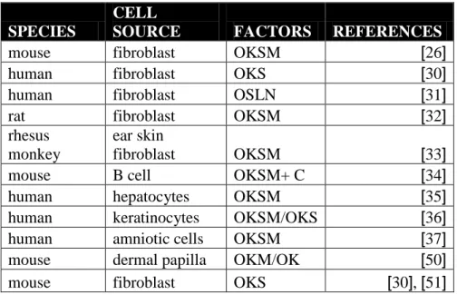
Table 1.1: IPS cells derived from different species and cell types by different factors. O: Oct4, S: Sox2, K:Klf4, M: cMyc, L: Lin28, C: C/EBPα
SPECIES
CELL
SOURCE FACTORS REFERENCES
mouse fibroblast OKSM [26]
human fibroblast OKS [30]
human fibroblast OSLN [31]
rat fibroblast OKSM [32]
rhesus monkey
ear skin
fibroblast OKSM [33]
mouse B cell OKSM+ C [34]
human hepatocytes OKSM [35]
human keratinocytes OKSM/OKS [36]
human amniotic cells OKSM [37]
mouse dermal papilla OKM/OK [50]
mouse fibroblast OKS [30], [51]
iPS cells are promising candidates to be used in therapy since they can be isolated from the patient himself and therefore immune rejection of the transplant is not an issue. In 2007 iPS cells generated from fibroblast of a sickle cell anemia murine model was used to cure the disease. Before the fibroblast were redifferentiated, the
8
genetic defect in the hemoglobin gene was treated in vitro. The engineered iPS cells were differentiated to hematopoietic progenitor cells and these cells were given to the donor mouse. It was observed that the mature erythrocytes had no defect in hemoglobin gene [52]. Another group used iPS cells to restore liver function in FAH deficient mice model, in which they had to have the drug, 2-(2-nitro-4-fluoromethylbenzoyl)-1,3-cyclohexanedione (NTBC), in order to prevent liver failure. After injection of the iPS cells to the blastocyst, the drug NTBC was withdrawn. It was observed that the iPS cells contributed to the formation of chimera and the mice had functional liver with FAH expressing hepatocytes [53]. Although iPS cells are promising candidates in cell therapy there is still a long way before this could happen. However, a more realistic approach of iPS cells in therapy is the use of disease specific iPS cells to mimic the disease and provide a patient spesific cell line to study the pathology in the culture. Several disease spesific iPSC have been established. A group of scientist generated a large number patient spesific iPS cell lines from patients with Adenosine deaminase deficiency-related severe combined immunodeficiency, Gaucher disease type III, Duchenne and Becker muscular dystrophy, Parkinson disease , Huntington disease , juvenile-onset, type 1 diabetes mellitus [54], spinal muscular atrophy [55], familial dysautonomia [56], and fanconi anemia [57]. Some of the iPS cell lines generated from this patients showed the disease profile in the culture and provided new opportunities to study the molecular mechanism of he disease and to search for drugs [58].
9
MULTIPOTENT ADULT STEM CELLS
1.2
Adult stem cells (ASC) are stem cells that are found in the adult organ or tissue of an organism. Unlike the ESCs or the iPSCs, ASCs are not pluripotent. Many of the ASCs are multipotent and a few are unipotent. ASCs can be thought of a reservoir of cells, which normaly stay in an undifferentiated state in the niche which is the environment where the ASCs reside. Like all stem cells ASCs possess two universal features; the ability to self renew and to differentiate [59], which is called stemness [60]. The maintenance of ASC pool is not clear, but the most accepted view is that ASCs divide in an assymetric fashion, giving rise to two different daughter cells with different fates. The one that is still in the niche becomes the self renewing sister for protecting the reservoir, while the other sister cell leaves the niche and differentiates into the desired cell type [61, 62]. Different ASCs have been identified. The first identified and so far the best charecterized ASCs are hematopoietic stem cells (HSCs). HSCs were first mentioned in the 60s by Till and McCulloch [63], and Siminovitch et al. [64]. These HSCs were able to reconstitude all of the blood cells [65, 66]. Two subsets of the HSCs have been classified; the Long term and short term reconstitutive cells (LT-HSC and ST-HSC, respectively) [65, 67]. In mouse in addition to these two subsets, there are also two other progenitor derived from the HSCs, Common Lyphocytes Progenitors (CLPs) and the Common Myeloid Progenitors (CMPs) [68, 69]. Different surface markers are used to separate these subtypes from each other [69].
10
Table 1.2: The location of different kinds of ASCs in the body and their differentiated progeny. (adopted from [70]).
CELL TYPE TISSUE SPECIFIC
LOCATION CELLS OR TISSUE PRODUCED
Hematopoietic stem cells
bone marrow, peripheral blood
bone marrow and blood lymphohematopoietic cells Mesenchymal
stem cells
bone marrow, peripheral blood
bone, cartilage, tendon, adipose tissue, muscle, marrow stroma, neural cells
Neural stem cells
ependymal cells,
subventricular zone of the central nervous system
neurons, astrocytes, oligodendrocytes Hepatic stem
cells
in or near the terminal bile ductules
oval cells that subsequently generate hepatocytes and ductular cells Pancreatic
stem cells
intraislet, nestin positive
cells, oval cells, duct cells beta cells
Skeletal-muscle stem cell/satellite cells
muscle fibers skeletal muscle fibers
stem cells of the skin
basal layer of the
epidermis, bulge zone of the hair follicles
epidermis, hair follicles
Epithelial stem cells of the lung
tracheal basal and mucus secreting cells,
bronchiolar Clara cells, alveolar type II
pneumocyte
mucous and ciliated cells, type I and II pneumocystis
Stem cells of the intestinal epithelium
epithelial cells located around the base of each crypt
paneths cells, brush border
enterocytes, mucus secreting goblet cells, enteroendocrine cells of the villi
Other than HSCs, intestinal stem cells, neuronal stem cells, hepatic stem cells, skin stem cell, mammary stem cells, pancreatic stem cells, and mesenchymal stem cells are other well characterized ASCs. These ASCs have the capacity to differentiate into different types of cells upon tissue damage or any other means of requirement
11
to maintain their homeostasis [70]. A list of the ASCs and their tissue spesific localization together with the spesific cell or tissue produced from are given in table 1.2.
MESENCHYMAL STEM CELL
1.2.1
MSCs) were first isolated from rat bone marrow and described in 1966 by Friedenstein et al [71] and further characterized by Pittenger et al. [72]. MSC contribution in the bone marrow is only 0.001% to 0.01%. These small number of cells can be isolated and cultured in vitro and induced to differentiate into bone, fat, cartilage, muscle, cardiomyocytes, neurons, endothelial cells and blood vessel as reviewed by Barry and Murphy [73]. Cell cycle analysis revealed that most of the MSCs stay at G0/G1 phase and do not enter the cell cycle [74]. There is variation on the proliferative capacity of MSCs in culture between the research groups. This difference could be due to the isolation procedure, source of MSCs, culture conditions and donor age [75]. It has been also shown that depending on the culture condition, MSCs retain their telomerase activity and karyotype in vitro [72]. However culturing these cells for long time can result in spontenous apoptosis [74] and senescence [75]. MSCs are promising tool for regenerative medicine due to easy harvesting and culturing, high differentiation potential and their nonimmunogenic nature. However, their rareness is a major roadblock. Therefore new strategies should be developed to increase the number of MSCs for further use without increasing their in vitro culturing time. Another important feature of MSCs
12
which makes it easy to work, is their diverse source in the body. Several groups successfully isolated MSCs from adipose tissue, periosteum, synovium, skeletal muscle, umblical cord blood, amniotic fluid, placenta, preipheral blood, lung, dental pulp, tendon and salivary gland [73, 75-77].
1.2.1.1 CHARACTERIZATION OF MSC
For the characterization of MSCs several aspects have been used. These include their morphology, their surface markers and capacity to differentiate into specific cells.
MSCs showed fibroblastic like appearance and formed colonies in petri dish. [78, 79]. The colony numbers has ben shown to vary in different organisms and in different culture conditions [77]. Although the colonies are derived from one precursor cell, studies have shown that the colonies may also contains heterogenous cell mixtures. Several groups have defined the cells in a colony as small spindle shaped cells which proliferate at high rate, large cuboidal shape cells which proliferate at a slower rate and a much smaller cell type which has the highest self renewing rate [80-82]. This variety in colonies makes it necessary to look for other characteristics for MSCs.
MSCs isolated from bone marrow are positive for the expression of CD44, CD105, CD106, CD90, CD166, CD29, CD73, CD117, CD146, CD157, STRO-1, Sca-1, integrins α1, α5 and β5, ICAM-1,ICAM-2, LFA-3 and L-selectin and negative for
13
CD11b, CD14, CD31, CD33, CD34, CD133 and CD45 [72, 74, 83-86]. However no unique marker has been identified yet to characterize MSC. Several studies have shown that a combination of some of the markers listed above are sufficient and necessary to determine MSCs [77]. The source of the MSC, the isolation procedure and the culture condition can effect the surface marker profile of the MSC [77]. Therefore the Mesenchymal and Tissue Stem Cell Committee of the International Society for Cellular Therapy proposed minimal criteria for MSC definition. According to this definition, MSCs must adhere to plastic surfaces, express CD105, CD73 and CD90 and not express CD45, CD34, CD14 or CD11b, CD79α or CD19 and HLA-DR; and differentiate to osteoblasts, adipocytes and chondroblasts in vitro [87].
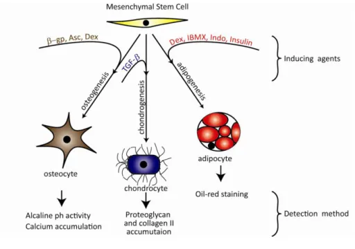
Besides differentiation into the mesenchymal lineage, MSCs have been also shown to differentiate to other cell types like cardiomyocytes, astrocytes, endothelial cells and epithelial cells such as retinal pigment cells, skin cells, sebaceous duct cells and epithelial cells in the kidney and to tenocytes [73, 88]. The differentiation procedure to osteoblasts, adipocytes and chondrocytes is well established [72, 73, 76, 77, 89-92]. Figure 1.1 summarizes the differentiation of MSCs to osteogenic, chondrogenic and adipogenic lineages, indicating the agents that are required to induce the differentiation and the detection methods to test the result.
14
Figure 1.1: The differentiation potential of MSCs into the mesenchymal lineage. Adapted from [77]. Dex= dexamethasone, IBMX= isobuthyl methyl xanthine, Indo= indomethacine, Asc= ascorbic acid, β-gp= beta glycerophosphate.
1.2.1.2 MESENCHYMAL STEM CELL HOMING
Upon damage or aging of somatic cells, stem cells leave the niche and migrate to the site of action and differentiate into the specific cell type of demand [72, 93, 94]. The homing capacity of MSCs decreases with long term culture [73, 77]. The homing mechanism of MSCs is not known for sure however the chemokine receptors on the surface of the MSCs are thought to be responsible for this mechanism. Several groups, although with a high variety among them, have identified different chemokine receptors on MSCs [95]. The presence of different chemokine receptors on the MSCs suggests a role for trafficking the MSCs through different paths to reach various tissues in the organism. Besides the chemokine
15
receptors, the adhesion molecules also play a role in MSC homing. P-selectin was shown to be involved in the rolling of intravenously administered human MSCs in the blood vessels. In P-selectin deficient mice, the administered MSCs were not able to roll along the walls of the blood vessels [96]. Since the MSCs do not express the known ligands for selectin, it was suggested that MSCs have a novel P-selectin ligand. The same study showed that VLA-4/VCAM-1 are required for hMSCs to adhere to endothelial cells [96]. Increased number of MSCs were in the peripheral blood after vascular injury in mice compared to noninjured ones [97]. Also the mobilization of MSCs to the blood after acute burns, skeletal muscle injury and in response to chronic hypoxia has been shown [95]. Besides mobilization to the peripheral blood, MSCs also migrate from blood to the damaged tissue, which was shown by different in vivo studies [98, 99]. Studying the molecular mechanism of the entire process of MSC homing, may provide new knowledge to develop more effective strategies in the field of regenerative medicine. Exploring the mechanism of stem cell homing could help in reducing the needs for high number of MSCs, by developing new methods for the enhanced recruitment of ex-vivo cultured MSCs to the injured tissue [100].
It has been shown that there are two types of homing; long-distance traffic which is via the blood stream and short- distance homing which is a local movement [75]. The short distance homing of MSCs to an injured site was shown in different cases like cartilage, muscle and heart injury, bone repair, migration throughout forebrain
16
and cerebellum [75]. MSC when injected through the intravenous infusion was capable of migrating to a specific site of injury like in the case of bone fracture, cerebral ischemia and myocardial infarction [77].
Besides all the research being done on MSC homing there are typical problems faced by the researchers. One problem regarding the MSC research in general is the lack of a universal definition, thus the heterogeneity of MSCs used in different studies. To overcome this, a particular MSC characterization should be performed. Since there aren’t any standart protocols for determining the homing efficiency and correlation, with functional effect, a direct comparison based on the outcome of the researches can not be done. A recommended action for this problem is that every paper should compare or contrast their results with previously published results. This comparison should include the number of cells that were administered, the infusion site, the time passed between MSC administration and assessment of homing, methods used to determine homing, homing efficieny and functional outcome of the experiment. Another problem that the researchers face with is the difficulty to determine the cell type that actualy has homed. The individual MSCs should be labelled in culture and tracked after administration to the organism [100]. To get a more accurate result in MSC homing research these problems should be solved and standart methods should be developed to compare different results. As the whole protocol of the experiments can effect the functional outcome every step should be explained clearly. MSCs are promising agents in stem cell therapy,
17
however their rareness and the high number of MSC needed for infusion makes it difficult to use them. After clearifiying the MSC homing mechanism, the MSCs could be used more efficiently in regenerative medicine. Direct targeting of the MSCs to the site of action could eliminate the demand for high number of cells to be administered.
1.2.1.3 MESENCHYMAL STEM CELLS IN REGENERATIVE MEDICINE The discovery of stem cells opened a new era in the field of therapy. Currently there are over 3000 clinical trials involving stem cells and 174 of them are with MSCs (http://www.clinicaltrials.gov). The beneficial effects of MSCs in the treatment of different cases are via different properties of the MSCs. MSCs can support the survival of endogenous cells [101, 102], induce angiogenesis [103], inhibit immune response [104], reduce apoptosis [105] , home to the site of injury, directly differentiate to the cell of the tissue, and release some chemicals which act on the near by cells to stimulate them [106].
Two features of MSCs made them a popular candidate in stem cell therapy. One is the ability to differentiate into various types of cells as mentioned above and the other is their immunosuppressive nature. It has been shown that MSC prevent T-cell activation either by direct contact or via secreted molecules [95]. Other studies have also shown that MSCs can hinder dendritic cell and B-cell maturation [95].
18
The immunosuppressive nature of MSCs was used in a case study in which the patient developed Graft versus Host Disease (GvHD) after HSC transplantation. MSC treatment in this patient resulted in the healing of the gut epithelium tissue and the patient was in a good condition after 1 year of follow up [107]. MSCs have also been used in ischemic heart diseases, pancreatic regenaration, neurological disorders, hepatic cirrhosis, limb ischemia, skin regenaration, rheumatoid arthritis and bone related diseases [106]. The ability of MSC to differentiate into cardiomyocytes made them a candidate for the treatment of heart failures. After bone marrow transplantion bone marrow derived cardiomyocytes were shown to be engrafted into the heart of the patient [108]. In another study it was shown that bone marrow derived cells delivered to the infarcted heart and resulted in a dramatic increase in the heart function [109]. MSCs are also prominent candidates for the treatment of muscular dystrophy [110], and environmental induced lung disease [111], Since MSCs are able to promote remyelineation [112], and to differentiate into neurons after transplantation [113, 114], and partially recover their function [115] they can be used in neuronal injuries. Clinical trials for osteogenesis imperfecta have shown an improve in the bone formation after MSC transplantation. The bone mineral content and the growth velocity increased, whereas the bone fracture frequencies reduced [116]. Treatment of severe idiopathic aplastic anaemia with MSCs showed improvement of marrow stromal function [117].
19
Besides being a promising tool as regenerative agents, there are some handicaps in the therapeutic application of MSCs. Although clinical trials show promising results, long term effect of MSC transplantation is not known. Most of the knowledge of MSCs come from in vitro studies. However how these cells act in
vivo is not very clear. What is the safe duration of transplanted MSCs, in other
words for how long do they reside in the organism and function properly [106]? The amount of MSCs needed for regenerative medicine is high. How can we increase the number of MSCs without increasing the culture time? Short preparation times are important for urgent cell based therapies. Lastly, sufficient MSCs should reach the site of action after infusion to the organism in short time periods. These problems must be solved before using MSCs in routine therapeutic applications. The proliferative and antiapoptotic effects of estrogen makes it a promising candidate in solving the last two problems.
ESTROGEN
1.3
Estrogen is one of the steroid hormone that is produced in the ovaries from cholesterol. 17β-estradiol, estrone and estriol are found in humans and 17β-estradiol is the most abundant one among them [118]. It does not only have an effect on the reproductive system but also on many other organs and systems like, brain, bone and organs of the cardiovascular system [119, 120] in female and male. Depending on the cell type, estrogen can have a proliferative, antiapoptotic or apoptotic effect
20
by changing the activity of cell cycle related or cell death related genes [121-125]. After menapouse, when estrogen levels decrease, increase in osteoporosis and coronary heart diseases occur [126, 127]. This suggests a role for estrogen in MSC biology. Determining the effects of estrogen on MSCs can provide insights for development of new strategies in MSC based therapies.
Estrogen exert its effects via the estrogen receptors alpha or beta (ERα or ERβ). These receptors are members of the nuclear receptor superfamily which are ligand induceable transcription factors [128].
ESTROGEN RECEPTOR PATHWAY
1.3.1
Two types of pathways are known so far that are involved in the action of estrogen. The genomic or classical pathway and the non genomic pathway. In the classical pathway, the hormone estrogen activates estrogen receptor (ER) in the cytosol and the receptor forms homodimer. Activated ER is translocated to the nucleus and binds to the estrogen response element (ERE) located mostly at the promoter region of the target gene. Subsequently, coactivators are also recruited to the receptor– DNA complex, which results in the expression of the target gene [120, 129]. Estrogen induced genes do not always contain EREs yet they are activated by ERs.
The non ERE dependent genomic pathway involves the action of other transcription factors which bind both to the ER-ligand complex and to the target gene promoter. In this way ligand activated ER activates the transcription factor and the target gene
21
is expressed. It has been shown in various studies that ER enhance the activity of several genes that do not have an ERE but contains AP-1 site, CRE-like element, electrophile response element, GC-rich promoter site. Several binding partners of ER on such non-ERE sites such as Jun/Fos, Jun/ATF, ATF, SP-1, NFκB, C/EBPβ are known but there are also unknown ones [118, 129].
A third form of genomic pathway involves growth factors instead of estrogen and is called as ligand independent genomic pathway. In this model the nuclear ERs are activated via phosphorylation by protein kinases which then become active upon growth factor stimulation. The activated ERs bind to ERE sites to start the expression of the target genes.
The second type of ER signaling is the nongenomic pathway. In this pathway estrogen activates the membrane bound ER and this starts a cascade of kinase signaling pathways such as MAPK, PI3/AKT, JAK/STAT pathway [130]. The nongenomic pathway does not involve transcription or translation but translational modification for the activity, thus it results in a very rapid cell type spesific response [118]. Figure 1.2 summarizes the ER signaling pathway.
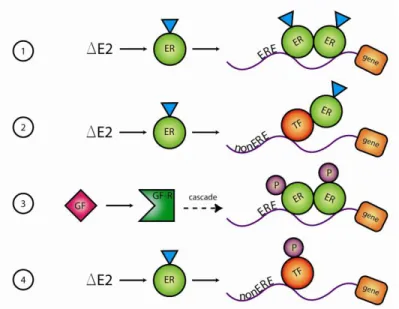
22
Figure 1.2: The ER signaling. 1. ligand dependent genomic pathway, 2. non-ERE dependent genomic pathway, 3. ligand independent genomic pathway and, 4. non genomic pathway
ESTROGEN AND MESENCHYMAL STEM CELL
S
1.3.2
Gender specific responses to various stimuli by MSCs have been attributed to sex hormones. For example MSCs from female donor express less pro-inflammatory cytokines, TNF-α and IL-6, compared with MSCs from male donor in response to acute LPS and hypoxia [131]. Also the hypoxia induced apoptosis was different in male MSC compared to female ones. The MSCs from male donor underwent a significantly higher apoptosis compared to the female MSCs [132]. Another difference between female and male MSC was in the treatment of heart ischemia. After transplantation of MSCs from female donors, heart exhibited improved contractility compared to MSCs from male donors [133]. Estrogen also have an
23
effcet on the proliferation of MSC in a gender dependent fashion. MSCs from female donors shows an increase in proliferation upon estrogen treatment, while MSC from male donors do not [134]. Another relationship between estrogen and MSC is that estrogen increases the telomerase activity and hTERT expression via ERα [135]. Estrogen has also an effect on the differentiation and proliferation of MSCs. It stimulates the osteogenic differentiation [134] which can be assessed by the increase of calcium deposition and the expression of the osteogenic markers. Estrogen also increases the adipogenic differentiation evidenced by the increase in the lipid droplets [136]. However, the molecular mechanism of estrogen action on MSCs is not known. Understanding the molecular mechanism of their interaction is important in regulating MSCs and obtaining the maximum efficiency from this colloboration. Our group has previously shown that estrogen had no effect on genes related to cell cycle progression and senescence on rat bone marrow derived MSCs (Scientific and Technological Research Council of Turkey, TUBITAK, SBAG-3200(105S393)). Since estrogen also has an effect on apoptosis, apoptotic pathways are important candidates in the regulation of MSCs. After explaining apoptosis, the relation between estrogen and apoptosis will be illustrated with examples.
24
CELL DEATH AND APOPTOSIS
1.4
Cell death can occur through two distinct pathways called necrosis and apoptosis.
Until recently necrosis was described as cell death which is not apoptosis or autophagy. However, latest research findings have provided more clear explanation for necrosis. Defined signaling pathways and catabolic mechanisms were found to have crucial role in necrotic cell death [137, 138]. Increased intracellular Ca2+, or Reactive oxygen species (ROS), decreased ATP generation [139], tumor necrosis factor- α (TNF-α) [140], poly-ADP-ribose polymerase-1 (PARP-1) [141] were shown to be important mediators of necrosis.
Apoptosis was first described by Kerr and friends upon typical morphological changes that occur in the dying cell [142]. According to the Nomenclature Committee on Cell Death (NCCD) cell death can be defined as apoptosis when it possesses all or some of the following characteristics. i) rounding-up and shrinkage of the cell, ii) retraction of pseudopods, iii) condensation of the chromatin, iv) nuclear disruption, v) preservation of the organelles, vi) apoptotic body formation while maintaining the membrane integrity and vii) phagocytosis by neighboring cells. The NCCD proposes that, programmed cell death (PCD) should not be used to exclusively define apoptosis [143] since cell death that occurs during development is programmed but not apoptotic [144, 145]. One should also not think of apoptosis merely as caspase dependent process [146]. It can also occur in a
25
caspase independent manner, although in some cases the outcome has a slight variation in the cytological features as compared to known apoptotic structures [146].
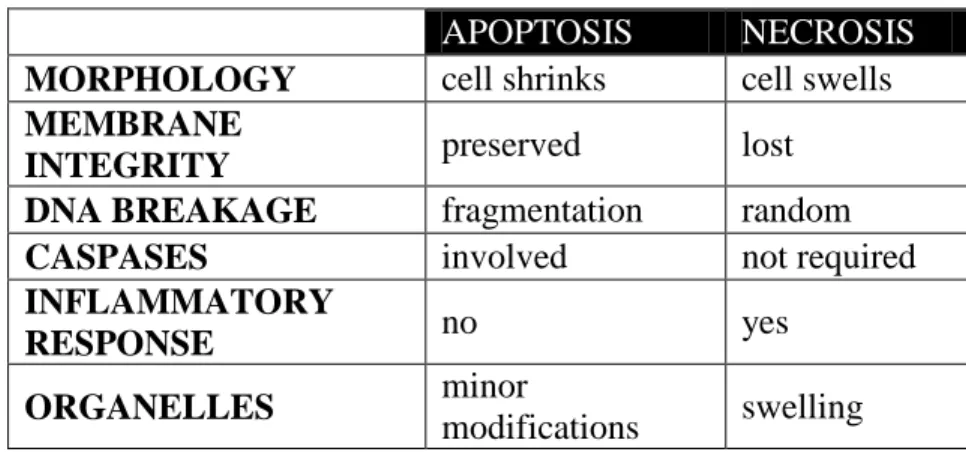
The molecular events that occur during necrosis and apoptosis are different as is the morphology (Table 1.3). While cells and the cytoplasmic organelles swell during necrosis, apoptotic cells shrink and the organelles undergo a minor change. Necrotic cell membrane ruptures, but the apoptotic cell membranes integrity is preserved. DNA breakage is random and caspases are not involved in necrosis but, DNA fragmentation occurs and caspases are required in apoptosis. Also necrosis leads to an inflammatory response but apoptosis does not [143, 147].
Table 1.3: Characteristics of apoptosis and necrosis
APOPTOSIS NECROSIS
MORPHOLOGY cell shrinks cell swells
MEMBRANE
INTEGRITY preserved lost
DNA BREAKAGE fragmentation random
CASPASES involved not required
INFLAMMATORY
RESPONSE no yes
ORGANELLES minor
modifications swelling
EXTRINSIC AND INTRINSIC PATHWAY
1.4.1
Well defined signaling pathways have roles in the apoptotic cell death. These pathways can be divided into two: the extrinsic (death receptor) and the intrinsic (mitochondrial) pathway [148]. Figure 1.3 represents the extrinsic and intrinsic
26
apoptotic pathways. After explaining the pathways, and the role of mitochondria in apoptosis, special focus will be given to the Bcl-2 family members and their function in apoptosis. The relation between estrogen and apoptosis will be explained with known examples.
The extrinsic pathway is activated when death ligands, TNF-α, TRAIL and FasL bind to their cognate receptors in the plasma membrane. This binding induces receptor oligomerization and recruitment of an adaptor protein. These adaptor proteins are Fas associated death domain (FADD) and TNF associated death domain (TRADD). After the joining of pro-caspase-8 or pro-caspase-10 the complex death inducing signaling complex (DISC) is formed. The initiator caspase is activated via proteolytic cleavage. This activation triggers the effector caspases, caspase-3 or caspase-7 and apoptosis occurs [149, 150].
The intrinsic pathway occurs mainly through the mitochondria. Various stimuli can trigger the mitochondrial pathway. Hypoxia, growth factor deprivation, reactive oxygen species, DNA damage, UV/ gamma irradiation are some of the apoptotic signals activating the intrinsic pathway. Mitochondria are crucial for cell survival due to their vital role in many metabolic activities [151]. Recent studies have established important roles for mitochondria during different stages of the cell death process. They are involved in energy generation for the cell death process, calcium homeostasis, ROS generation, pro-apoptotic proteins releasing, apoptosome formation and caspase activation [151].
27
Figure 1.3: The extrinsic and intrinsic apoptotic pathways. [149]
Normally mitochondria have a transmembrane potential which creates an electrochemical gradient across the inner membrane that is essential for ATP
28
generation. The disruption of this transmembrane potential due to the permeabilization of the mitochondrial membranes results in cell death [151, 152]. Mitochondrial membrane permeabilization can occur through lipids [153-155], mitochondrial permeability transition pore (MPTP) [156, 157], or Bcl-2 family of proteins. The induction of the mitochondrial membrane permeabilization results in the release of apoptotic factors from the mitochondria to the cytosol or nucleus and initiates apoptosis.
The pro-apoptotic members, BCL2-antagonist/killer (Bak) and Bcl2-associated X protein (Bax) of the Bcl-2 family of proteins permeabilize the outer membrane of the mitochondria, which results in the release of pro-apoptotic proteins such as cytochrome c (Cyt c), apoptosis inducing factor (AIF) and endonuclease G from the mitochondria to the cytosol. While AIF and endonuclease G goes to the nucleus and induce DNA fragmentation [158, 159] in a caspase independent form [159, 160], Cyt c stays in the cytoplasm. In the cytosol, Cyt c together with apoptotic protease-activating factor (Apaf-1), dATP and pro-caspase-9 forms the apoptosome complex. After proteolytic cleavage of pro-caspase-9 in the apoptosome, it is activated and can activate the effector caspase, caspase-3 [161].
BCL-2 FAMILY MEMBERS
1.4.2
According to their function, Bcl-2 family of proteins is divided into two types, anti-apoptotic and pro-anti-apoptotic members. The pro-anti-apoptotic ones are further categorized as the multi-domain proteins and the BH3 only proteins due to their
29
structural and functional difference. More than 25 members of this family are known [162, 163]. (Figure 1.4)
All of the members share one or more conserved Bcl-2 homology (BH) domains. The four BH domains which are specific to the family members are BH1, BH2, BH3, and BH4 [164]. These domains are required for the function of the proteins [165-168]. Many of the Bcl-2 family proteins also have a carboxy terminal hydrophobic domain. The localization of the members to the mitochondria is directed through this domain [162, 169, 170].
Figure 1.4: The structure of the Bcl-2 family members. BH= Bcl-2 homology domain and TM= transmembrane domain.[170]
Among anti-apoptotic members of the family,B cell lymphoma like X (Bcl-xL) and
30
generally integrated mostly to the mitochondrial outer membrane but also can be found in the endoplasmic reticulum membrane or the cytosol [171, 172]. Bcl-2 prevents cell death by inhibiting free radical production, suppressing caspase activation, regulating calcium sequestration, and by preventing the pro-apoptotic factors from inducing apoptosis [173]. Bcl-2 is widely expressed during embryogenesis but it decreases and becomes much more restricted postnatally in the nervous system [174]. Bcl-2 and Bcl-xL prevent apoptosis either by segmenting
caspases or by preventing the release of mitochondrial apoptogenic factors such as cytochrome c and AIF into the cytoplasm [175].
The multi domain pro-apoptotic members have the BH1-3 in common and lack the BH4 domain. The localizations of the proteins are different in that in healthy cells, Bak is in the outer mitochondrial membrane whereas Bax is mostly in the cytosol and travels to the mitochondria upon apoptotic stimuli [176-179]. The presence of either Bax or Bak, which are thought to function mainly at the mitochondria, seem to be crucial for apoptosis in many cell types since most death stimuli converge on their activation [180, 181]. Bax heterodimerizes with Bcl-2 and homodimerizes with itself. When Bax is overexpressed, apoptotic death in response to a death signal accelerates, but when Bcl-2 is overexpressed, it heterodimerizes with Bax and apoptosis is repressed [182]. Therefore the ratio of Bax and Bcl-2 is important in regulating apoptosis.
31
The BH3 only proteins possess only the conserved BH3 domain. The BH3 only proteins are known to function in two distinct mechanisms. Some, like Bid, can either directly activate the multidomain pro-apoptotic members while others, like Bad and NOXA, inactivate the anti-apoptotic members [183, 184].
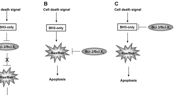
The interaction between the anti-apoptotic, pro-apoptotic and BH3 only proteins determines apoptosis [171]. Several distinct models have been proposed to explain the relationship between the anti-apoptotic, the pro-apoptotic and the BH3 only proteins during regulating apoptosis (Figure 1.5). In the first model the pro-apoptotic members are sequestered and hold in an inactive state, by the anti-apoptotic ones when there is no need for apoptosis. Upon anti-apoptotic stimulus the BH3 only proteins bind to the anti-apoptotic members and set free the pro-apoptotic ones. This release results in activation of the pro-apoptotic members and subsequently apoptosis. In the second proposed model again the anti-apoptotic ones prevent the apoptotic ones from inducing apoptosis, however this time the pro-apoptotic members are activated by the BH3 only members via direct interaction between these two members. In the third model the anti-apoptotic members hinder the BH3 only members to activate the pro-apoptotic ones. Upon apoptotic stimuli the BH3 only proteins overcome the hurdle and activate the pro-apoptotic members [170].
32
Figure 1.5: Regulation of Bcl-2 proteins. A)BH3-only proteins sequester anti-apoptotic proteins and allow the pro-anti-apoptotic ones to initiate apoptosis. B) BH3-only proteins directly activate pro-apoptotic members. C)the anti-apoptotic proteins sequester the BH3-only proteins and thereby preventing apoptosis. [170]
1.4.2.1 SIGNALING PATHWAY
After activating the intrinsic pathway through various stimuli like growth factor deprivation or DNA damage, the signaling pathway starts. The first reaction involves the activation of the multi-domain pro-apoptotic proteins. This activation is mainly at post translational level where the inactive protein becomes activated through conformational changes. Upon apoptotic stimuli, the C-terminal and N-terminal domains of Bax undergo a conformational modification, which results in the translocation of the protein from the cytosol to the mitochondrial outer membrane where it forms oligomers [176, 185, 186]. Since Bak is already in the mitochondrial outer membrane, there is no need for its translocation. However,
33
studies have shown that also at the N-terminus of Bak a conformational alteration occurs after an apoptotic stimulus [187] suggesting that there are other consequences of the modification than translocating to the mitochondria. Pro-apoptotic proteins, Bak and Bax form channels on the outer mitochondrial membrane by homo- or hetero-oligomerization [151]. These channels cause the release of the pro-apoptotic activators from the mitochondria to the cytosol. There are also other models to explain the role of Bak and Bax oligomers on inducing apoptosis. One of these models is that, Bak and Bax cause a change in the lipid bilayer of the outer mitochondrial membrane. This change facilitates the release of the apoptotic factors like Cyt c to translocate to the cytosol [188, 189]. Other mechanism by which Cyt c is released includes the interaction of Bax with the mitochondrial protein voltage dependent anion channel (VDAC) [190] but the detailed function of VDAC in this process is not known [152]. Once in the cytosol, Cyt C, binds to APAF-1. As discussed above Cyt C, APAF-1 and dATP forms a complex called the apoptosome. In this complex APAF-1 undergoes a conformational change and as a result of this, its caspase recruitment domain (CARD) becomes exposed. The pro-caspase 9 is recruited to the apoptosome and proteolytic cleavage occurs to activate the caspase. The active caspase activates the effector caspases, caspase 3 or 7, which leads to apoptosis [161, 191]. Other mitochondrial proteins which are involved in this caspase dependent cell death pathway, is Smac/DIABLO and HtrA2/Omi. Both proteins bind and sequester the
![Figure 1.3: The extrinsic and intrinsic apoptotic pathways. [149]](https://thumb-eu.123doks.com/thumbv2/9libnet/6002136.126314/46.918.222.761.187.886/figure-extrinsic-intrinsic-apoptotic-pathways.webp)
![Figure 1.4: The structure of the Bcl-2 family members. BH= Bcl-2 homology domain and TM= transmembrane domain.[170]](https://thumb-eu.123doks.com/thumbv2/9libnet/6002136.126314/48.918.251.735.546.883/figure-structure-family-members-homology-domain-transmembrane-domain.webp)
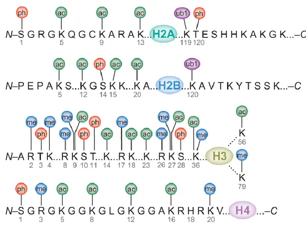
![Table 1.4: The regulatory roles of several histone marks. (Adopted from [218])](https://thumb-eu.123doks.com/thumbv2/9libnet/6002136.126314/58.918.177.806.225.484/table-regulatory-roles-histone-marks-adopted.webp)
![Table 1.5: Demethylases and their substrates. (Adopted from [217]).](https://thumb-eu.123doks.com/thumbv2/9libnet/6002136.126314/60.918.211.765.261.516/table-demethylases-substrates-adopted.webp)
