Ankara Üniversitesi Tıp Fakültesi Mecmuası 2008, 61(4)
CERRAHİ BİLİMLER / SURGICAL SCIENCES Olgu Sunumu / Case Report
Subdural Hematoma Associated With An Arachnoid Cyst After
Head Trauma: A Case Report
Kafa Travması Sonrası Gelişen Araknoid Kist ile İlişkili Subdural Hematom: Olgu Sunumu
Ayşe Karataş, Mehmet Akgül, Feyzullah Akyüz, Osman Akgül, Murat Döşoğlu
Düzce Üniversitesi Tıp Fakültesi, Nöroşirürji Anabilim Dalı
220
Received: 28.07.2008 • Accepted: 16.10.2008
Corresponding author
Yrd. Doç. Dr. Ayşe Karataş
Düzce Üniversitesi Tıp Fakültesi, Nöroşirürji Anabilim Dalı Phone : (533) 48 044 12
E-mail address : [email protected]
Arachnoid cysts are collections of cerebrospinal fl uid within the arachnoid membrane. They are benign, congenital lesions, and are usually asymptomatic. Rarely, they may become acutely symptomatic, due to cyst enlargement, subdural or the presence of the intracystic hemorrhage. Intracranial arachnoid cyst associated with subdural hematoma is an uncommon after minor head trauma or spontaneous. The damage to the bridging veins within the cyst, or on the wall of the cyst, is thought to be a source of possible bleeding. We report here the case of a 29-year-old man who, after a minor head injury, developed subacute subdural hematoma associated with an underlying arachnoid cyst. It is important to keep in mind that arachnoid cysts can be associated with subdural hematomas.
Key Words: arachnoid cyst, subdural hematoma, intracystic hemorrhage
Araknoid kistler, araknoid membran içinde gelişen beyin omurilik sıvısı içeren birikimlerdir. Bunlar, doğumsal, selim seyirli lezyonlardır ve genellikle asemptomatiktirler. Nadiren, kistin genişlemesi, subdural veya kist içerisine kanama nedeni ile akut olarak semptomatik hale gelebilirler. Araknoid kistin spontan veya minor kafa travma sonrası kanaması ile gelişen subdural hematom olguları nadirdir. Kist duvarı veya içerisindeki köprü venlerinin hasarlanması muhtamel kanama kaynağı olarak düşünülmektedir. Bu yazıda, 29 yaşında erkek hastada minor kafa travmasından sonra ge-lişen altta yatan araknoid kist ile ilişkili subakut subdural hematom olgusu bildirilmiştir. Araknoid kistlerin subdural hematomlarla ilişkili olabilirliğinin akılda tutulması önemlidir.
Anahtar Kelimeler: Araknoid kist, subdural hematom, intrakistik kanama
Intracranial arachnoid cysts are cer-ebrospinal fluid-filled collections between the arachnoid layers, which are benign developmental anomalies accounting for 1% of all atraumatic intracranial mass le-sions (1). They are located in the middle cranial fossa in 25-80% of cases. Almost all of these cysts are unilateral, with a slight predilec-tion for the left side and the male gender (1,2). They are often di-agnosed in childhood as inciden-tal findings in imaging. They usu-ally remain fairly stable in size. However, 60-80% of cases ulti-mately become symptomatic (3). Seizures, signs of raised intracra-nial pressure, neurological
defi-cits, and, in children, macrocra-nia and developmental retarda-tion are the main symptoms (2). However, very rarely they can be symptomatic due to hemorrhage of the cyst. Subdural hematomas are infrequent complications of arachnoid cysts, with or without preceding trauma (2). This report describes a patient with left mid-dle fossa arachnoid cyst that pre-sented as a subacute subdural he-matoma after minor closed head injury.
Journal of Ankara University Faculty of Medicine 2008, 61(4)
221 Ayşe Karataş, Mehmet Akgül, Feyzullah Akyüz, Osman Akgül, Murat Döşoğlu
Case Report
A 29-year-old man was admitted to our emergency department with a severe headache and nausea. Results of a neurological exami-nation were normal. His clinical history revealed a fall from a bi-cycle 2 months earlier. The pa-tient had visited a private hospi-tal 4 weeks after the initial injury, complaining of headache. A cra-nial computed tomography (CT) scan showed a left middle cranial fossa hypodense lesion in the re-gion of the sylvian fissure, which was a type II arachnoid cyst, ac-cording to the Galassi classifica-tion, and thinning of the overly-ing calvaria at that time (Figure 1). The cyst was considered asympto-matic and the patient was given analgesic drugs.
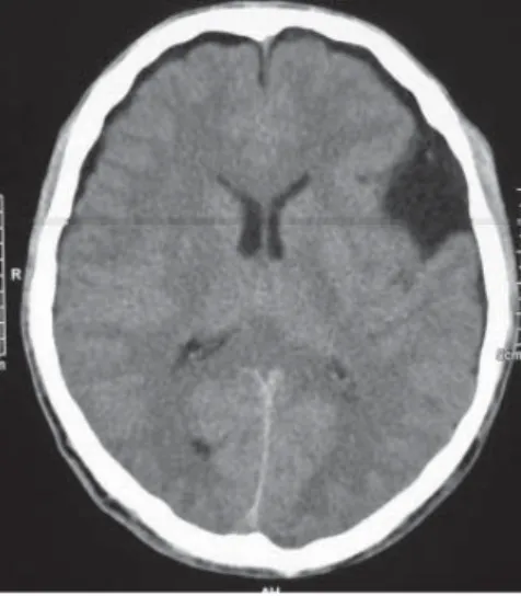
However, the patient’s headache worsened and he came to our hos-pital. A cranial CT scan revealed a left frontoparietal isodence su-bacute subdural hematoma, with a maximum thickness of 1.4 cm, compressing the left lateral ventri-cle, a 1.1-cm midline shift, and left hemispheric edema (Figure 2). The patient underwent a
two-burr-hole surgery, on the frontal and parietal bones. The subacute sub-dural hematoma was confirmed and evacuated. Postoperatively, the patient reported headache relief. A control cranial CT study after evacuation of the hemato-ma revealed an arachnoid cyst in the middle cranial fossa. The pa-tient was asymptomatic and cra-nial magnetic resonance imaging (MRI) revealed that the arachnoid cyst was the same size 5 months later.
Discussion
The natural history of arachnoid cysts is still not well defined. While many are silent, in some cases they become symptomatic if there is acute cyst enlargement, subdural effusions after rupture of the cyst, or subdural or intra cystic hemorrhage. Hemorrhage into or around an arachnoid cyst is primarily precipitated by minor trauma and, very rarely, can be spontaneous. Several reports have been published on the association of arachnoid cysts with subdural hygroma and subdural hematoma after minor head injuries. There
is no clear explanation for the pathogenesis. However, damage to the bridging veins within the cyst, or on the wall of the cyst, is thought to be a source of possible bleeding (2). Additionally, the cyst can be ruptured, then returned to a hygroma, and transformed into a hematoma. Intra-cystic or sub-dural hemorrhage can mask the presence of an arachnoid cyst and can make it difficult to distinguish an arachnoid cyst on CT. The su-bacute intracytic or subdural he-matoma is typically isodence on CT. MRI is useful in such cases. The management of arachnoid cysts
is still controversial; it includes both conservative non-operative and operative treatments. Opera-tive treatments include cyst sur-gery (fenestrating, resection or extirpation of cyst membranes), cyst shunting, or combinations of the two methods (2,3). Asympto-matic arachnoid cysts do not re-quire treatment and no evidence exists to suggest that operative treatment prevents bleeding (2). The annual hemorrhage risk for chronic subdural hemorrhage in patients with a middle cranial fos-sa cyst remains about 0.04% (2). In our case, the cyst was
asympto-matic until the injury, and the patient, were treated by irrigat-ing the hematoma through burr holes; this was because his symp-toms were considered to be pri-marily due to the increased intrac-ranial pressure caused by the sub-dural hematoma. The patient’s symptoms resolved immediately after the operation; thus, no cyst surgery was performed. The pa-tient was followed up using mag-netic resonance imaging. No sub-dural hematoma recurred dur-ing the postoperative period of 5 months, and the arachnoid cyst was not enlarged.
Figure 1. On an initial computed tomography
image, the arachnoid cyst showed a well-defined cystic mass, 3.2×4 cm, in the left middle fossa.
Figure 2. On a subsequent CT image, a left
frontoparietal isodence subdural hematoma was seen, 1.4 cm thick, as was a 1.1-cm midline shift and left hemispheric edema.
Ankara Üniversitesi Tıp Fakültesi Mecmuası 2008, 61(4)
222 Subdural Hematoma Associated With An Arachnoid Cyst After Head Trauma: A Case Report
Arachnoid cysts of the middle cra-nial fossa, revealed with subdur-al hematomas, can be operated on in two ways: first, the subdur-al hematoma should be evacuated as an emergency treatment, as in our case, and delayed cyst surgery should be performed if the cyst
has become symptomatic on fol-low up (2). However, if a craniot-omy is performed to evacuate the space-occupying collection, cyst surgery can be done during such a procedure.
Conclusion
It is important to keep in mind that arachnoid cysts can be associat-ed with subdural hematomas. We recommend irrigation of the sub-dural hematoma as the initial pro-cedure of choice in such cases. REFERENCES
1. Iaconetta G, Esposito M, Maiuri F, et al. Arachnoid cyst with intracystic haemorrhage and subdural haema-toma: case report and literature revi-ew. Neurol Sci 2006; 26: 451-455.
2. Parsch CS, Krau J, Hofmann E, et al. Arachnoid cysts associated with subdural hematomas and hygro-mas: Analysis of 16 cases, long-term follow-up, and review of the literatu-re. Neurosurgery 1997; 40: 483-490.
3. Donaldson JW, Edwards-Brown M, Luerssen TG. Arachnoid cyst rupture with concurrent subdural hygroma. Pediatric Neurosurg 2000; 32: 137-139.