Yazışma Adresi/Address for Correspondence: Dr. Alev Arslan, Başkent University Faculty of Medicine, Adana Dr. Turgut Noyan Training and Research Center, Division of Pediatric Cardiology, Adana, Turkey E-mail: [email protected] , [email protected]
Geliş tarihi/Received: 05.01.2018 Kabul tarihi/Accepted: 29.03.2018
ARAŞTIRMA / RESEARCH
Cardiac effects of hemodialysis in children with chronic kidney disease
Kronik böbrek yetersizliği olan çocuklarda hemodiyalizin kardiyak etkileri
Alev Arslan
1, Osman Küçükosmanoğlu
1, Aytül Noyan
2, Nazan Özbarlas
11Çukurova University Faculty of Medicine Department of Pediatrics, Divison of Pediatric Cardiology, Adana, Turkey 2Başkent University Faculty of Medicine, Dr.Turgut Noyan Training and Research Center, Department of Pediatrics, Divison of Pediatric Nephrology, Adana, Turkey
Cukurova Medical Journal 2018;43(4):1015-1022
Abstract Öz
Purpose: The aim of the study was to evaluate cardiac
functions before and after hemodialysis in children with chronical renal failure.
Materials and Methods: The study comprised 24
children undergoing hemodialysis more then six months. Conventional and tissue Doppler echocardiography was performed, NT-proBNP levels were measured before and after hemodialysis. Echocardiographic results were compared regard to hydration status which measured with body composition monitor and hypotension event during dialysis.
Results: Myocardial performance indices which gained
with tissue Doppler echo were high in 22 patient. Mitral E/A ratio significantly decreased after hemodialysis. NT-proBNP levels were high but no difference was determined after dialysis. Nine patient experienced hypotension but nonsignificant difference was detected regarding echocardiographic findings and NT-proBNP levels.
Conclusion: Diastolic dysfunction is frequent in chronical
renal failure. Tissue Doppler echocardiography and myocardial performance index supply a more proper evaluation of global and regional cardiac function in chronical renal failure due to less hydration affectability. NT-proBNP should be monitored for cardiac dysfunction in chronical renal failure. Body composition monitor should be used in children for setting the ultrafiltration volume.
Amaç: Çalışmamızda kronik böbrek yetersizliği nedeniyle
hemodiyaliz yapılan hastalarda hemodiyaliz öncesi ve sonrası kardiyak fonksiyonların değerlendirilmesi planlanmıştır.
Gereç ve Yöntem: Çalışmaya altı aydan daha uzun süredir
düzenli hemodiyaliz yapılan 24 hasta dahil edildi. Diyaliz öncesi ve sonrası konvansiyonel ve doku Doppler ekokardiyografi uygulandı, NT-proBNP düzeyleri kontrol edildi. Ekokardiyografi sonuçları, vücut komposizyon monitörü ile ölçülen hidrasyon durumuna gore ve diyaliz sırasında hipotansiyon gelişmesine gore karşılaştırıldı.
Bulgular:. Doku Doppler ekokardiyografi ile hesaplanan
miyokardiyal performans indeksi 22 hastada normalden yüksekti. Mitral E/A oranları hemodiyalizden sonra istatistiksel olarak daha düşüktü. NT-proBNP düzeyleri normalden yüksekti ancak diyaliz öncesi ve sonrası düzeyleri arasında fark bulunmadı. Dokuz hastada diyaliz sırasında hipotansiyon gelişti. Hipotansiyon gelişen hastaların ekokardiyografik bulguları ve NT-proBNP düzeyleri arasında, hipotansiyon gelişmeyen hastaların sonuçları ile karşılaştırıldığında anlamlı fark saptanmadı.
Sonuç: Kronik böbrek yetersizliğinde kalpte diyastolik
disfonksiyon sıktır. Hidrasyon durumundan daha az etkilendikleri doku Doppler ekokardiyografi ve miyokardiyal performans indeks ölçümleri ile bütünsel ve bölgesel kalp fonksiyonları daha doğru değerlendirilir. Kronik böbrek yetersizliğinde NT-proBNP ile kalpteki fonksiyon bozukluğu takip edilebilir. Çocuklarda ultrafiltrasyon sıvı hacmini belirlemek için vücut kompozisyon monitörü kullanılabilir.
Key words: Body composition monitor, chronic kidney
disease, myocardial performance index, tissue Doppler echocardiography
Anahtar kelimeler: Vücut kompozisyon monitörü, kronik
böbrek yetersizliği, miyokardiyal performans indeksi, doku Doppler ekokartdiyografi
1016
INTRODUCTION
Cardiovascular risk factors are one of the most common causes of mortality in children with chronic kidney disease1. Cardiovascular changes are
primarily associated with volume and pressure overload2,3. The cardiovascular disease should be
subclinical at early stages of the disease. Major cardiovascular risk factors that increase preload (anemia, hypertension, volume overload) and those that increase afterload (deregulated metabolism of calcium/phosphate) are responsible for cardiac structural and functional changes. Insufficient ultrafiltration volume in dialysis leads to fluid retention and hypertension, while excessive amounts should lead hypotension during the dialysis session. Such these conditions would exacerbate systolic and diastolic heart dysfunctions. Overhydration volume should be measured quantitatively with body composition monitor.
Tissue Doppler echocardiography is an accurate method for global and regional myocardial function assessment. It can be performed at the same cardiac cycle with less volume dependent measurement error when compared conventional pulse wave Doppler echocardiography.
The purpose of this study was to test the hypothesis that the incidence of hypotension during hemodialysis should be correlated diastolic heart dysfunction and quantitative hydration statuses before the session.
MATERIALS AND METHODS
Patients
Twenty four patient (11 female, 13 male) who were treated with hemodialysis for chronic renal replacement therapy at least five months were included the study. Consents were obtained from all parents. Hemodialysis protocol was similar to all patient as 3.5-4 hours with standard bicarbonate dialysate solution, three times a week. Hemodialysis durations were min 5–max 144 months. The mean age of patients was 14.2 ±3.2 years (min 8- max 19). The mean body weights were 32.8 ±9 kg (17-49), lengths 142 ±14 cm (118-171). Resting brachial systolic and diastolic blood pressure was measured using the right arm after 5 min rest in the sitting position. Children and adolescents were considered hypertensive if their systolic or diastolic blood
pressure was above the 95th percentile of normal values. Baseline blood pressure and body weight were recorded. Ultrafiltration volumes were determined through individual dry weights which are determined by the same Pediatric Nephrologist.
NT-BNP measurement
NT-proBNP levels were studied from the 2 mL venous blood. Quantitative NT-proBNP levels were analyzed by using Elecsys proBNP II test via Roche Cobas E411 (Roche Diagnostics GmbH D-68298 Mannheim-Germany) device before hemodialysis and after 30 minutes of session.
Body Composition Monitor
Overhydration status and extracellular liquid volume were measured via body composition monitor (Body composition monitor, Fresenius Medical Care). Electrodes were placed on one hand and one foot at the supine position. Patients data including gender, length (cm), weight (kg), systolic and diastolic blood pressures loaded the device. Overhydration (OH) volume, the extracellular fluid amount (ECF) were calculated. Relative overhydration (OH rel.) status was calculated as % ratio of OH/ECF. Relative overhydration status was accepted as hypervolemia ≥ 15% and normovolemia <15% 4.
Echocardiography
Pediatric Cardiologist performed echocardiography before hemodialysis and repeated 30 minutes after hemodialysis at chairside. Conventional M-mode, pulsed-wave (PW) Doppler examinations and Tissue Doppler echocardiography (TDE) were performed in all subjects on a portable device, Vivid I (General Electronics) with 3 and 6 MHz probes. The following parameters were measured on M-mode echocardiography: interventricular septum thickness in diastole (IVS), left ventricle posterior wall thickness (PWT) in diastole, left ventricle end diastolic diameter (LVDd), left ventricle systolic diameter, left ventricular shortening fraction (LVSF) and left ventricular ejection fraction (LVEF) as recommended American Society of Echocardiography guideline. The LVEF was considered normal if ≥ 55%. Left ventricle mass (LVM) was calculated as described Devereux Formula5. Left ventricle mass index (LVMI) was calculated as LVM/length(m)2.7. LVMI >39.36
1017
gr/m2.7 at boys, >36.88 gr/m2.7 at girls was accepted as left ventricle hypertrophy. Relative wall thickness ((RWT): (IVS+ PWT)/ LVDd) was calculated and >0.41 was accepted abnormal. Left ventricle geometric shapes were estimated according to RWT and LVMI as normal, concentric remodeling, eccentric left ventricle hypertrophy and concentric left ventricle6.
Diastolic mitral inflow velocity was measured with pulse wave Doppler echocardiography in the apical four-chamber view, early (E), late (A) diastolic mitral peak velocities, deceleration time and E/A ratio were calculated. Isovolumic relaxing time and pulse wave myocardial performance index (PD MPI) was calculated with Pulse wave Doppler at apical five chamber view7. All study participants were examined with tissue Doppler echocardiography. At least five cardiac cycles were recorded and stored. The standard apical four-chamber view for the long-axis motion of both ventricles was used for echocardiographic evaluation. The velocity curves were taken from the base of the mitral annulus at the insertion of the mitral leaflets in left ventricle free wall and insertion of the tricuspid leaflet in right ventricle nonseptal walls. MPI, which indicates global ventricular (systolic and diastolic) function, was calculated for each ventricle as right ventricle MPI (RV MPI) from tricuspid valve and left ventricle MPI (LV MPI) from the mitral valve. Tissue Doppler velocities early diastolic wave (E') were recorded. Mitral E/E' ratios were calculated. Diastolic function is determined as normal, abnormal relaxation, pseudonormal pattern or restrictive physiology according to mitral inflow velocities and Mitral E/E' ratios8,9. Normal values of PD MPI was accepted as 0.36±0.07, LV MPI as 0.38±0.06 and RV MPI as 0.37±0.05 10,11.
Body weight, blood pressure, hydration status and all echocardiographic studies were repeated by the same Pediatric Cardiologist 30 minutes after hemodialysis. Study is approved by the ethical committee of Cukurova University Medical Faculty (20 January 2011, Meeting number: 11) and each subject was informed about the study and asked to fill in the signed informed consent form. All procedures performed in studies involving human participants were in accordance with the ethical standards of the institutional and/or national research committee and with the 1964 Helsinki Declaration and its later amendments or comparable
ethical standards.
Statistical analysis
Statistical analysis was performed using the statistical package SPSS software (Version 14.0, SPSS Inc., Chicago, IL, USA). If continuous variables were normal, they were described as the mean±standard deviation (p>0.05 in Kolmogorov-Smirnov test or Shapira-Wilk (n<30)). Comparisons between groups were applied using Mann Whitney U Test were used for the data not normally distributed. Pre- Post hemodialysis measures were analyzing Wilcoxon Test. While investigating the associations between non-normally distributed variables, the correlation coefficients and their significance were calculated using the Spearman test. Values of p < 0.05 were considered statistically.
RESULTS
Mean blood pressure before dialyzes were systolic 127±21 mmHg (min 90- max 180) and diastolic 78±10 mmHg (min 60-max 100). Mean pressure after dialysis were systolic 114±16 mmHg (min 80- max 140) and diastolic 71±11 mmHg (min 60-max 100). According to instantaneous baseline blood pressure, 9 patient had systemic hypertension and 4 remained after dialysis. The difference between body weight and dry weight is determined as weight difference and it was calculated mean 2045±956 gr (min 0 - max 3800). Ultrafiltration volume determined by dry weight was calculated mean 2047±1043 mL (0-4000). Overhydration (OH) amounts that measured with BCM was 1402±6290 mL (0-2600). The relative fluid load was >%15 at sixteen patient. Hypotension developed and dialysis ended at nine patient.
Laboratory results
NT-proBNP levels were median 10972 pg/mL (min 1160-max 35000) before dialysis and median 10917 pg/mL (min 1397-max 35000) after dialysis. A nonsignificant difference was determined between pre-post NT-proBNP levels.
Echocardiography results
M-mod measurements and pulse-wave Doppler velocities that gained before and after hemodialysis were summarized in Table 1. Relative wall thickness was 0.54 ±0.11 mm (min 0.35- max 0.74), left ventricle mass index was 64±34 g/m2.7 (min 22- max
1018
170). Cardiac geometric shapes were estimated through RWT and LVMI; 3 were normal, 4 had concentric remodeling, 15 had concentric left ventricle hypertrophy, 2 had eccentric left ventricle hypertrophy. Two patient with eccentric left ventricle hypertrophy had prior peritoneal dialysis history. PD MPI was 0.53±0.09 (min 0.42-max 0.76), LV MPI was 0.52±0.09 (min 0.40- max 0.83) , RVMPI was 0.52±0.07 (0.40-0.72). All patients except two had higher PD MPI, LV MPI and RV MPI values than normal. A nonsignificant difference was detected regard to pre- and post dialysis ejection fraction, fractional shortening values and NT-proBNP levels. Postdialysis deceleration time, isovolumic relaxing time, Mitral E velocity measurements were shorter and mitral A velocities were longer but not statistically significant. Mitral E/A ratios were significantly lower after dialysis (p<0.03). Also, there were nonsignificant differences regard to pre- and post dialysis Mitral E/E' ratio, PD MPI, LV MPI, RV MPI values (Table 1). There was 7 patient with normal diastolic function, 2 with abnormal relaxation, 12 with pseudonormal pattern
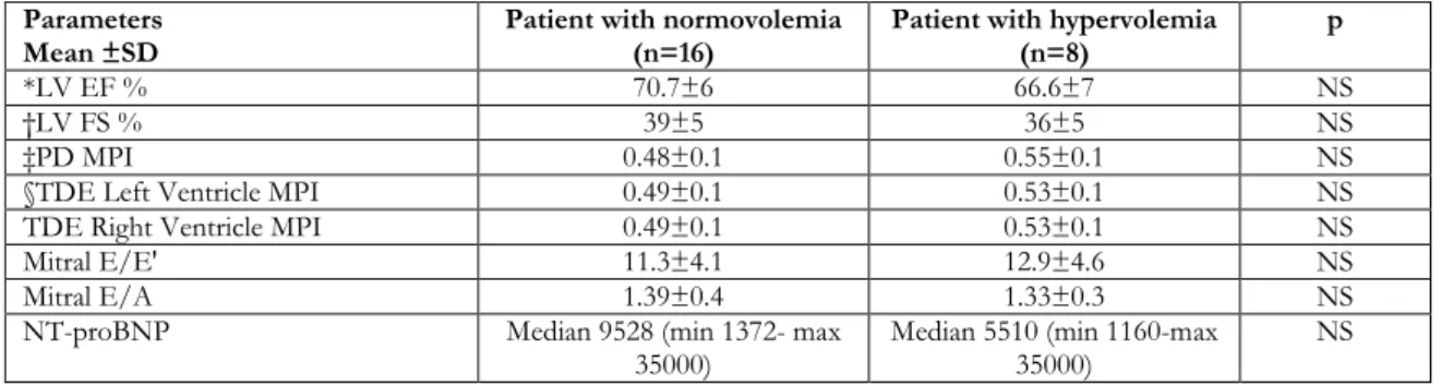
and 3 with restrictive physiology. The median NT-proBNP values were 4576 pg/mL at normal, 3660 pg/mL at abnormal relaxation, 11106 pg/mL at pseudonormal pattern and 28483 at restrictive physiology. Diastolic functions of nine patients who experienced hypotension during dialysis were evaluated and 5 had a pseudonormal pattern, 2 had restrictive physiology, 1 had an abnormal relaxation pattern and 1 with normal function. Baseline systolic and diastolic heart functions and Doppler findings were compared patients with normovolemia and hypervolemic status which gained through OH/ESS ratios >15%. A nonsignificant difference was detected regarding LVEF, LVSF, PD MPI, LV MPI, RV MPI values (Table 2).
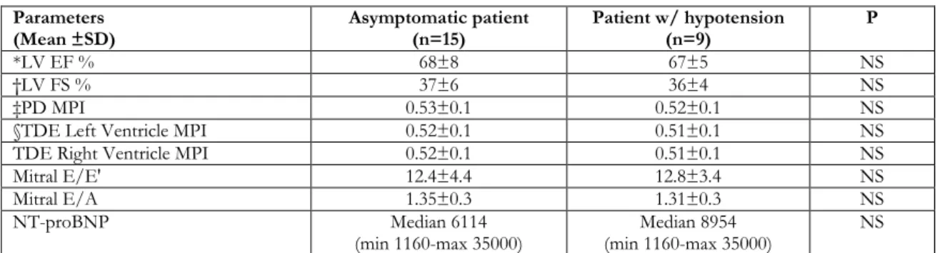
Baseline systolic and diastolic heart functions and Doppler findings were compared with patients who experienced hypotension during dialysis and asymptomatic. A nonsignificant difference was detected regarding LVEF, LVSF, PD MPI, LV MPI, RV MPI values (Table 3).
Table 1. Comparison of echocardiography findings. Parameters Before hemodialysis
mean±SD (min-max) After hemodialysis mean±SD (min-max) p
*LV EF % 68±7 (58-80) 67±6 (58-79) NS †LV FS % 37±6 (29-49) 37±6 (24-48) NS Mitral E (m/sn) 0.92±0.17 (0.58-1.2) 0.88±0.2 (0.52-1.4) NS Mitral A (m/sn) 0.71±0.2 (0.38-1.28) 0.76±0.22 (0.43-1.19) NS Mitral E/A 1.35±0.36 (0.8-2.08) 1.16±0.25(0.74-1.73) <0.03 Deceleration time (msn) 106±29 (60-160) 95±29 (59-155) NS
Isovolumic relaxation time (msn) 83±21 (48-130) 77±13 (55-96) NS
‡PD MPI 0.53±0.09 (0.34-0.76) 0.53±0.1 (0.32-0.86) NS
§TDE Left Ventricle MPI 0.52±0.09 (0.40-0.83) 0.51±0.05 (0.44-0.63) NS TDE Right Ventricle MPI 0.52±0.07 (0.40-0.72) 0.51±0.06 (0.42-0.65) NS
Mitral E/E' 12.4±4.5 (5-20) 11.9±3.7 (6.5-20) NS
*LV EF; left ventricle ejection fraction, †LV FS; left ventricle fractional shorthening, ‡PD MPI; pulse wave Doppler myocardial performance index, §TDE; tissue Doppler echocardiography, || NS; nonsignificant (p>0.05)
Table 2. Comparison of patient regard to hydration status Parameters
Mean ±SD Patient with normovolemia (n=16) Patient with hypervolemia (n=8) p
*LV EF % 70.7±6 66.6±7 NS
†LV FS % 39±5 36±5 NS
‡PD MPI 0.48±0.1 0.55±0.1 NS
§TDE Left Ventricle MPI 0.49±0.1 0.53±0.1 NS
TDE Right Ventricle MPI 0.49±0.1 0.53±0.1 NS
Mitral E/E' 11.3±4.1 12.9±4.6 NS
Mitral E/A 1.39±0.4 1.33±0.3 NS
NT-proBNP Median 9528 (min 1372- max
35000) Median 5510 (min 1160-max 35000) NS
*LV EF; left ventricle ejection fraction, †LV FS; left ventricle fractional shorthening, ‡PD MPI; pulse wave Doppler myocardial performance index, §TDE; tissue Doppler echocardiography, || NS; nonsignificant (p>0.05)
1019
Table 3. Comparison of patient regard to symptom of hypotension during dialysis Parameters
(Mean ±SD) Asymptomatic patient (n=15) Patient w/ hypotension (n=9) P
*LV EF % 68±8 67±5 NS
†LV FS % 37±6 36±4 NS
‡PD MPI 0.53±0.1 0.52±0.1 NS
§TDE Left Ventricle MPI 0.52±0.1 0.51±0.1 NS
TDE Right Ventricle MPI 0.52±0.1 0.51±0.1 NS
Mitral E/E' 12.4±4.4 12.8±3.4 NS
Mitral E/A 1.35±0.3 1.31±0.3 NS
NT-proBNP Median 6114
(min 1160-max 35000) (min 1160-max 35000) Median 8954 NS
*LV EF; left ventricle ejection fraction, †LV FS; left ventricle fractional shorthening, ‡PD MPI; pulse wave Doppler myocardial performance index, §TDE; tissue Doppler echocardiography, || NS; nonsignificant (p>0.05)
DISCUSSION
Cardiovascular risk factors are major mortality reasons in CRF. Left ventricle hypertrophy (LVH) due to volume and pressure overload is the most powerful determinant of mortality in CRF. LVH prevalence in pediatric chronical renal disease was reported 20-30% in stage II-IV and 85% in ESRD12.
In the study, twenty patient had concentric hypertrophy or concentric remodeling and twenty-one had systemic hypertension. Mitsnefes and colleagues reported that diastolic dysfunction is more frequent in chronical dialysis and associated with anemia, hyperphosphatemia and LVH13.
Individual proper ultrafiltration amount detection is so valuable for hydration status management in CRF. Due to lack of a simple and feasible method for extracellular fluid volume measurement, hydration status is usually predicted via clinical signs. In children, peripheral edema appears at excessive over hydration statuses and clinical signs would appear later with more complications. The aim of hemodialysis is to ultrafiltrate excessive extracellular fluid and supply normal hydration status (as known dry weight). Dry weight should be reconsidered especially if one complicates cramps and hypotension during dialysis. Therefore quantitative extracellular and intracellular fluid volume calculation should be done before dialysis in such cases. Bioimpedance spectroscopy is noninvasive, simple, cheap method and provides quantitative data about intracellular, extracellular and total body fluid amounts.
The aim of renal replacement treatment is to hold the patients in normal hydration status. In some
studies >15% OH/ECF amount is accepted as hypervolemia and associated with mortality14.
Machek et colleagues were detected extracellular fluid volume via BCM in 70 adult CRF patient, the group with hypervolemia experienced a decrease in systolic blood pressure 25 mmHg and decrease antihypertensive usage by 35% after proper ultrafiltration volumes. Also, they reported when dry weights were corrected through BCM, ultrafiltration volumes increased up to 1.3 liters, side effect incidence decreased by 70% and blood pressures didn't increase15.
The mechanism of hypotension during dialysis had not been clarified exactly. Usually when hypotension occurs patients require volume replacement and this would decrease the hemodialysis effectiveness. Hypotension incidence was reported as 15-50% before16. Overhydration is mostly in interstitial and
less in the intracellular compartment in the body but volume is ultrafiltrated with hemodialysis from central circulation that leads a decrease in venous return. Cardiac dynamics need left ventricle diastolic function to supply enough cardiac filling. Decreased left ventricle compliance due to left ventricle hypertrophy in CRF leads insufficient cardiac filling that reflects as hypotension in dialysis. Alterations in left ventricular filling should be concealed due to hypervolemia before dialysis and reflects the pseudonormal diastolic pattern in transmitral velocities.
Tissue Doppler echocardiography should be done especially patients with pseudonormal pattern due to less preload influenceability. Left ventricle and left atrium dimensions decrease after hemodialysis due to the reduction in intravascular fluid amount
1020
(preload). As preload decreases, Pulse Doppler echocardiographic measurements such as mitral early inflow velocities (E) and mitral E/A ratios decrease. Mitral E/A ratio should be reflected left ventricle diastolic impairment accurately when the patient had dry weight. In our study, nine patient had experienced hypotension during dialysis and five of them had had the pseudonormal pattern and two with restrictive type diastolic dysfunction. Ultrafiltration was stopped, venous drainage was increased with elevation of lower extremities. When hypotension proceeds, intravenous liquid administration was applied. There was no significant difference regard to systolic and diastolic heart functions, mitral inflow velocities, deceleration times, mitral E/A, mitral E/E' ratios when compared between patient experienced hypotension and not (Table 3).
In the study, a nonsignificant correlation was found between the hypotension event and hydration status that measured with BCM. But when dry weight and OH amounts checked, patients who experienced hypotension had lower OH amounts then dry weights and ultrafiltration volumes. This can be explained as ultrafiltration amount should be determined with OH volume rather than dry weights especially patients who experience frequent hypotension events during dialysis. Dry weights should be predetermined with consecutively liquid volume distribution measurements at different dialysis sessions.
Left ventricular systolic dysfunction is frequent in adults with end-stage renal disease, but it is usually preserved in the child17. But left ventricle diastolic
dysfunction is frequent. Left ventricle mass index increases in early renal disease. Diastolic functions are usually evaluated with pulsed Doppler echocardiography measurements from mitral inflow (Mitral E velocity and mitral E/A ratio) in common practice. Goren and colleagues reported left ventricle relaxation impairment with pulsed Doppler echocardiography in children on dialysis when compared to healthy controls18. But transmitral
Doppler velocities are influenceable to multiple factors; especially preload and left atrium pressure. Tissue Doppler echocardiographic measurements should be more valuable for ventricle relaxation in CRF due to variable preload. Also TD myocardial peak velocities are more specific than Mitral E/A ratio19,20.
Nagueh et colleagues suggested that mitral E/E' is a more proper measurement for cardiac evaluation in CRF8. In the study, ten patients mitral E/ E' ratio
were >10 but mitral E/A ratio were <1 in just six. This could be explained as mitral E velocities were mismeasured due to high preload so mitral E/A ratios were >1 at remaining four despite diastolic impairment. Another important point to determine diastolic dysfunction with Doppler is the time of the study. It should be done just before and 30 minutes after the hemodialysis session and data should be compared each other in CRF.
MPI which gained with pulse Doppler echocardiography is a simple, reproducible test and reflects global ventricle function. But it is affected by heart rate variabilities. Tissue Doppler MPI provides direct myocardial contraction and relaxation velocities in the same heartbeat cycles. Twenty-two patients PD MPI and TD MPI values were higher than normals. This should be reflected subclinical myocardial damage of heart. Hence nonsignificant difference was detected between pre and post dialysis MPI values MPI indices (especially tissue Doppler studies) should be used for evaluating cardiac functions even early stages of the chronical renal disease. Peak early mitral annular velocity (E') is less affected by preload when compared peak early transmitral velocity (E). Uçar et al. reported a correlation between left ventricle mass index and global left ventricle function determined with tissue Doppler echocardiography on 25 peritoneal dialysis patient21. In our study, there was a
significant difference between mitral E/A ratios before and after hemodialysis but a nonsignificant difference was detected regarding mitral E/E' ratios. Significant decreases in myocardial tissue Doppler velocities were reported after hemodialysis in some studies22,23. Hayashi et al determined nonsignificant
differences between heart functions after hemodialysis24. In our study nonsignificant
differences detected before and after hemodialysis measurements regard to PD MPI, early peak mitral transannular velocities (E' ), left and right ventricle TD MP indices.
High plasma BNP levels were reported as a risk factor for cardiovascular morbidity and mortality in children with end-stage renal disease undergoing hemodialysis. Significant correlation was detected between BNP concentrations and left ventricle functions from Ouali et al25. Liu and colleagues were
1021
adults undergoing hemodialysis with normal EF value26. In the current study, all NT-proBNP levels
were >125 pg/mL. Nonsignificant difference was detected between NT-proBNP levels before and after hemodialysis. There were statistically significant correlations between NTproBNP levels and transmitral A velocities (p=0.004, r=0.408), mitral E/E' ratios (p=0.001,r=0.471), PD MPI (p=0.007,r=0.383), right ventricle MPI (p=0.019,r=0.338), respectively.
Our study suggests that diastolic dysfunction with normal systolic function is determined in CRF. Diastolic heart dysfunction should be evaluated carefully with pulse and tissue Doppler echocardiographic measurements. Body composition monitor should be used for setting the optimal ultrafiltration volumes especially symptomatic patients during and after dialysis. Patients have evaluated two different dialysis session at most. Hence over hydration volumes should be variable each session, individual dry weight determination via BCM should be done more repetitive sessions.
REFERENCES
1. Chavers BM, Herzog CA. The spectrum of cardiovascular disease in children with pre-dialysis chronic kidney disease. Adv Chronic Kidney Dis. 2004;11:319-27.
2. Flynn JT. Cardiovascular disease in children with chronic renal failure. Growth Horm IGF Res. 2006;16:84–90.
3. Scharer K, Ulmer HE. Cardiovascular complications of renal failure. In Pediatric Nephrology, 2nd ed (Eds MA Holliday, TM Barratt, RL Vernier):887-96. Baltimore, Williams and Wilkins, 2002.
4. Wizemann V, Rode C, Wabel P. Whole-body spectroscopy (BCM) in the assessment of normovolemia in hemodialysis patients. Contrib Nephrol. 2008;161:115-8.
5. Lang RM, Bierig M, Devereux RB, Flachskampf FA, Foster E, Pellikka PA et al. Recommendations for chamber quantification, Eur J Echocardiogr. 2006;7:79-108.
6. Mitsnefes MM. Cardiovascular complications of pediatric chronic kidney disease. Pediatr Nephrol. 2008;23:27-39.
7. Cui W, Roberson DA. Left ventricular Tei index in children: comparison of tissue Doppler imaging, Pulsed Wave Doppler, and M-Mode echocardiography normal values. J Am Soc Echocardiogr. 2006;19:1438-45.
8. Nagueh SF, Middleton KJ, Kopelman HA. Doppler tissue imaging: A noninvasive technique for evaluation of left ventricular relaxation and estimation of filling pressures. J Am Coll Cardiol. 1997;30:1527-33.
9. Appleton CP, Hatle LK. The natural history of left ventricular filling abnormalities: assessment by two-dimensional and Doppler echocardiographic study. Echocardiography. 1990;9:453-62.
10. Cui W, Roberson DA. Left ventricular Tei index in children: comparison of tissue Doppler imaging, Pulsed Wave Doppler, and M-Mode echocardiography normal values. J Am Soc Echocardiogr. 2006;19:1438-45.
11. Roberson DA, Cui W. Right ventricular Tei index in children: Effect of method, age, body surface area, and heart rate. J Am Soc Echocardiogr. 2007;20:764-70.
12. Mitsnefes M, Flynn J, Cohn S, Samuels J, Blydt-Hansen T, Saland J et al. Masked hypertension associates with left ventricular hypertrophy in children with CKD. J Am Soc Nephrol. 2007;21:137-44.
13. Mitsnefes MM, Kimball TR, Border WL, Witt SA, Glascock BJ, Khoury PR et al. Impaired left ventricular diastolic function in children with chronic renal failure. Kidney Int Apr. 2004;65:1461-66. 14. Wizemann V, Wabel P, Chamney P, Zaluska W,
Moissl U, Rode C et al. The mortality risk of overhydration in haemodialysis patients. Nephrol Dial Transplant. 2009;24:1574-79.
15. Machek P, Jirka U, Moissl U, Chamney P, Wabel P. Guided optimization of fluid status in haemodialysis patients. Nephrol Dial Transplant. 2010;25:538-44. 16. Maggiore Q, Dattolo P, Piacenti M, Morales MA,
Pelosi G, Pizzarelli F et al. A pathophysiological overview of dialysis hypotension. Contrib Nephrol. 1996;119:182-8.
17. Johnstone LM, Jones CL, Grigg LE, Wilkinson JL, Walker RG, Powell HR. Left ventricular abnormalities in children, adolescents and young adults with renal disease. Kidney Int. 1996;50:998-1006.
18. Goren A, Glaser J, Drukker A. Diastolic function in children and adolescents on dialysis and after kidney transplantation: An echocardiographic assessment. Pediatr Nephrol. 1993;7:725-8.
19. Harada K, Tamura M, Toyono M, Oyama K, Takada G. Assessment of global left ventricular function by tissue Doppler imaging. Am J Cardiol. 2001;88:927-32.
20. Su HM, Lin TH, Voon W, Lee KT, Chu CS, Yen HW et al. Correlation of Tei index obtained from tissue Doppler echocardiography with invasive measurements of left ventricular performance. Echocardiography. 2007;24:252-7.
21. Ucar T, Tutar E, Yalçınkaya F, Çakar N, Özçakar Z, Atalay S. Global left-ventricular function by tissue
1022 Doppler imaging in pediatric dialysis patients. Pediatr Nephrol. 2008;23:779-85.
22. Dincer I, Kumbasar D, Nergisoglu G, Atmaca Y, Kutlay S, Akyurek O et al. Assessment of left ventricular diastolic function with Doppler tissue imaging: effects of preload and place of measurements. Int J Cardiovasc Imaging. 2002;18:155-60.
23. Agmon Y, Oh JK, McCarthy JT, Khandheria BK, Bailey KR, Seward JB. Effect of volume reduction on mitral annular diastolic velocities in hemodialysis patients. Am J Cardiol. 2000;85:665-8.
24. Hayashi SY, Brodin LA, Alvestrand A, Lind B, Stenvinkel P, Mazza do Nascimento M et al. Improvement of cardiac function after
haemodialysis: auantitative evaluation by color tissue velocity imaging. Nephrol Dial Transplant. 2004;19:1497-506.
25. Ouali S, Bougmiza I, Abroug S, Omezzine A, Ben Salem H, Neffeti E et al. Relationship of brain natriuretic peptide concentrations to left ventricular function and adverse outcomes in children with end-stage renal disease undergoing hemodialysis. Pediatr Cardiol. 2011;32:568-77.
26. Liu H, Zhang YZ, Gao M, Liu BC. Elevation of
B-type natriuretic peptide is a sensitive marker of left ventricular diastolic dysfunction in patients with maintenance haemodialysis. Biomarkers. 2010;15:533-7.