Abstract
©Copyright 2018 by Turkish Society of Dermatology Turkish Journal of Dermatology published
by Galenos Publishing House.
Introduction
Dermatological examination is essential for the diagnosis of dermatological diseases. In the routine dermatological practice, many infectious and inflammatory diseases can be diagnosed and treated by clinical findings without any additional test. However, genetic and laboratory examinations should be performed before the treatment of some patients. For instance, although the diagnosis of the aphthae can be made clinically, some laboratory tests and the pathergy test are performed in case of recurrent aphthaes. However, if the Tzanck smear is not performed in these patients, they may be misdiagnosed or followed up with Behçet’s disease. The diagnostic tests that are used to determine the treatment of the dermatological diseases are reviewed in this article.
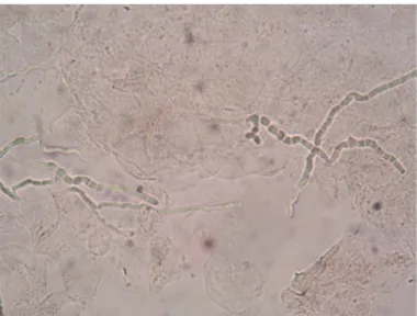
Potassium hydroxide examination: The
potassium hydroxide (KOH) examination, that is used especially for the differential
diagnosis of the erythematous and scaly lesions, is also important in the follow up of patients and in the therapeutical approach (1). In steroid-resistant eczema and psoriasis lesions, the presence of dermatophytical infections must be investigated by the KOH examination and/or the patch test must be performed to show allergic-contact dermatitis due to the topical agents in use (Figures 1, 2) (2). Parasitic agents can also be seen in the direct microscopical examination besides the KOH examination. If blood and allergy tests are performed before the direct microscopic examination in patients with scabies, not only will patient’s itching continue, but also the patient risks infecting additional people with parasites. Dermatoscopic examination can also be used for the follow up of the patients whose itching persist after the therapy besides the microscopic examination (3).
Tzanck smear: The Tzanck smear, which is
a cheap, rapid and repeatable diagnostic
Öz
Dermatolojik hastalıkların tanısında klinik muayene yanında sitoloji, dermatoskopi, histopatolojik incelemeler, bazı laboratuvar testler ve radyolojik incelemeler gerekir. Ancak bu testler sadece tanı anında değil, hasta takiplerinde ve tedavi yaklaşımını belirlemede de oldukça önemlidir. Bu makalede dermatolojik hastalıkların tedavisini belirlemede dermatologlara yardımcı olabilecek tanısal testler derlenmiştir.
Anahtar kelimeler: Dermatoskopi, histopatoloji, KOH inceleme, tanısal testler, Tzanck
yayma, Wood ışığı
The diagnosis of dermatological diseases requires cytological, dermatoscopic, histopathological examinations, some laboratory tests, and radiology imaging in addition to clinical examination. However, these tests are quite important, not only in time of diagnosis, but also in patient follow ups and determining the therapeutic approach. In this article, the diagnostic tests are reviewed that may help to dermatologists in detecting the treatment of dermatological diseases.
Keywords: Dermatoscopy, histopathology, KOH examination, diagnostic tests, Tzanck
smear, Wood’s light
Correspondence/ Yazışma Adresi: Şırnak State Hospital, Clinic of
Dermatology, Şırnak, Turkey E-mail: [email protected] ORCID-ID: orcid.org/0000-0002-1498-1167 Submitted/Geliş Tarihi: 05.06.2018 Accepted/Kabul Tarihi: 13.06.2018
Şırnak State Hospital, Clinic of Dermatology, Şırnak, Turkey
*Başkent University, Adana Dr. Turgut Noyan Application and Research Center, Clinic of Dermatology, Adana, Turkey
Ali Haydar
Eskiocak,
Murat Durdu*
Tedaviyi Belirlemede Tanısal Yaklaşımların Rolü
The Role of Diagnostic Approaches to
Determine the Treatment
method, should be a part of the dermatological examination in the follow up of some diseases besides the diagnosis of many erosive-vesiculobullous, pustular, granulomatous and tumoral diseases (4-8). Cytology is very important in approaching new lesions developed especially in the mucosal regions of pemphigus patients. If conjunctival erythema develops during follow up of pemphigus patient,
conjunctival involvement should be distinguished from bacterial conjunctivitis and allergic contact dermatitis. According to this separation, the dose of systemic steroid should be increased if there is conjunctival involvement; steroid eye drops should be recommended in case of allergic contact dermatitis; or antibiotic eye drops should be used in case of bacterial conjunctivitis. For this distinction, biopsy is not practical for the diagnosis of every recurrent lesion. Initiation of therapy without these distinctions can cause significant complications in patients who use immunosuppressive drugs. The increase of the steroid dose may aggravate the infection in the pemphigus patient who developed bacterial conjunctivitis; on the other hand, the use of antibiotic eye drops will delay the healing of the recurrent pemphigus lesion (4).
Nowadays, in patient with pemphigus, the most common cause of mortality is severe infections. In order to exclude secondary infection, especially in the follow up of oral mucosal blisters, polymerase chain reaction and culture should be made in addition to the Tzanck smear test (9). If a Tzanck smear test is not performed on erosive mucosal lesions due to candidiasis or herpes simplex infections, new lesions are considered clinically as a relapse of pemphigus. In this case, the use of unnecessary systemical steroids causes an increase in lesions, sepsis, and ultimately death (10).
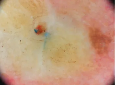
The Tzanck smear test is also important in the follow up and in the therapeutical approach of the tumoral diseases. After surgical treatment of malignant skin tumors, if a new lesion develops, recurrence of a tumor should be differentiated from other causes such as infections and foreign body granuloma. If a Tzanck smear test shows foreign body type multinucleated giant cell and foreign body materials, foreign body granuloma caused by the suture material is considered and intralesional steroid injection may be performed instead of surgical therapy (Figures 3-5) (6).
Although diagnosis of folliculitis can be made clinically, cytological examination should be made in order to detect
Figure 1. Steroid-resistant erythematous scaly plaques on the dorsa of hands and forearm of a patient with psoriasis vulgaris
Figure 2. The potassium hydroxide test shows the hyphae structures in the samples taken from the steroid-resistant plaques of a patient with psoriasis (potassium hydroxide
a therapeutic approach and causes of folliculitis. Despite this fact, such patients are usually treated with topical and/ or systemic antibiotics because the most common cause of folliculitisis is Staphylococcus aureus. Thence, fungal, parasitic
and viral folliculitis are tried to be treated with antibiotics for years. If the literature is reviewed, it is demonstrated that the mean duration of diagnosis of the fungal, parasitic and viral folliculitis are 114, 197 and 285 days, respectively (6). Even a case of viral folliculitis that has been mistakenly treated for 20 years with antibiotics has been reported because only a a bacterial culture was made instead of cytology. Although the treatmentof herpes simplex folliculitis and zona zoster folliculitis is different, their distinction can not be made by routine cytological examination. For this purpose, a direct immunofluorescence study on smears should be performed. In a direct immunofluorescence examination, if the positivity is detected with anti-herpes simplex virus monoclonal antibody, it supports the herpes folliculitis; however, if the positivity is detected with the anti-herpes zoster monoclonal
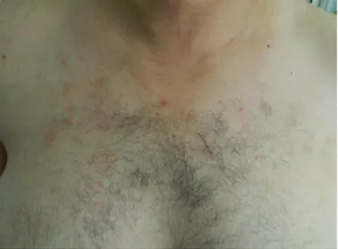
antibody, herpes zoster folliculitis should be considered (6). Finally, if there is resistance to antibiotics in acne vulgaris patients, cytologic examination is quite important to exclude Gram-negative folliculitis and Malassezia folliculitis (Figures 6-8) (11).
Dermatoscopy: Dermatoscopy, which is known as a part of
the dermatological examination, is mostly used to follow up pigmented lesions and melanocytic nevi. Dermatoscopical findings are quite important for the decision of the treatment of nevi. However, the dermatoscopy may also exhibit misleading findings in some diseases. A decision of excision of a non-melanocytic lesion may be made because it may be supposed to be an atypical melanocytic lesion or malignant skin tumours may not be excised because they are considered as non-melanocytic lesions. It is reported that using the dermatoscopy and the cytology together rises the
Figure 4. A dermatoscopy shows blue suture material
Figure 5. A Tzanck smear test reveals suture materials in a foreign-body giant cell in a patient with a suture reaction (May-Grünwald Giemsa 1000x)
Figure 7. A Tzanck smear examination of papulopustular lesions caused Malassezia folliculitis shows footprint-shaped budding spores (May-Grünwald Giemsa 1000x) Figure 6. Erythematous papules, pustules and nodules on the frontal chest of a patient with Malassezia folliculitis, who had previously received steroid-cream treatment for considering the drug reaction
rate of the right therapeutical decision. A melanocytic nevus in which the use of both diagnostic methods is important is epidermolysis bullosa nevus. Although melanocytic nevi that occur in patients with epidermolysis bullosa are considered as malignant both clinically and dermatoscopically, cytologic examination reveals non-atypical melanocytes. If cytology is not performed, unnecessary excisional treatment
is recommended at every control of the patients with epidermolysis bullosa nevus (Figures 9, 10) (8).
Dermatoscopy can be used not only for a diagnosis of psoriasis, but also for follow up of the therapy. When the treatment response of psoriasis patients is evaluated, not only clinical improvement, but also dermatoscopic improvement is important. After treatment, hemorrhagic dots are indicative of the response to the treatment while the persistence of the dotted vessels is the sign of an incomplete response to the therapy or a relapse (Figure 11) (12). Overuse of topical steroids leads to an increase in linear telengiectasias. High-frequency ultrasound and confocal microscopy can also be
used in the evaluation of the response to the treatment of psoriatic lesions (13,14). However, the confocal microscopy is an expensive and non-practical method for routine dermatological examination (14).
In the follow up of vitiligo patients, the dermatoscopy can also be used, as well as Wood’s light examination. In the
dermatoscopical examination, perifollicular repigmentation indicates the good response to the therapy, whereas starburst appearance and micro-Koebner sign suggests disease progression (15).
The dermatoscopy can be used both for the diagnosis and follow up of patients with scabies. The typical dermatoscopical findings of scabies are the intraepidermal tunnels and parasitic triangular structures seen in the tunnels. The positivity of this dermatoscopic (40x) finding in scabiesis is reported as 93% (16). These dermatoscopical findings are important in distinguishing the active infestation from eczema and
Figure 8. The clinical improvement of the lesions of the Malassezia folliculitis after systemic antifungal therapy
Figure 9. Asymmetrical melanocytic nevi after bullous
lesions in a patient with congenital epidermolysis bullosa Figure 11. The hemorrhagic dots developing in the psoriatic lesion after topical steroid therapy Figure 10. Dermatoscopy of epidermolysis bullosa nevus shows irregular pigment network, blue-whitish veil, and asymmetry
parasitophobia in patients with persistent pruritus after antiparasitic treatment. If this discrimination cannot made, the patients may take unnecessary therapies. Detection of the parasites and tunnels via the confocal microscopy and optic coherans tomography is possible. However, use of these methods is difficult in routine follow up because of the cost and difficulty of practical usage (17).
In decision of treatment of the patients of folliculitis, dermatoscopy can also provide some additional findings besides cytology. Firstly, U-shaped ingrown hairs, scar-like appearance caused by fibrosis, blue color change related to deeply located hairs, linear vessels with occasional areas of hemorrhage are the signs of pseudofolliculitis seen on the dermatoscopical examination. Thence, epilation of the hairs or retinoic acid ointments can be used in these patients with pseudofolliculitis (18). Second, in the follow up of patients receiving treatment for tinea capitis, the presence of broken and curly hairs indicate the persistence of the disease. Besides KOH examination, dermatoscopy is important for the determination of antifungal therapy which is to be started before the result of the fungal culture. In the patients of tinea capitis caused by
Trichophyton tonsurans, because the endothrix spores of fungi
mostly cause the breaking of hairs, they give rise to black dots, comma, and corkscrew hairs. However, in the cases of tinea capitis caused by Microsporum canis, the spores outside the hairs (ectothrix) do not break the hairs completely and so that Morse alphabet-like appearance occurs (19,20). In the cases of tinea capitis caused by M. canis, a bright green color is detected on the hair roots via an ultraviolet dermatoscopy. Barcode-like white structures are detected with polarized light on the same areas (21). Third, apart from the superficial skin biopsy, a follow up of the bulges of demodex parasites can be made to evaluate the response of the treatment by the dermatoscopy, in addition to the diagnosis of demodicosis. There are grayish-brown plugs inside the dilated follicules that has demodex and there is an erythematous halo around the follicle (22). Last, tufted hair is a sign of folliculitis decalvans. However, tinea capitis mimicking folliculitis decalvans has been reported with videodermatoscopy so that the cytological examination should be the first method for the diagnosis of folliculitis (11,23). Another group of diseases in which dermatoscopy is important to make a decision about treatment is the cicatricial and non-cicatricial alopecias. In the follow up of a patient with alopecia areata, the broken hairs, cadaveric hairs, tapered hairs and exclamation mark hairs indicate the active disease, while the new developed vellus hairs, pigmentation and thickening of the proximal part of the hair, are the signs of a positive response to the treatment (24,25). Furthermore, in a recent study, it is reported that development of atypical red vessels also negatively correlate with active disease (26). In the patients of frontal fibrosing alopecia, the presence of pruritus along with the perifollicular erythema and the perifollicular hyperkeratosis in the dermatoscopical examination are the signs of histopathological lichenoid infiltration and activity of the disease (27).
In addition to the early detection of melanoma, the dermatoscopy provides some details about the presence
of the BRAF mutation that has changed the therapeutical approach in recent years. It is reported that the presence of blue-white veil is highly associated with BRAF mutation (28). The appearance of blue-gray colored peppering is more common in the melanomas that showed MAPK (BRAF and NRAS) mutation (29). In a study performed with the confocal microscopy and dermatoscopy, it is reported that ulcerations and irregular peripheral streaks are signs of with BRAF-mutated melanoma, while dotted vessels are indicative of wild-type melanoma. However, there was no association between the confocal microscopic findings and BRAF mutation (30).
Wood’s light: The Wood’s light is used for the diagnosis and
the therapeutical approach of various pigment diseases, bacterial and fungal infections, and tumors (31).
The bacterial infections that the Wood’s light is important for following up, are erythrasma and pseudomonal infections. Because erythrasma and fungal infections are common; especially in the patients with flexural dermatitis, Wood’s light examination should be performed in addition to KOH
examination. Coral red fluorescence on Wood’s light is a sign of erythrasma on the intertriginous areas of the patients using a long time steroid cream due to atopic eczema and psoriasis. In a study conducted in Turkey, erythrasma was detected via the Wood’s light examination in 46.7% of all patients that were clinically considered as tinea pedis. Moreover, findings of fungal infection were detected with KOH examination in 31.6% of these patients. If a Wood’s light examination is negative, a Gram staining and bacterial culture should be performed (32).
A Wood’s light examination is very important in a follow up, as well as the diagnosis of the pityriasis versicolor. In the diagnosis of pityriasis versicolor, the positivity of yellow-green fluorescence on Wood’s light was found to be higher (88%) than the positivity of KOH examination (82%). For this reason, patients with hypopigmented lesions after antifungal therapy should be examined with Wood’s light to distinguish postinflammatory hypopigmentation from active disease (33).
The efficiency of antifungal therapy varies according to the agent causing tinea capitis. The Wood’s light examination gives important findings in terms of determining antifungal therapy to be started before the culture results. Dull green fluorescence on Wood’s light is observed in the patients of tinea capitis caused by Microsporum spp. The rate of mycological cure with griseofulvin is higher than the rate of mycological cure of terbinafine in the cases of tinea capitis caused by M. canis (34).
Wood’s light examination should be performed to distinguish the pruritic papulopustular acne lesions that are especially worse in summer and with hot weather from the
Malassezia folliculitis. The patients of Malassezia folliculitis
may unnecessarily take antibiotic therapies for years if this discrimination is not made. In the Wood’s light examination of papulopustular lesions, the yellow-green reflection indicates the Malassezia folliculitis, while the red reflection indicates
acne lesions infected with Propionibacterium acnes (11). The importance of a Wood’s light examination for distinguishing vitiligo from post inflammatory hypopigmentation, pityriasis versicolor alba, mycosis fungoides, nevus depigmentosus and tuberous sclerosis, is well-known. In case this method is not performed, other diseases will be treated with the misdiagnosis of vitiligo for years. Wood’s light examination is also quite important after the diagnosis of vitiligo in the evaluation of the response to the therapy by observing the dotted repigmentation. Furthermore, it is important for the detection of newly developed lesions that can not be recognized by naked eye (35).
Wood’s light may be used for testing patients who are suggested to use sunscreen ointments if they use them sufficiently. Moreover, it is used for detection of the areas that the topical medications are applied (36).
As Wood’s light can be used to determine the surgical border in lentigo malignant melanomas, it can also be used to detect the subclinic lesions of actinic keratosis after application of 5-aminolevulinic acid (ALA). It is reported that, because there is an increase of epidermal melanin in the lesions of lentigo malignant melanomas, the surgical borders may be determined easier via the Wood’s light. However, in a prospective study, it is reported that the lesions that could not be seen by naked eye were detected by Wood’s light in only 11.7% of the cases (37,38). This last procedure of fluorescence diagnosis, in which 5-ALA and Wood’s light are used together, can also be used for determination of the border of the lesions of basal cell carcinoma and squamous cell carcinoma before surgery or radiotherapy. The sensitivity and specificity rates of fluorescence diagnosis were found to be 79% and 100%, respectively (39).
Other diseases in which the Wood’s light is important for following up, are porokeratosis, progressive macular hypomelanosis and morphea. Unlike the other diseases (e.g. annular lichen planus) that caused to annular lesions, porokeratosis lesions resemble a specific diamond necklace-like or ring-like structure on Wood’s light (40). A follicular red fluoroscence is seen with the Wood’s light in the hypopigmented lesions of progressive macular hypomelanosis. This fluoroscence is thought to be related to
P. acnes (41). The Wood’s light was also used for early detection
of morphea lesions (42).
Allergy tests: Detection of the responsible allergen in allergic
skin diseases may change the whole therapy. In patients with allergic contact dermatitis, detection of the allergen and avoiding the responsible agents will provide sparing topical steroids for a long time (43). In the therapy-resistant eczema patients, patch test is should be performed with products that are used to show the allergic response to topical steroids (44). In the patients of physical urticaria, detection of the physical factors that aggravate the disease, and bewaring of some points, can remove the need of therapy (45). In the patients developed drug allergy and who cannot safely use any drugs, performing an oral provocation test with hospitalization may change the therapeutical approach (46).
Histopathological examination: The pathological diagnosis
can be made in 82% of skin biopsies and clinicopathological correlation can be made only in 68% of them. Although some malignant tumoral diseases can be diagnosed by clinical findings and dermatoscopical examination, detection of the histopathological subtypes, depth, mitotic rate and differentiation grade of the disease are quite important for determination of the therapeutical approach (47).
Laboratory examination: In the dermatoses caused
by metabolical disease like xanthelasma, laboratory analysis is needed for the decision of the therapy. There are hyperlipidemia in about half of the patients who had xanthelasma and the first therapy for these patients should be given is the lipid-lowering drugs and diet recommendations in such patients. On the other hand, destructive therapies such as cryotherapy, cauterisation or laser is the first choice in the rest of the normolipidemic patients (48).
The lipid profile and liver function tests should be measured in the patients who are candidates for systemic retinoid acid therapy. The other therapies should be primarily considered in the patients with hyperlipidemia. If the retinoid therapy is absolutely indicated, it should be combinated with the lipid-lowering drugs (49).
The serologic tests of syphilis is valuable in the follow up of systemic lupus erythematous that may show false positivity, as it is used for the diagnosis and the follow up of syphilis. In syphilis serology, the treponemal tests (Treponema
pallidum haemagglutination test, fluorescent treponemal
antibody absorption test) that detect the antibodies against of treponemas and the non-treponemal tests (venereal disease research laboratory test, rapid plasma reagin test) are used. The specific treponemal antibodies turn positive on 1-2 weeks after the genital chancre appears. Because the positivity may continue for life, it can not be used for the follow up of the treatment. However, quantitative results can be obtained with the non-treponemal tests and so it can be used for evaluation of the response to the therapy (50). Another situation that the laboratory is important for, is determining the therapy of the patient who will be initiated immunosuppressive drugs. To determine the preferred therapeutical agent, especially in the treatment of patients with pemphigus, psoriasis, atopic dermatitis, lupus erythematous and vasculitis, some laboratory tests should be performed in terms of any comorbidities; for example, diabetes mellitus, viral hepatitis, hyperlipidemia and renal insufficiency. The systemic therapy may be changed depending on the results of the laboratory tests. Furthermore, thiopurine methyltransferase enzyme activity should be measured for the effect and side effect of azathioprine, and the level of glucose-6-phosphate dehydrogenase enzyme should be measured in addition to the routine examinations in the patients who will initiate to dapson therapy (51,52). In romatological diseases and vasculitis, some laboratory tests and autoantibodies are ordered to detect the internal involvement, and to evaluate the severity of the disease. In this cases, if there is involvement of the skin only, topical
treatments will be sufficient. But if there is an internal involvement, systemic therapies are should be added to the treatment (53).
Some serological tests are needed in addition to the clinical, histopathological and immunofluorescence studies to make the diagnosis of autoimmune bullous diseases. Some recently developed serological tests can also be used for the follow up of therapy because they can measure the antibody titrations (54).
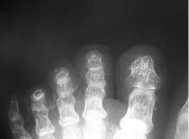
One of the most important triggering factors of generalized pustular psoriasis is the deficiency of calcium and vitamin D. If this deficiency is detected and cured in the patients of generalized pustular psoriasis, they can be treated without any need for systemic therapy. There has been reported a case of generalized pustular psoriasis improved with calcium therapy in the literature (Figures 12, 13) (55).
Radiological examinations: Although there are numerous
causes of nail dystrophy, if the KOH examination and other diagnostic tools are not used, about half of the patients with subungual exocytosis take unnecessary antifungal treatments. X-ray graphy should be performed in cases of nail distrophy that KOH examination is negative and particularly in cases whose distrophy is in a single nail. The surgical operation should be performed after the diagnosis is made (56).
Needful radiological examinations should be performed in the cases of skin cancer and lymphomas for staging, and also in the cases with systemic diseases to detect the internal involvements. Additionally, radiological examination is important for dermatoses that may be caused by cancers. The radiological examinations, in addition to laboratory tests, should be done for the disease that can make systemic involvement in order to determine whether the therapy will target the skin only, or will be systemic therapy (57). It is reported that some kind of pemphigoid, such as bullous pemphigoid, anti-laminin 332 pemphigoid and epidermolysis bullosa acquisita, may appear secondary to malignancy apart from paraneoplastic pemphigus. Moreover, it is reported that in some cases in which the bullae were regressed after the treatment of malignancy (58).
The most frequent method that is used to determine the therapy for peripheral vascular diseases, is the color Doppler evaluation. It can also be used for the follow up alongside the diagnosis. The increase of the vascularity with the color Doppler is a sign of activation of morphea, psoriasis, hidradenitis suppurativa, and human papillomavirus infections (59).
High-frequency ultrasounds are useful for the follow up and for evaluation of the objective response to the treatment of atopic dermatitis, psoriasis, morphea and some skin tumors (60). Furthermore, this non-invazive diagnostic method that provide a detailed evaluation of epidermis and dermis, can also be used for determination of the surgical border before the surgery (61).
The high-resolution optical coherence tomography that provides transvers and vertical images, can be used for the follow up of non-melanoma skin cancers, the evaluation of the response to the therapy and determination of the surgical border. Furthermore, it can be used for the discrimination between inflammatory diseases such as psoriasis and lichen planus and for evaluation of the response to the therapy (62).
Toluidine blue test: The affinity of toluidine blue to both DNA
and RNA is quite high. The toluidine blue test can be used for the detection of suspicious lesions in the oral mucosa. The procedure of this test is as following: 1) The oral cavity is firstly rinsed with water and then with 1% acetic acid, for 20 seconds for each. 2) The oral cavity is dried with gauze. 3) The 1% toluidine blue is applied by stick cotton swab. 4) The mouth is rinsed with 1% acetic acid and then with water. A dark blue stain is accepted as positive (63).
Acetic acid test: The 5% acetic acid test can be used for
detection of suspicious human papilloma virus lesions in genital region. However, physicians should keep in mind that
Figure 12. Lesions of generalized pustular psoriasis caused by pseudohypoparathyroidism
Figure 13. Calcifications detected on foot radiography in a patient with generalized pustular psoriasis caused by pseudohypoparathyroidism
this test may show false positivity with some inflammatory diseases and may show false negativity, particularly in areas that are not damp (64).
Iodine-starch test: In the patients with hyperhidrosis, the
iodine-starch test can be performed to detect the region that will be treated and to evaluate the response to the therapy. In this test, first, the application area is well-dried. Iodine solution is applied to the application area by a stick cotton swab and is waited to dry. After it dries, the area is covered with starch by a thick brush. After waiting for 5-10 minutes, the areas that turned blue-black are pointed by a water-resistant pen (65).
Ethics
Informed Consent: Approval was obtained for patient pictures.
Peer-review: Internally peer-reviewed. Authorship Contributions
Surgical and Medical Practices: M.D., Concept: M.D., Design: M.D., Data Collection or Processing: M.D., A.H.E., Analysis or Interpretation: M.D., Literature Search: M.D., Writing: A.H.E., M.D.
Conflict of Interest: No conflict of interest was declared by
the authors.
Financial Disclosure: The authors declared that this study
received no financial support.
References
1. Wilkison BD, Sperling LC, Spillane AP, Meyerle JH. How to teach the potassium hydroxide preparation: a disappearing clinical art form. Cutis 2015;96:109-12.
2. Al-Mutairi N, Abdalla TO, Nour TM. Resistant palmoplantar lesions in patients of psoriasis: evaluation of the causes and comparison of the frequency of delayed-type hypersensitivity in patients without palm and sole lesions. Med Princ Pract 2014;23:561-7.
3. Hicks MI, Elston DM. Scabies. Dermatol Ther 2009;22:279-92.
4. Sehgal VN, Sharma S, Sardana K. Unilateral refractory (erosive) conjunctivitis: a peculiar manifestation of pemphigus vulgaris. Skinmed 2005;4:250-2. 5. Durdu M, Baba M, Seçkin D. The value of Tzanck smear test in diagnosis of
erosive, vesicular, bullous, and pustular skin lesions. J Am Acad Dermatol 2008;59:958-64.
6. Durdu M, Ilkit M. First step in the differential diagnosis of folliculitis: cytology. Crit Rev Microbiol 2013;39:9-25.
7. Durdu M, Baba M, Seçkin D. More experiences with the Tzanck smear test: cytologic findings in cutaneous granulomatous disorders. J Am Acad Dermatol 2009;61:441-50.
8. Durdu M, Baba M, Seçkin D. Dermatoscopy versus Tzanck smear test: a comparison of the value of two tests in the diagnosis of pigmented skin lesions. J Am Acad Dermatol 2011;65:972-82.
9. Kumar S, De D, Handa S, et al. Identification of factors associated with treatment refractoriness of oral lesions in pemphigus vulgaris. Br J Dermatol 2017;177:1583-9.
10. Kalajian AH, Callen JP. Atypical herpes simplex infection masquerading as recalcitrant pemphigus vulgaris. Australas J Dermatol 2007;48:242-7. 11. Durdu M, Güran M, Ilkit M. Epidemiological characteristics of Malassezia
folliculitis and use of the May-Grünwald-Giemsa stain to diagnose the infection. Diagn Microbiol Infect Dis 2013;76:450-7.
12. Lallas A, Argenziano G, Zalaudek I, et al. Dermoscopic hemorrhagic dots: an early predictor of response of psoriasis to biologic agents. Dermatol Pract Concept 2016;6:7-12.
13. Micali G, Lacarrubba F, Santagati C, et al. Clinical, ultrasound, and videodermatoscopy monitoring of psoriatic patients following biological
treatment. Skin Res Technol 2016;22:341-8.
14. Başaran YK, Gürel MS, Erdemir AT, et al. Evaluation of the response to treatment of psoriasis vulgaris with reflectance confocal microscopy. Skin Res Technol 2015;21:18-24.
15. Thatte SS, Khopkar US. The utility of dermoscopy in the diagnosis of evolving lesions of vitiligo. Indian J Dermatol Venereol Leprol 2014;80:505-8. 16. Argenziano G, Fabbrocini G, Delfino M. Epiluminescence microscopy. A
new approach to in vivo detection of Sarcoptes scabiei. Arch Dermatol 1997;133:751-3.
17. Micali G, Lacarrubba F, Verzì AE, et al. Scabies: Advances in Noninvasive Diagnosis. PLoS Negl Trop Dis 2016;10:e0004691.
18. Kaliyadan F, Kuruvilla J, Al Ojail HY, Quadri SA. Clinical and Dermoscopic Study of Pseudofolliculitis of the Beard Area. Int J Trichology 2016;8:40-2. 19. Lekkas D, Ioannides D, Apalla Z, et al. Dermoscopy for discriminating
between Trichophyton and Microsporum infections in tinea capitis. J Eur Acad Dermatol Venereol 2018;32:234-5.
20. Schechtman RC, Silva ND, Quaresma MV, et al. Dermatoscopic findings as a complementary tool in the differential diagnosis of the etiological agent of tinea capitis. An Bras Dermatol 2015;90(3 Suppl 1):13-5.
21. Tang J, Ran X, Ran Y. Ultraviolet dermoscopy for the diagnosis of tinea capitis. J Am Acad Dermatol 2017;76:28-30.
22. Friedman P, Sabban EC, Cabo H. Usefulness of dermoscopy in the diagnosis and monitoring treatment of demodicidosis. Dermatol Pract Concept 2017;7:35-8.
23. Rudnicka L, Olszewska M, Rakowska A, Slowinska M. Trichoscopy update 2011. J Dermatol Case Rep 2011;5:82-8.
24. Inui S, Nakajima T, Nakagawa K, Itami S. Clinical significance of dermoscopy in alopecia areata: analysis of 300 cases. Int J Dermatol 2008;47:688-93. 25. Lacarrubba F, Dall’Oglio F, Rita Nasca M, Micali G. Videodermatoscopy
enhances diagnostic capability in some forms of hair loss. Am J Clin Dermatol 2004;5:205-8.
26. Kibar M, Aktan Ş, Lebe B, Bilgin M. Trichoscopic findings in alopecia areata and their relation to disease activity, severity and clinical subtype in Turkish patients. Australas J Dermatol 2015;56:1-6.
27. Fernández-Crehuet P, Rodrigues-Barata AR, Vañó-Galván S, et al. Trichoscopic features of frontal fibrosing alopecia: results in 249 patients. J Am Acad Dermatol 2015;72:357-9.
28. Armengot-Carbó M, Nagore E, García-Casado Z, Botella-Estrada R. The association between dermoscopic features and BRAF mutational status in cutaneous melanoma: Significance of the blue-white veil. J Am Acad Dermatol 2018;78:920-6.
29. Pozzobon FC, Puig-Butillé JA, González-Alvarez T, et al. Dermoscopic criteria associated with BRAF and NRAS mutation status in primary cutaneous melanoma. Br J Dermatol 2014;171:754-9.
30. Bombonato C, Ribero S, Pozzobon FC, et al. Association between dermoscopic and reflectance confocal microscopy features of cutaneous melanoma with BRAF mutational status. J Eur Acad Dermatol Venereol 2017;31:643-9.
31. Klatte JL, van der Beek N, Kemperman PM. 100 years of Wood’s lamp revised. J Eur Acad Dermatol Venereol 2015;29:842-7.
32. Inci M, Serarslan G, Ozer B, et al. The prevalence of interdigital erythrasma in southern region of Turkey. J Eur Acad Dermatol Venereol 2012;26:1372-6. 33. Shah A, Koticha A, Ubale M, et al. Identification and speciation of malassezia
in patients clinically suspected of having pityriasis versicolor. Indian J Dermatol 2013;58:239.
34. Gupta AK, Summerbell RC. Tinea capitis. Med Mycol 2000;38:255-87. 35. Wang YJ, Chang CC, Cheng KL. Wood’s lamp for vitiligo disease stability and
early recognition of initiative pigmentation after epidermal grafting. Int Wound J 2017;14:1391-4.
36. Jankowski M, Nowowiejska L, Czajkowski R. Wood’s lamp fluorescence of dihydroxyacetone treated skin. J Eur Acad Dermatol Venereol 2016;30:125-6.
37. Paraskevas LR, Halpern AC, Marghoob AA. Utility of the Wood’s light: five cases from a pigmented lesion clinic. Br J Dermatol 2005;152:1039-44. 38. Walsh SB, Varma R, Raimer D, et al. Utility of Wood’s light in margin
determination of melanoma in situ after excisional biopsy. Dermatol Surg 2015;41:572-8.
39. Neus S, Gambichler T, Bechara FG, et al. Preoperative assessment of basal cell carcinoma using conventional fluorescence diagnosis. Arch Dermatol Res 2009;301:289-94.
Photodermatol Photoimmunol Photomed 2017;33:114-6.
41. Pflederer RT, Wuennenberg JP, Foote C, et al. Use of Wood’s lamp to diagnose progressive macular hypomelanosis. J Am Acad Dermatol 2017;77:99-100. 42. Curtiss P, Singh G, Lo Sicco K, Franks AG Jr. Wood’s lamp as a tool in the
evaluation of morphea. J Am Acad Dermatol 2018;78:33-4.
43. Gündüz Ö, Aytekin A, Tutkun E, Yılmaz H. Comparison of European Standard Patch Test Results of 330 Patients from an Occupational Diseases Hospital. Dermatol Res Pract 2016;2016:9421878.
44. Zirwas M. Allergy to topical steroids. J Drugs Dermatol 2012;11:9-11. 45. Colgecen E, Ozyurt K, Gul AI, Utas S. Evaluation of etiological factors in
patients with chronic urticaria. Acta Dermatovenerol Croat 2015;23:36-42. 46. Zisa G, Riccobono F, Bommarito L, et al. Provocation tests with the
offending nonsteroidal anti-inflammatory drugs in patients with urticaria/ angioedema reactions. Allergy Asthma Proc 2012;33:421-6.
47. Paolino G, Donati M, Didona D, et al. Histology of Non-Melanoma Skin Cancers: An Update. Biomedicines 2017;5:71.
48. Wang KY, Hsu KC, Liu WC, et al. Relationship Between Xanthelasma Palpebrarum and Hyperlipidemia. Ann Plast Surg 2018;80(2S Suppl 1):84-6. 49. Lee YH, Scharnitz TP, Muscat J, et al. Laboratory Monitoring During
Isotretinoin Therapy for Acne: A Systematic Review and Meta-analysis. JAMA Dermatol 2016;152:35-44.
50. Pillay A. CDC Syphilis Summit - Diagnostics and Laboratory Issues. Sex Transm Dis 2018.
51. Wise M, Callen JP. Azathioprine: a guide for the management of dermatology patients. Dermatol Ther 2007;20:206-15.
52. Wozel G, Blasum C. Dapsone in dermatology and beyond. Arch Dermatol Res 2014;306:103-24.
53. Rawlings CR, Fremlin GA, Nash J, Harding K. A rheumatology perspective on cutaneous vasculitis: assessment and investigation for the non-rheumatologist. Int Wound J 2016;13:17-21.
54. Özkesici B, Akman Karakaş A. Otoimmün Büllöz Hastalıkların Serolojik Tanısı.
Turk J Dermatol 2016;10:1-8.
55. Durdu M, Baba M, Anaforoğlu İ. Psödohipoparatiroidizmin Eşlik Ettiği Bir Psoriazis Olgusu. Turkiye Klinikleri J Dermatol 2009;19:170-5.
56. Göktay F, Atış G, Güneş P, et al. Subungual exostosis and subungual osteochondromas: a description of 25 cases. Int J Dermatol 2018;57:872-881.
57. Palmucci S, Torrisi SE, Caltabiano DC, et al. Clinical and radiological features of extra-pulmonary sarcoidosis: a pictorial essay. Insights Imaging 2016;7:571-87.
58. Noguchi K, Kawamura H, Ishizu H, Okada K. Dramatic resolution of bullous pemphigoid after surgery for gastric cancer: A case report. Int J Surg Case Rep 2014;5:212-4.
59. Barcaui Ede O, Carvalho AC, Lopes FP, et al. High frequency ultrasound with color Doppler in dermatology. An Bras Dermatol 2016;91:262-73.
60. Polańska A, Dańczak-Pazdrowska A, Jałowska M, et al. Current applications of high-frequency ultrasonography in dermatology. Postepy Dermatol Alergol 2017;34:535-42.
61. Zeitouni NC, Rohrbach DJ, Aksahin M, Sunar U. Preoperative ultrasound and photoacoustic imaging of nonmelanoma skin cancers. Dermatol Surg 2015;41:525-8.
62. Odorici G, Losi A, Ciardo S, et al. Non-invasive evaluation of Secukinumab efficacy in severe plaque psoriasis with confocal microscopy and optical coherence tomography: A case report. Skin Res Technol 2018;24:160-2. 63. Allegra E, Lombardo N, Puzzo L, Garozzo A. The usefulness of toluidine
staining as a diagnostic tool for precancerous and cancerous oropharyngeal and oral cavity lesions. Acta Otorhinolaryngol Ital 2009;29:187-90. 64. Kumar B, Gupta S. The acetowhite test in genital human papillomavirus
infection in men: what does it add? J Eur Acad Dermatol Venereol 2001;15:27-9.
65. Maillard H, Lecouflet M. [Management of hyperhidrosis]. Ann Dermatol Venereol 2015;142:252-61.
1. Which of the following diagnostic tool is not used to determine the surgical borders in the tumoral diseases?
a. Wood’s light examination after application of 5-aminolevulinic acid
b. Laboratory tests
c. High frequency ultrasonography d. Histopathological examination e. Optical coherence tomography
2. Which of the following types of folliculitis is indicative of yellow-green fluorescence in the Wood’s light examination in a patient with antibiotic resistance acne?
a. Molluscum contagiosum folliculitis b. Gram-negative bacterial folliculitis c. Herpetic folliculitis
d. Demodex folliculitis e. Malassezia folliculitis
3. Which of the following clinical or/and dermatoscopical findings is accepted as an indication of good therapeutical response in the psoriatic lesions?
a. Hemorrhagic dots b. Dotted vessels
c. Increasing of linear vessels d. Atrophia and striae e. Hypertrichosis
4. Which of the following diagnostic methods is not used to distinguish the active infestation from the other pruritic diseases in a patient with persistent pruritus after antiparasitic treatment?
a. Direct microscopical examination b. Dermatoscopy
c. Acetic acid test d. Confocal microscopy
e. High frequency ultrasonography
5. Which of the following dermatoscopical findings is not indicative of pseudofolliculitis in a patient of folliculitic?
a. “U” shaped hairs
b. Perifollicular erythema and keratosis c. Scar-like appearance due to fibrosis d. Blue discoloration
e. Linear vessels and hemorrhage
6. Which of the following claims about the dermatoscopical findings of tinea capitis is incorrect?
a. The dermatoscopy provides important clues for determination of the antifungal treatment in patients with tinea capitis
b. Black dots, comma, and corkscrew hairs are seen in cases of tinea capitis caused by Trichophyton tonsurans c. In the cases of tinea capitis caused by Microsporum canis, a bright green color is detected on the hair roots via an ultraviolet dermatoscopy
d. In the cases of tinea capitis caused by M. canis, the endothrix located spores break the hairs completely and so that Morse alphabet-like appearance occurs
e. The presence of broken and curly hairs indicate of the persistence of the disease
7. Which of the following dermatoscopical findings of alopesia areata is not considered as an indication of active disease?
a. Broken hairs b. Cadaveric hairs c. Tapered hairs
d. Exclamation mark hairs e. Atypical red vessels
8. Which of the following dermatoscopical findings is incorrect in terms of determining the treatment of a patient with melanoma?
a. Blue.white veil is highly associated with BRAF mutation b. Blue.gray peppering appearance is more common in
the melanomas with MAPK mutation
c. Ulcerations and irregular peripheral streaks are less common in the melanoma with BRAF mutation d. Dotted vessels are more common in the non-mutated melanoma
e. Dotted vessels is a negatively predictive finding for BRAF mutation
9. Which of the following matches is incorrect in terms of disease and diagnostic test?
a. The level of thiopurine methyltransferase enzyme- dermatitis herpetiformis
b. The level of calcium and vitamin D-generalized pustular psoriasis
c. High frequency ultrasonography-atopic dermatitis d. Color doppler examination-hidradenitis supurativa e. Optical coherence tomography-non-melanoma skin tumors
10. The Wood’s light is not used in which following dermatological diseases? a. Morphea b. Actinic keratosis c. Acne vulgaris d. Vitiligo e. Furunculosis Answ er key 1.b, 2.e , 3.a, 4.c, 5.b , 6.d, 7.e , 8.c, 9.a, 10.e Quiz