993
http://journals.tubitak.gov.tr/medical/ © TÜBİTAK
doi:10.3906/sag-1803-235
Value of shear wave elastography by virtual touch tissue imaging quantification in
patients with diffuse thyroid gland pathology
Feride KURAL RAHATLI1,*, Hale TURNAOĞLU1, Nazlı Gülsoy KIRNAP2, Özlem TURHAN İYİDİR2,
Kemal Murat HABERAL1, Aresh SOUDMAND1, Nihal USLU1 1Department of Radiology, Faculty of Medicine, Başkent University, Ankara, Turkey 2Department of Endocrinology, Faculty of Medicine, Başkent University, Ankara, TURKEY
* Correspondence: [email protected]
1. Introduction
Hashimoto’s thyroiditis (HT) and Graves’ disease (GD) are frequently occurring autoimmune thyroid diseases that diffusely affect the thyroid gland. Both HT and GD are characterized by lymphocyte infiltration of the thyroid gland (1). The peak incidence of HT is from 30 to 50 years of age, and most affected patients are female (2). GD is characterized by diffuse goiter, orbitopathy, and hyperthyroidism. Its peak incidence is from 30 to 60 years of age, and it is 5 to 10 times more frequently seen in female than male patients (3).
The diagnosis of HT and GD is based on clinical symptoms, laboratory results, and ultrasonography findings. Ultrasonography is a useful diagnostic tool for the evaluation of autoimmune thyroid diseases. Both HT and GD are characterized by heterogeneous to hypoechogenic thyroid parenchyma and elevated or decreased vascularity on conventional color Doppler ultrasonography. Stiffness of the thyroid gland increases due to inflammation and fibrosis. The stiffness of the thyroid gland can be evaluated
with elastography (4). Elastography is a new dynamic technique that provides information about tissue hardness using ultrasound waves (5).
The two basic types of elastography are strain elastography and shear wave elastography (SWE). Both of these techniques have been demonstrated for measurement of the stiffness of different tissues in several studies. Strain elastography enables prediction of tissue elasticity by measuring the tissue deformation parallel to the direction of the external force. The stiffness is viewed as an elasticity color map superimposed on grayscale images. This technique allows the clinician to obtain a qualitative or semiquantitative measurement by calculating the strain ratio, but the interpretation of qualitative strain information and operator-dependent freehand compression lead to interobserver variability (6).
SWE is an operator-independent quantitative elastography method. It utilizes acoustic radiation force impulse (ARFI) imaging to measure the shear wave velocity (SWV) of the target area inside the region of interest
Background/aim: This study was performed to assess the diagnostic accuracy of shear wave elastography by virtual touch tissue imaging
quantification in patients with diffuse thyroid gland pathology and to differentiate Graves’ disease from Hashimoto’s thyroiditis.
Materials and methods: Thirty patients with Hashimoto’s thyroiditis and 22 patients with Graves’ disease were evaluated with
conventional ultrasonography and shear wave elastography by virtual touch tissue imaging quantification. Thirty healthy participants formed the control group. Ten elastographic measurements of each thyroid lobe were performed in all three study groups. The mean shear wave velocity was calculated and compared between the patients and controls.
Results: The mean shear wave velocity of the controls (1.92 ± 0.14 m/s) was significantly lower than that of the patients with Graves’
disease (2.71 ± 0.22 m/s) and Hashimoto’s thyroiditis (2.50 ± 0.20 m/s). Patients with Graves’ disease had significantly higher shear wave velocities than those with Hashimoto’s thyroiditis (P < 0.001).
Conclusion: Shear wave elastography by virtual touch tissue imaging quantification can be used to discriminate a normal thyroid gland
from diffuse thyroid gland pathology after conventional sonography and may assist in the diagnosis of Graves’ disease and Hashimoto’s thyroiditis.
Key words: Shear wave elastography, virtual touch tissue imaging quantification, thyroid, Hashimoto’s thyroiditis, Graves’ disease Received: 31.03.2018 Accepted/Published Online: 08.09.2018 Final Version: 31.10.2018
(ROI) without external compression and can estimate the minor differences inside the ROI. The target tissue is mechanically stimulated by short-duration acoustic pulses to produce small (1- to 10-mm) localized tissue displacements; this tissue displacement results in shear wave dissemination, which is tracked with the probe and measured as SWV (m/s) (7). Virtual touch tissue imaging quantification (VTQ) is a new technique that involves the use of SWE to quantify tissue elasticity by calculating the SWV (8,9).Previous studies have shown that VTQ is useful for evaluation of the extension of liver fibrosis, diagnosis of chronic autoimmune thyroiditis (CAT), differentiation of benign and malignant breast lesions, and differentiation of benign and malignant thyroid nodules (7,8,10,11).
In this study, we assessed the diagnostic accuracy of SWE by VTQ in patients with diffuse thyroid gland pathology and the usefulness of this technique in the differentiation of GD from HT.
2. Materials and methods 2.1. Patients
In total, 30 patients with HT (2 male, 28 female), 22 patients with GD (7 male, 15 female), and 30 healthy controls (6 male, 24 female) were enrolled in this prospective nonrandomized study from January to July 2016. The study population included patients being followed for either GD or HT in our endocrinology department. None of the patients had thyroid nodules according to their previous ultrasonography reports. The diagnosis of GD was based on the presence of thyrotoxicosis at the initial examination, a high thyroid-stimulating hormone (TSH) receptor antibody titer, and a hypoechoic to heterogeneous pattern with increased vascularity in the thyroid gland on ultrasonography. Twelve of the patients with GD were undergoing antithyroid therapy and had a low TSH concentration, and 10 were not receiving therapy and had normal thyroid function test results. The diagnosis of HT was based on the presence of a high antithyroglobulin antibody titer, high antithyroid peroxidase antibody titer, or both as well as a hypoechoic to heterogeneous pattern with diffuse goiter and increased or normal vascularity in the thyroid gland on ultrasonography. All patients with HT were receiving L-thyroxine therapy and had normal thyroid function test results. Participants in the control group had normal thyroid ultrasonography findings and normal serum TSH concentrations. The exclusion criteria were a history of thyroid surgery, radioactive iodine therapy, radiation therapy to the neck, and calcification or cystic lesions in the thyroid gland.
2.2. Conventional ultrasonography and SWE by VTQ All conventional ultrasonographic and elastographic examinations were performed by two radiologists
with 10 years of experience in sonography and 4 years of experience in elastography. Both radiologists were blinded to the patients’ diagnoses. All of the patients were examined by both radiologists. All patients underwent a sonographic examination of the thyroid gland including color Doppler ultrasonography using a Siemens ACUSON S3000 diagnostic ultrasound system (Siemens Medical Solutions, Erlangen, Germany). A Siemens 9L4 linear probe with a frequency range of 4 to 9 MHz was used in both the conventional ultrasonographic and elastographic examinations. The patients were placed in the supine decubitus position on the examination couch with their neck in hyperextension. The sonographic pattern and volume of the thyroid gland were recorded for all patients. After the B-mode sonography examination, color Doppler ultrasonography was performed to record the vascular pattern of the gland.
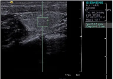
During the B-mode ultrasonography examination, SWE by VTQ was performed by switching to the elastographic mode. The probe was gently placed on the gel with light pressure, and the patients were asked to hold their breath. VTQ was performed on B-mode ultrasonographic images with a fixed 5- × 6-mm ROI placed in the thyroid parenchyma. The SWV of the tissue, which was proportional to the square root of the tissue elasticity, was then measured (Figures 1a and 1b). The SWV ranged from 0 to 9 m/s. Ten successful measurements per thyroid lobe were performed with the ROI placed at different points on the thyroid gland. The mean values of the measurements were calculated (7,9,12).The study was performed in accordance with the ethical guidelines of the Declaration of Helsinki and was approved by the local ethics committee.
2.3. Statistical analysis
The mean ± standard deviation and median, minimum, and maximum values were used as descriptive statistics for the quantitative variables, whereas frequency and percentage were used for the qualitative variables. Normality assumption was analyzed by the Shapiro–Wilk test. For normally distributed measurements, analysis of variance was used to compare the three groups. Tukey’s multiple comparison test was used as a post hoc test after analysis of variance. The Kruskal–Wallis test was used to compare nonnormally distributed variables among the three groups. The Conover–Dunn multiple comparison test was used as a post hoc test after the Kruskal–Wallis test. The chi-square test was used to compare qualitative variables among the groups. SPSS 21.0 for Windows (IBM Corp., Armonk, NY, USA) was used for all statistical analyses. P < 0.05 was considered statistically significant. Interobserver correlation agreement between the two radiologists was tested using an interclass correlation coefficient test.
3. Results
In total, 82 patients (15 male, 67 female; median age, 40 years; age range, 21–67 years) underwent thyroid ultrasonography and SWE by VTQ. Thirty patients with HT (median age, 39 years), 22 patients with GD (median age, 44 years), and 30 healthy controls (median age, 37 years) were examined. The demographic and biochemical characteristics of the study population are shown in the Table. The groups were similar in terms of age and sex.
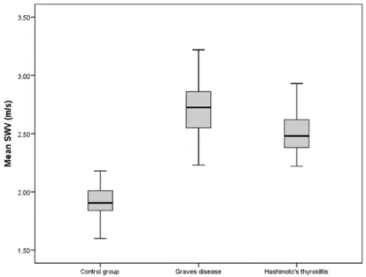
In both the HT and GD groups, the thyroid glands were hypoechoic to heterogeneous by B-mode ultrasonography and showed increased vascularity by Doppler ultrasonography. The mean SWV was 2.71 ± 0.22 m/s in the GD group (range, 2.23–3.22 m/s), 2.50 ± 0.20 m/s in the HT group (range, 2.22–3.04 m/s), and 1.92 ± 0.14 m/s in the control group (range, 1.60–2.18 m/s) (Table ). The mean SWVs in the GD and HT groups were significantly higher than that in the control group (P <
0.001). The mean SWV in the GD group was significantly higher than that in the HT group (P < 0.001) (Figures 1a, 1b, and 2).
The mean SWV of patients with diffuse thyroid pathology (GD + HT groups) was 2.59 ± 0.23 m/s (range, 2.22–3.22 m/s). The difference between the control group and patients with diffuse thyroid pathology was statistically significant (P < 0.001).
In the GD group, the mean TSH concentration of patients who were and were not undergoing antithyroid therapy was 0.08 ± 0.10 and 1.81 ± 0.13 µIU/mL, respectively (P < 0.001). Patients who were undergoing antithyroid therapy and had a low TSH concentration showed a significantly higher SWV than patients who were not undergoing antithyroid therapy (2.81 ± 0.18 vs. 2.59 ± 0.22 m/s, respectively; P = 0.023) (Figure 3).
The interobserver agreement was almost perfect for the two radiologists (0.99).
Figure 1a. Shear wave velocity (2.47 m/s) of a 42-year-old woman with
Hashimoto’s thyroiditis.
Figure 1b. Shear wave velocity (2.80 m/s) of a 35-year-old woman with
Figure 2. Box plot of the mean shear wave velocity (m/s) in the control group,
Graves’ disease group, and Hashimoto’s thyroiditis group.
Figure 3. Box plot of the mean shear wave velocity (m/s) in patients with Graves’ disease according to the presence or absence of
antithyroid therapy.
Table. Age, SWV, serum TSH concentration, and sex of patients in HT, GD,
and control groups.
HT GD Control P-value
Age (years)* 39 (25–65) 44 (25–63) 37 (21–67)
Sex (M/F) 2/28 7/15 6/24
SWV (m/s) 2.50 ± 0.20 2.71 ± 0.22 1.92 ± 0.14 <0.001 TSH (µIU/mL) 2.88 ± 0.13 1.09 ± 0.10 2.25 ± 1.30 <0.001 *Median (min–max). M: Male, F: female, SWV: shear wave velocity, TSH: thyroid-stimulating hormone.
4. Discussion
The major finding of the present study is that the SWV of the patient group (HT + GD) was significantly higher than that of the control group (Figure 2). This indicates that the stiffness of the thyroid gland increases in the presence of diffuse thyroid gland pathology, which can be explained by replacement of the parenchyma by fibrous tissue and lymphocyte infiltration. Most of the previous studies did not separate CAT patients; they evaluated all forms of CAT. Our study comprised patients with only one form of CAT, who were diagnosed with Hashimoto’s thyroiditis specifically. The diagnosis of HT and GD was based on clinical and laboratory findings consistent with high titers of antithyroid antibodies and diffuse hypoechoic hypervascular pattern of thyroid tissue on ultrasonography. We did not use pathological examination in the diagnosis of HT and GD.
Ruchala et al. used sonoelastography to evaluate patients with acute, subacute, and chronic thyroiditis and found that the strain ratios of patients with chronic thyroiditis were significantly higher than those in the healthy control group (13). In another study, Menzilcioglu et al. found significantly higher strain ratios in patients with CAT than in the healthy controls (14). Although they used strain ratios rather than SWV, our findings are compatible with these two studies. Fukuhara et al. compared SWVs between patients with CAT and a healthy control group using SWE by ARFI (11). They found that the mean SWV in patients with CAT was 2.47 ± 0.57 m/s, which was significantly higher than that in the control group (1.59 ± 0.41 m/s). Friedrich-Rust et al. reported that the median SWV of normal thyroid glands was 1.98 m/s, which was very similar to our results (1.92 ± 0.14 m/s) (15). Hekimoglu et al. used SWE by ARFI to evaluate 50 patients with CAT and 40 control subjects and found that the SWV of patients with CAT (2.56 ± 0.30 m/s) was significantly higher than that in the control group (1.63 ± 0.12 m/s) (5). In the current study we also found that the SWV of the patient group (HT + GD) was significantly higher than that of the control group.
We also compared SWVs of patients with HT and GD and found that the SWVs in patients with GD were significantly higher than that in patients with HT. Sporea
et al. compared the SWV among 29 patients with GD, 22 patients with CAT, and 23 control subjects and found that the mean SWV of patients with CAT and GD was significantly higher than that of healthy controls (16). They also found a significantly higher SWV in the GD than in the CAT group (2.82 ± 0.47 vs. 2.49 ± 0.48 m/s, respectively). In the current study, the SWV in the GD group was significantly higher than that in the HT group (Figure 2). Clinical examination of patients with HT reveals hard, diffuse goiter, whereas that of patients with GD reveals elastic goiter upon palpation (17,18). We expected the HT group to have a higher SWV than the GD group. However, the higher SWV in the GD group could have occurred because of the limited number of patients, especially in the GD group, or the irregular distribution of fibrosis in the thyroid gland. This result might also be explained by the course of inflammation, which is chronic in HT but fulminant in GD. Further studies with larger sample sizes are needed.
We also compared the SWVs of patients in the GD group who were undergoing antithyroid therapy with a low TSH concentration and patients under remission with normal TSH levels who received antithyroid therapy previously but it was stopped because of the normalization of thyroid function tests. We also found that in the GD group, patients with a low TSH concentration had a higher SWV than patients with a normal TSH concentration (P = 0.023) (Figure 3). Low SWV could be because of the decreased inflammation during the remission of the disease. It may be caused by the immunosuppressive effect of the previous antithyroid therapy. Sporea et al. also found significantly higher SWVs in patients with abnormal TSH concentrations (16).
The main limitations of our study were the small sample size, especially in the GD group, and the female predilection. We also could not find cut-off values because of the overlaps between the GD and HT groups.
In clinical practice, SWE by VTQ can be used to discriminate normal thyroid glands from diffuse thyroid gland pathologies after conventional ultrasonography. This technique may also be useful to distinguish between HT and GD.
References
1. Cipolla C, Sandonato L, Graceffa G, Fricano S, Torcivia A, Vieni S, Latteri S, Latteri MA. Hashimoto thyroiditis coexistent with papillary thyroid carcinoma. Am Surg 2005; 71: 874-878. 2. Sakiyama R. Thyroiditis: a clinical review. Ann Fam Physician
1993; 48: 615-621.
3. Hemminki K, Li X, Sundquist J. The epidemiology of Graves’ disease: evidence of a genetic and an environmental contribution. J Autoimmun 2010; 34: 307-313.
4. Dudea SM, Botar-Jid C. Ultrasound elastography in thyroid disease. Med Ultrason 2015; 17: 74-96.
5. Hekimoglu K, Yildirim Donmez F, Arslan S, Ozdemir A, Demir C, Yazici C. The role of shear wave elastography in the diagnosis of chronic autoimmune thyroiditis. Med Ultrason 2015; 17: 322-326.
6. Cantisani V, Lodise P, Grazhdani H, Mancuso E, Maggini E, Di Rocco G, D’Ambrosio F, Calliada F, Redler A, Ricci P et al. Ultrasound elastography in the evaluation of thyroid pathology. Current status. Eur J Radiol 2014; 83: 420-428. 7. Zhang H, Shi Q, Gu J, Jiang L, Bai M, Liu L, Wu Y, Du L.
Combined value of Virtual Touch tissue quantification and conventional sonographic features for differentiating benign and malignant thyroid nodules smaller than 10 mm. J Ultrasound Med 2014; 33: 257-264.
8. Cebi Olgun D, Korkmazer B, Kılıç F, Dikici AS, Velidedeoğlu M, Aydoğan F, Kantarcı F, Yılmaz MH. Use of shear wave elastography to differentiate benign and malignant breast lesions. Diagn Interv Radiol 2014; 20: 239-244.
9. Han R, Li F, Wang Y, Ying Z, Zhang Y. Virtual touch tissue quantification (VTQ) in the diagnosis of thyroid nodules with coexistent chronic autoimmune Hashimoto’s thyroiditis: a preliminary study. Eur J Radiol 2015; 84: 327-331.
10. Sporea I, Sirli R, Bota S, Vlad M, Popescu A, Zosin I. ARFI elastography for the evaluation of diffuse thyroid gland pathology: preliminary results. World J Radiol 2012; 28: 174-178.
11. Fukuhara T, Matsuda E, Izawa S, Fujiwara K, Kitano H. Utility of shear wave elastography for diagnosing chronic autoimmune thyroiditis. J Thyroid Res 2015; 2015: 164548.
12. Bai M, Du L, Gu J, Li F, Jia X. Virtual touch tissue quantification using acoustic radiation force impulse technology: initial clinical experience with solid breast masses. J Ultrasound Med 2012; 31: 289-294.
13. Ruchala M, Szczepanek-Parulska E, Zybek A, Moczko J, Czarnywojtek A, Kaminski G, Sowinski J. The role of sonoelastography in acute, subacute and chronic thyroiditis: a novel application of the method. Eur J Endocrinol 2012; 166: 425-432.
14. Menzilcioglu MS, Duymus M, Gungor G, Citil S, Sahin T, Boysan SN, Sarica A. The value of real-time ultrasound elastography in chronic autoimmune thyroiditis. Br J Radiol 2014; 87: 20140604.
15. Friedrich-Rust M, Romenski O, Meyer G, Dauth N, Holzer K, Grünwald F, Kriener S, Herrmann E, Zeuzem S, Bojunga J. Acoustic radiation force impulse imaging for evaluation of the thyroid gland. Ultrasonics 2012; 52: 69-74.
16. Sporea I, Vlad M, Bota S, Sirli RL, Popescu A, Danila M, Sendroiu M, Zosin I. Thyroid stiffness assessment by acoustic radiation force impulse elastography (ARFI). Ultraschall Med 2011; 32: 281-285.
17. Dayan CM, Daniels GH. Chronic autoimmune thyroiditis. N Engl J Med 1996; 335: 99-107.
18. Volpe R. Autoimmune thyroiditis. In: Braveman LE, Utiger RD, editors. Werner and Ingbar’s The Thyroid: A Fundamental and Clinical Text. 6th ed. Philadelphia, PA, USA: J.B. Lippincott; 1991, p. 921.