Ankara Üniv Vet Fak Derg, 54, 1-5, 2007
Gross morphological features of the nasal cavity in the Japanese quail
Aysun ÇEVİK-DEMİRKAN1, İbrahim KÜRTÜL2, R. Merih HAZIROĞLU31 Department of Anatomy, Faculty of Veterinary Medicine, Kocatepe University, Afyon, 03200, Turkey; 2 Department of Anatomy, Faculty of Veterinary Medicine, Kafkas University, Kars, 36200, Turkey; 3 Department of Anatomy, Faculty of Veterinary Medicine,
Ankara University, Ankara, 06110, Turkey.
Summary: This study aimed to document the detailed features of the nasal cavity in Japanese quail (Coturnix coturnix
japonica) by the use of ten mature quails. The heads of the birds were sectioned slightly paramedially and transversally in
rostro-caudal sequence. Then, sections were placed in 0.1% methylene blue for 15 min followed by 50% and 70% ethyl alcohol series, respectively. The nasal cavity was formed rostrally by the nostrils which were two narrow longitudinal openings located at the upper part of the base of the beak. They were Nares perviae, a kind of open nares type. The choana possessed two openings communicating the nasal cavity with the oral cavity. The rostral nasal concha was present lying opposite the nostrils, showing, in transverse sections, C-shaped appearance in form with 6.23±0.1 mm long and 3.85±0.2 mm wide dorso-ventrally at its base. The middle nasal concha situated obliquely between the rostral nasal concha and the caudal nasal concha was the largest of all, being 8.32±0.21 mm long and 2.54±0.12 mm wide dorso-ventrally. In cross section, it exhibited a scroll-like structure with one-half turning ventro-laterally. The caudal nasal concha, the smallest one, resembled a hemisphere of 3.2±0.15 mm in diameter with its caudal border attaching to the olfactory region of the nasal cavity, and its cranial edge being free. The septal nasal concha was absent and the infraorbital sinus was highly developed in the Japanese quails examined.
Key words: Japanese quail, morphology, nasal cavity.
Japon bıldırcınlarında cavitas nasalis’in morfolojik özellikleri
Özet: Bu çalışma, on ergin Japon bıldırcını kullanarak (Coturnix coturnix japonica) cavitas nasalis’in özelliklerini detaylı olarak incelemeyi amaçladı. Kuşların başlarına paramedian ve rostro-caudal yönde sırasıyla transversal kesitler yapıldı. Daha sonra kesitler, sırasıyla 15 dakika %0,1’lik metilen blue içerisinde bekletilmesini takiben %50 ve %70’lik etil alkolden geçirildi. Burun boşluğu rostralde ibiğin tabanının üst kısmında yerleşmiş olan iki dar longitudinal açıklık olan burun delikleri tarafından şekillendirilmişti (açık tip burun deliği olan nares perviae). Choana burun boşluğu ve ağız boşluğu arasındaki ilişkiyi sağlayan iki açıklığa sahipti. Concha nasalis rostralis burun boşluklarının karşı tarafında yerleşmiş, transversal kesitlerde dorso-ventral olarak tabanda 6.23±0.1 mm uzunluğunda ve 3.85±0.2 mm genişliğinde, C harfi şekline bir yapıdaydı. Concha nasalis rostralis ve concha
nasalis caudalis arasında oblik olarak yerleşmiş olan ve dorso-ventral 8.32±0.21 mm uzunluğunda ve 2.54±0.12 mm genişliğinde olan concha nasalis medialis concha’lar arasında en büyüğüydü. Enine kesitlerde yarısı ventro-lateral’e dönük külah benzeri bir yapıdaydı. En küçüğü olan concha nasalis caudalis’in arka sınırı burun boşluğunun olfactorik bölgesine yapışmakta ve cranial sınırı serbest, 3.2±0.15 mm yarıçapında bir yarımküreye benzemekteydi. İncelenen Japon bıldırcınlarında concha nasalis septalis yoktu, sinus infraorbitalis ise oldukça gelişmişti.
Anahtar sözcükler: Burun boşluğu, Japon bıldırcını, morfoloji.
Introduction
Coturnix coturnix japonica (Japanese quail) was
begun to be reared as a song bird at 12th century A.D. and
to be egg producing bird in 1910, in Japan. Over the years, it was distributed to the other countries (17). Japanese quail is one of the essential species that has been extensively focused by the researchers for easy housing, reproduction and maintenance. It has recently been used widely in vivo studies in laboratories (9, 12, 14, 18).
The nasal cavity of avian species extends from the nostrils to the choana, possessing several anatomical
features such as chonchas and meatuses. There are principally three conchas with broad variations in forms in most avian species; the rostral, middle, and caudal nasal conchas. They imply homology to those of the mammals and other vertebrates. There is also an accessory concha, the septal nasal concha, which is very unique to Petrels (1). While the three common conchas are present in most avian species particularly domestic birds, the rostral one is, at times, absent in Sulidae (1) and quail (13). Likewise, the middle nasal concha lacks in Phalacrocoracids (2), and the caudal nasal concha in some taxa including Collocolia (1) and Psittacus (16).
The form of the meatuses naturally differs greatly in the birds lacking one of the chonchas.
Recent increasing usage of different avian species rather than domestic chicken as models for biological research leads the anatomists to focus their studies on the morphology of these species (3, 8, 14). Furthermore, morphology of Coturnix genus in general was described by Fitzgerald (9). The Japanese quail is also among the most frequently used bird of Coturnix genus in scientific research. Therefore, this study aimed at augmenting the knowledge on the gross morphology of the nasal cavity of the Japanese quail in particular and to compare it with the literature. Thus, the findings of this study might be a valuable contribution to the avian anatomy literature.
Materials and Methods
Six mature male Japanese quails from Faculty of Agriculture, Ankara University and four females from Animal Research Center, Afyon Kocatepe University, Turkey, were obtained. Birds were deeply anesthetized by intramuscular administration of a combination of ketamine, 60 mg/kg, (Parke-Davis) and xylazine, 6 mg/kg, (Bayer) in order to achieve euthanasia (10, 17). Animal welfare and directory of ethical committee of Afyon Kocatepe University were considered. They were gently decapitated after the euthanasia. The heads were sectioned slightly paramedially and transversally in rostro-caudal sequence. Then, the sections were placed in 0.1 % methylene blue for 15 min to acquire clear vision, followed by 50% and 70% ethyl alcohol series, respectively. Finally, structures in the nasal cavity were observed and photographed using a stereomicroscope (Olympus, Optical Co Ltd).
Nomina Anatomica Avium was used for the anatomical nomenclature (3).
Results
The nasal cavity (NC) was formed rostrally by the nostrils which were two narrow longitudinal openings located at the upper part of the base of the beak. They were Nares perviae, a kind of open nares type. The size of the nostrils, however, greatly reduced by the nasal operculum (NO) dorsally (Figs 1/a and 2/a), and by the vertical lamella ventrally (Fig 2/d). Both were highly developed. The longitudinal nasal septum (NS), a caudally continuous part, divided the NC into two equal parts (Figs 2/c, 3/c and 4/c). The floor of the NC was formed cranially by the rostral, caudally by the caudal parts of the choana. The choana (Fig 4/d) communicating the NC with the oral cavity, possessed a slit-like rostral part and a triangular caudal part, namely interpalatine cleft in the Japanese quails observed. It was separated by the ventral portion of the NS; however, this separation
was not the case at the most caudal part, forming a common opening into the pharynx.
Figure 1. Medial view of a sagittal section through the head. a- NO, b- RNC, c- MNC, d- CNC, e- IDM, f- DM. (Bar: 2mm). Şekil 1. Başın sagital kesitinin medial’den görünümü. a- NO, b- RNC, c- MNC, d- CNC, e- IDM, f- DM. (Ölçü çubuğu: 2mm).
Figure 2. Transverse section through the RNC. a- NO, b- RNC, c- NS, d-Vertical lamella of nostril. (Bar: 2mm).
Şekil 2. RNC’nin transversal kesiti. a- NO, b- RNC, c- NS, d- Burun deliklerinin vertikal lamella’sı. (Ölçü çubuğu: 2mm).
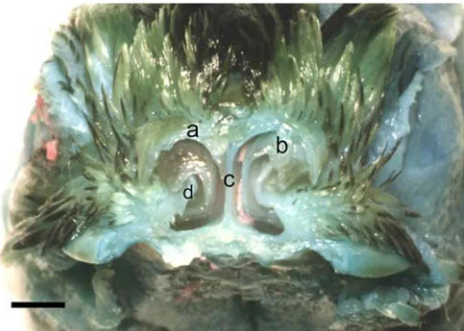
Figure 3. Transverse section through the MNC. a- Opening of infraorbital sinus, b- MNC, c-NS, d- Choanal opening, e- VM, f- Nasolacrimal duct. (Bar: 2mm).
Şekil 3. MNC’nin transversal kesiti. a- Sinus infraorbitalis’in açıklığı, b- MNC, c-NS, d- Choana, e- VM, f- Ductus nasolacrimalis. (Ölçü çubuğu: 2mm).
The rostral nasal concha (RNC) was present lying opposite the nostrils (Figs 1/b and 2/b). It was triangular in outline with a rostrally pointing cone like process. Its cranial edge projected through the nostril. The caudal border of the concha attached ventrally to the floor of the NC while it fastened dorsally to the dorsal border of the NC, completely closing the related area. As far as gross examinations were concerned, there was no air passage observed from inside space of the RNC to outer spaces. In transverse sections, the concha showed a C-shaped appearance in form with 6.23±0.1 mm long and 3.85±0.2 mm wide dorso-ventrally at its base.
The middle nasal concha (MNC) was situated obliquely between the RNC and CNC (Figs 1/c and 3/b). It was the largest of all, being 8.32±0.21 mm long and 2.54±0.12 mm wide dorso-ventrally. Its cranial edge attached to the dorsal wall of the NC while its caudal edge projected into the choana. In cross section, it exhibited a scroll-like structure with one-half turning ventro-laterally. This type of formation provided communication of the lumen of the concha with NC.
The caudal nasal concha (CNC) (Figs 1/d and 4/a), the smallest one, resembled a hemisphere of 3.2±0.15 mm in diameter with its caudal border attaching to the olfactory region of the NC, and its cranial edge being free. It was located dorso-caudal to the MNC. It was formed constantly with its cavity connecting with the infraorbital sinus, but not the NC.
The septal nasal concha (SNC) was absent in the Japanese quails examined.
The infraorbital sinus was highly developed triangular paranasal cavity situated beneath the orbit and in the lateral region of the upper jaw (Figs 3/a and 4/b). It extended cranially through the nostril and the orbit and
caudally to a region behind the orbit. The sinus had two openings; one located dorsally and leading into the CNC and the other into the ceiling of the NC above the MNC.
The common meatus (CM) was the largest part of the air passages (Fig 4/f). It was situated between the conchas and the NS, communicating freely with the other meatuses. The dorsal meatus (DM) served as an air passage between the dorso-lateral wall of the NC and the CNC (Fig 1/f). The intermedio-dorsal meatus (IDM) lied between the RNC and MNC (Fig 1/e) while the intermedio-caudal meatus (ICM) was present between the CNC and MNC. The ventral meatus (VM) was also seen between the RNC and MNC dorsally and the floor of the NC ventrally (Fig 4/e). All the other meatuses were directly in contact with the CM.
Figure 4. Transverse section through the CNC. a- CNC, b- Opening of infraorbital sinus, c-NS d- Choanal opening, e- VM. (Bar: 4mm).
Şekil 4. CNC’nin transversal kesiti. a- CNC, b- Sinus infraorbitalis’in açıklığı, c-NS d- Choana, e- VM. (Ölçü
çubuğu: 4mm). No differences were noted between male and females.
Discussion and Conclusion
Morphology of the NC in several avian species is well documented such as domestic fowl (14), Denizli cock (18) and domestic ducks (6). However investigations on the Japanese quail appear to be limited even though morphology of NC in Coturnix genus in general was described perfectly by Fitzgerald (9). Our study revealed the detailed features of the NC in the Japanese quail in particular.
The nostrils of the Japanese quails observed were Nares perviae type as in most birds including domestic chickens, composing of two narrow longitudinal openings located at the upper part of the base of beak (3). However, Das et al (6) reported that the oval nostrils were located on the dorsolateral aspect of the caudal one-third of the bill in the domestic duck. The size of the nostrils was reduced by the NO in Gallus (8, 9, 15). It was also the case in the nostrils of the Japanese quail. Formation of the nostrils and NO was mainly similar to that of other Gallus.
The choanal opening is generally a median elongated opening consisting of a triangular caudal part and a slit-like rostral part in avian species (11). In certain groups of birds including Galliformes; however, it remains unfused forming a cleft connecting the NC to the oral cavity (3). Our study, on the other hand, found that each choana displaying the slit-like rostral part and the triangular caudal part, namely interpalatine cleft, possessed two openings communicating the NC with the oral cavity and pharynx. This was also documented in Coturnix genus by Fitzgerald (9). Although the caudal end of the choana in Coturnix genus joins the pharynx through a common opening in the roof of the mouth (9) this common opening was very short in Japanese quail.
Although King and McLelland (13) and McLelland (14) reported that the RNC was absent in quails, it was observed in the Japanese quail of our study, which had also been documented in Coturnix genus by Fitzgerald (9). The moist mucous membrane and the rich blood structure of the NC help in warming and filtering the inhaled air. The absence or presence of the one of the NC’s might only be speculated at this stage as the less or more warming or filtering requirements in the NC of the quail.
The caudal border of this structure in Coturnix genus (9) attaches to the lateral wall of the nasal fossa and the dorsal border of the nostril, and its free dorsal and ventral edges were scrolled approximately three-fourths of a revolution laterally. We observed in the Japanese quail that the caudal border of the RNC attached ventrally to the floor of the NC while it fastened dorsally to the dorsal border of the NC. Therefore, it uniquely formed a completely closed structure. Gross examinations indicated that there was no communicating air ways observed from inside space of the RNC to outer spaces. This suggests that air transfer hereby, if there is, might occur at subgross and histological levels. According to King and McLelland (13) in transversal sections of the RNC of domestic fowl, appeared a simple, branched, T- or scroll-like shape with an additional vertical lamella of cartilage that arisen from the ventral border of the nostril. On the contrary, our study displayed a C-shaped RNC in the Japanese quails observed in transverse section. As to our knowledge, there are no other reports displaying the shape of the RNC in different avian species in cross section. Fitzgerald (9) also did not mention that in Coturnix genus. Due to that, it can’t be speculate whether this kind of shape is a specific feature of the Japanese quail or not.
The MNC has been documented to be the largest of the three conchas, possessing a scroll resemblance which is a specific feature of the MNC (13, 18), and situated obliquely between the dorsal and ventral turbinates in the Coturnix genus (9). It was displayed to exhibit, in cross section, a scroll 1-1.5 turns ventro-laterally. Moreover, Bittner (5), in cross sections, noted that the MNC was a scroll-like structure with at most two turns in domestic birds including chicken. In our study, the MNC in Japanese quail was also seen to exhibit one-half turns ventro-laterally, and positioned obliquely between the RNC and CNC, which is similar to those indicated in the literature.
The CNC in Coturnix genus, according to Fitzgerald (9), was the smallest nasal concha. Our study also indicated that in Japanese quail it was also the
smallest nasal concha, as in the pigeon (7, 15). However, this concha is a fairly constant features in birds, but is occasionally missing as in some falconiformes and swifts (13). Moreover, the SNC, a very unique structure of Petrels (1), was also not observed in the Japanese quail.
Naming of the meatuses is different in avian species that in mammals since the three main conchas of them lie in a rostro-caudal series so do the meatuses (3). The mammalian terms, dorsal, middle, and ventral nasal meatuses are not suitable for the avian species. The terms used in this study was derived from Fitzgerald (9).
The infraorbital sinus, as reported in quails by Fitzgerald (9), was developed into a triangular paranasal cavity situated beneath the orbit and in the lateral region of the upper jaw in Japanese quails.
In conclusion, this study documented the gross morphology of the NC in the Japanese quail, thus, comparing it to some other avian species. These may be useful findings for the researchers who are interested in gross morphology and possibly surgery of Japanese quails for the treatment of operable diseases or disorders.
Acknowledgements
We would like to thank Assoc Prof Dr Ibrahim Demirkan of Department of Surgery, Faculty of Veterinary Medicine, Kocatepe University, Afyon, Turkey, for his valuable discussion on the manuscript.
References
1. Bang BG (1971): Functional anatomy of the olfactory
system in 23 orders of birds. Acta Anat, 79, 1-76.
2. Bang BG, Wenzel BM (1985): Nasal cavity and olfactory
system. pp. 195-225. In: Form and Function in Birds (A.S.
King and J. McLelland, eds.) Vol. 3. Academic Press, London.
3. Baumel JJ, King AS, Breazile JE, Evans HE, Vanden Berge JC (1993): Nomina Anatomica Avium. Published by The Nuttall Ornithological Club. No: 23, Cambridge, Massachusetts.
4. Bellairs A, Jenkin CR (1960): Biology and Comparative
Physiology of Birds. Vol. 1. New York Academic Press.
5. Bittner H (1925): Nasenböhle und ihre Nebenböhle belm
Hausgeflügel. Berl. 41, 576-579.
6. Das LN, Mishra DB, Biswal G (1965): Comparative
anatomy of the domestic duck (Anas boscas). Ind Vet J, 42:
320-326.
7. Doğuer S, Erençin Z (1964): Evcil Kuşların Komparativ
Anatomisi. Ankara Universitesi Basımevi, Ankara,
Türkiye.
8. Dyce KM, Sack WO, Wensing CJG (1996): Avian
Anatomy. pp. 824-828. In: Textbook of Veterinary
Anatomy, 2nd ed. W.B. Saunders, Philadelphia.
9. Fitzgerald CT (1970): The Coturnix Quail Anatomy and
10. Flecknell PA (1992): Laboratory Animal Anesthesia. Academic Press Limited London.
11. Getty R (1975): Sisson and Grossman’s The Anatomy of
the Domestic Animals 5th ed. W.B. Saunders Company,
New York.
12. Howes JR, Ivey WD (1961): Coturnix quail for avian
research. Feedstuffs, May 12, 1-2.
13. King AS, McLelland J (1984): Birds: Their Structure and
Function. 2nd ed. Bailliere Tindall, London.
14. McLelland J (1990): A Colour Atlas of Avian Anatomy. Wolfe Publishing Ltd. England.
15. Nickel R, Schummer A, Seiferler E (1977): Anatomy of
domestic birds. Verlag Paul Parey. Berlin-Hamburg.
16. Pohlmeyer K, Kummerfeld N (1989): Morphologie der
Nasenhöhle und der nasen Nebenhöhlen sowie ihre klinische Bedeutung bei Grosspapageien. Kleintierpraxis.
34, 127-133.
17. Samuel OP, Sanches-Halman A, Goslaw Jr GE (1997):
Wing upstroke and the evolution of flapping flight. Nature.
387, 799-802.
18. Taşbaş M, Hazıroğlu RM, Çakır A, Özer M (1994):
Denizli horozunun solunum sisteminin morfolojisi I. Cavitas nasalis. Ankara Univ Vet Fak Derg, 41, 63-80.
19. Yamashina Y (1965): Haltung der Wachtelin in Japan. Deutsche Geflügelwirtschaft. 17, 415-416.
20. Zucker H, Gropp J, Peh J, Zentz Ch (1967):
Erfahrungen mit der japanischen Wachtel (Coturnix coturnix japonica) als Labortier sowie einige Ergebnisse von Nährstoffbedarfsuntersuchungen. Tierärtzliche
Umschau. 8, 416-435.
Geliş tarihi: 16.05.2006 / Kabul tarihi: 22.06.2006
Adress of Correspondance:
Dr A Çevik-Demirkan
Department of Veterinary Anatomy, Faculty of Veterinary Medicine, Afyon Kocatepe University, Afyonkarahisar, 03200, Turkey Tel: + 90 272 214 9309 Fax: + 90 272 228 1349 e-mail: [email protected]