Department of Emergency Medicine, Muğla Sıtkı Koçman University Faculty of Medicine, Muğla-Turkey 2Department of Radyology, Muğla Sıtkı Koçman University Faculty of Medicine, Muğla-Turkey
3Department of Thoracic Surgery, Muğla Sıtkı Koçman University Faculty of Medicine, Muğla-Turkey 4Department of Computer Hardware, Muğla Sıtkı Koçman University Faculty of Technology, Muğla-Turkey
ABSTRACT
BACKGROUND: There is still no agreed radiographic rule for the evaluation of blunt thoracic trauma. Emergency physicians want radiography according to their experience and examination findings. Various studies have been carried out on this subject and some of these studies have reached findings that can support the initial steps of the rules of radiography. One of them is the rule of Nexus thorax radiography rules. In this study, we aim to determine the accuracy of nexus thorax radiography rules.
METHODS: Our study was a prospective cohort study performed in the emergency department of our University Hospital. In this study, 690 patients were evaluated.
RESULTS: As a result of our study, we observed that patients were asked for more thoracic trauma because of chest pain, palpation tenderness in the thorax and sudden deceleration mechanism and pathology was found in approximately 25% of all imaging. The most common pathology we observed was rib fracture. Approximately 45% of the patients underwent thorax CT, and thorax CT was the most frequently requested for the detailed examination. When we evaluate the patients according to nexus thorax radiography rules, it was seen that the mechanism of sudden deceleration, intoxication and the disturbing, painful injury was more important than other parameters. The overall sensitivity and specificity of Nexus thorax radiographs were found to be 98% and 38%, respectively.
CONCLUSION: In the evaluation of blunt thoracic trauma, the rules of nexus thorax radiography are considered useful concerning pathological detection.
Keywords: Blunt thoracic trauma; emergency department; nexus rules.
vical vertebra radiography rules are used when evaluating cervical traumas. However, there is still no clinically accepted radiographic rule in the evaluation of thoracic trauma. Physi-cians request radiography according to examination findings and their own experience. Various studies have been car-ried out on this subject and some of these studies have ob-tained results that can support the initial steps of the rules of radiography. One of these is the rule of Nexus thorax radiography. However, these rules have not been included in the guidelines yet, and further studies are needed[6,8] In our
INTRODUCTION
Trauma is one of the most important causes of mortality and morbidity in adult patients. Thoracic, head, vertebral and abdominal traumas frequently result in death.[1–3] Currently, there are accepted imaging rules in abdominal and extremity trauma except head and cervical traumas.[4–7] For example, when evaluating head trauma, Glasgow coma score (GCS), as well as Canadian Brain Computed Tomography (CT) rules or New Orleans Brain CT rules, are used, and Nexus
cer-Cite this article as: Acar E, Demir A, Yıldırım B, Kaya G, Alataş ÖD, Kılınç RM, et al. Evaluation of the Nexus X-ray rules in blunt thorax trauma. Ulus Travma Acil Cerrahi Derg 2020;26:920-926.
Address for correspondence: Ethem Acar, M.D.
Muğla Sıtkı Koçman Üniversitesi Tıp Fakültesi, Acil Tıp Anabilim Dalı, Muğla, Turkey Tel: +90 252 - 212 75 27 E-mail: [email protected]
Ulus Travma Acil Cerrahi Derg 2020;26(6):920-926 DOI: 10.14744/tjtes.2020.55594 Submitted: 09.12.2019 Accepted: 11.02.2020 Online: 26.10.2020 Copyright 2020 Turkish Association of Trauma and Emergency Surgery
study, we aim to determine the accuracy of nexus thorax radiography rules.
MATERIALS AND METHODS
Our study is a prospective cohort study conducted in Mug-la Sıtkı Kocman University Education and Research Hospital Department of Emergency. Ethics committee permission was obtained for this study. Patients who were admitted to the emergency department with blunt thoracic trauma between May 2018 and May 2019 were included in this study. Inclusion criteria were: 1) Patients over 18 years of age, (2) patients who applied to the emergency department within 24 hours after blunt trauma (3) were asked to perform thorax imaging. The exclusion criteria were: (1) Patients under the age of 18 (2) who did not want to take part in this study (3) who ap-plied to the emergency department due to penetrating injury mechanism (4) who applied to our emergency department more than 24 hours after trauma (5) X-ray untreated or un-reached patients.
Before this study, a form was constructed, including the crite-ria and additional critecrite-ria of the NEXUS chest radiography rule (Table 1). Emergency medicine specialists who participated in this study were informed about this study and the questioning of the criteria. Emergency physicians were asked to evaluate the trauma according to the patient examination findings and their own experience and to question the criteria in the post-form. If the physicians underwent X-ray/CT in the presence of thoracic pathology, they were asked to mark the presence or absence of the causes in the above-mentioned form before the X-ray/CT evaluation (Table 1b). The form was also re-quested to mark whether there was a thoracic injury on the radiograph/CT. Hemothorax, pneumothorax, aortic and large vessel injury, rib fracture, diaphragmatic injury and pulmonary contusion were determined as thoracic injuries.
In the form, the indications of thoracic CT request of the physicians were questioned (Table 1c). In addition, whether the patient had any additional injuries to the form, if any (1) did not require follow-up (2) whether there was a pathology
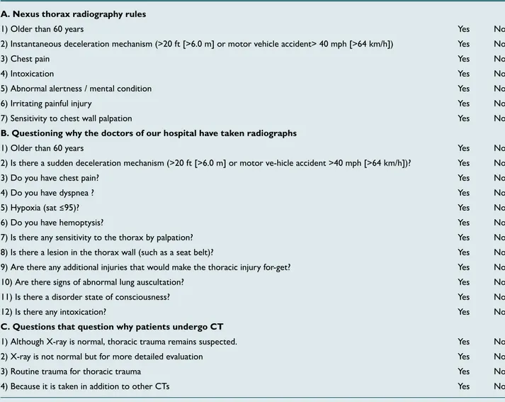
Table 1. Patient information collection form
A. Nexus thorax radiography rules
1) Older than 60 years Yes No
2) Instantaneous deceleration mechanism (>20 ft [>6.0 m] or motor vehicle accident> 40 mph [>64 km/h]) Yes No
3) Chest pain Yes No
4) Intoxication Yes No
5) Abnormal alertness / mental condition Yes No
6) Irritating painful injury Yes No
7) Sensitivity to chest wall palpation Yes No
B. Questioning why the doctors of our hospital have taken radiographs
1) Older than 60 years Yes No
2) Is there a sudden deceleration mechanism (>20 ft [>6.0 m] or motor ve-hicle accident >40 mph [>64 km/h])? Yes No
3) Do you have chest pain? Yes No
4) Do you have dyspnea ? Yes No
5) Hypoxia (sat ≤95)? Yes No
6) Do you have hemoptysis? Yes No
7) Is there any sensitivity to the thorax by palpation? Yes No
8) Is there a lesion in the thorax wall (such as a seat belt)? Yes No
9) Are there any additional injuries that would make the thoracic injury for-get? Yes No
10) Are there signs of abnormal lung auscultation? Yes No
11) Is there a disorder state of consciousness? Yes No
12) Is there any intoxication? Yes No
C. Questions that question why patients undergo CT
1) Although X-ray is normal, thoracic trauma remains suspected. Yes No
2) X-ray is not normal but for more detailed evaluation Yes No
3) Routine trauma for thoracic trauma Yes No
4) Because it is taken in addition to other CTs Yes No
whether the patient had thoracic trauma or whether other trauma had priority in patients with multiple traumas. In de-ciding which priority area it concerns, it was asked to decide which of the treatments were more prioritized and that were more serious injuries. In case of any uncertainty at this point, the situation was decided together with the physician caring for the patient together with two associate professors (EA, BY) who were in the same study. To minimize the margin of er-ror in this study, 10% of the patients were randomly selected. Radiographs of these patients were interpreted to the radiolo-gy and thoracic surgery specialists in the study independently. If radiological and thoracic surgeons were found to have signif-icant differences between the first physician and the physician who checked the patient, all radiographs were planned to be evaluated by the radiologist and thoracic surgeon. In line with these data, each parameter in the study form was recorded in the SPSS program and statistical analyzes were performed.
Statistical Analysis
All data obtained from this study were saved in the standard program of Statistical Package for Social Sciences for Windows 20. Numerical variables were summarized as mean ± SD (stan-dard deviation), and categorical variables were summarized as numbers and percentages. Frequency analysis was performed. To determine the weights of the attributes, the Evolutionary Weighting algorithm was used in Model-1 and Model-2. Here, the presence or absence of pathology on X-ray radiography as model 1 and the presence and absence of pathology on thorax CT as model 2 are expressed. Evolutionary algorithms (EAs: Evolutionary Algorithms) are among the most common-ly used algorithms for feature selection (FS). The basic mech-anism of such algorithms is to optimize the defined objective function for FS and to obtain the best subset of features.[9] This algorithm includes the application of the evolutionary algorithm for feature weighting. The weights obtained pro-vide quantitative information about the relative importance of the properties.[10] The optimization weight (evolutionary) operators calculate the weights of the attributes in the given data set using the genetic algorithm. The higher the weight of an attribute, the more relevant it is. Sensitivity or recall and specificity metrics were used to compare the performance of models created for feature selection. Sensitivity is the ratio of true positives correctly classified by the model. Specificity is the proportion of true negatives correctly classified by the
385 (55.8%) were injured due to in-vehicle traffic accidents,
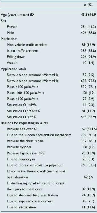
Table 2. Demographic characteristics of the patients (n=690)
n (%)
Age (years), mean±SD 45.8±16.9
Sex
Female 284 (41.2)
Male 406 (58.8)
Mechanism
Non-vehicle traffic accident 89 (12.9)
In-car traffic accident 385 (55.8)
Falling down 206 (29.9)
Assault 10 (1.4)
Application vitals
Systolic blood pressure ≤90 mmHg 52 (7.5)
Systolic blood pressure ≥90 mmHg 638 (92.5)
Pulse ≤100 pulse/min 532 (77.1) Pulse: 100–120 pulse/min 131 (19) Pulse ≥120 pulse/min 27 (3.9) Saturation O2 ≤89% 16 (2.3) Saturation O2: 90-94% 81 (11.7) Saturation O2 ≥95% 593 (85.9)
Reasons for requesting an X-ray
Because he’s over 60 169 (524.5)
Due to the sudden deceleration mechanism 209 (30.3)
Because the chest is pain 332 (48.1)
Because dyspnea 131 (19)
Because hypoxia (sat ≤95) 75 (10.9)
Due to hemoptysis 23 (3.3)
Due to thorax sensitivity by palpation 258 (37.4)
Lesion in the thoracic wall (such as seat
belt. abrasion) 62 (9)
Disturbing injury which cause to forget
the injury to the thorax 89 (12.9)
Due to abnormal lung auscultation 74 (10.7)
Due to impaired consciousness 49 (7.1)
Due to intoxication 11 (11.6)
206 (29.9%) were injured due to falls and 449 (65.1%) pa-tients applied to our emergency department by ambulance. It was found that 52 (7.5%) of the patients had systolic blood pressure below 90 mmHg at the time of admission to the emergency department, and 16 (2.3%) had oxygen saturation below 89%. Radiological examination indications for trauma were reported as chest pain in 332 patients (48.1%), thoracic sensitivity by palpation in 258 patients (37.4%), and radio-logical examination in 209 patients (30.3%) due to serious injury mechanism. The demographic values of the cases are given in Table 2. Radiological imaging of the trauma revealed 181 (26.2%) patients with chest trauma. Traumatic thoracic findings are presented in the table (Table 3).
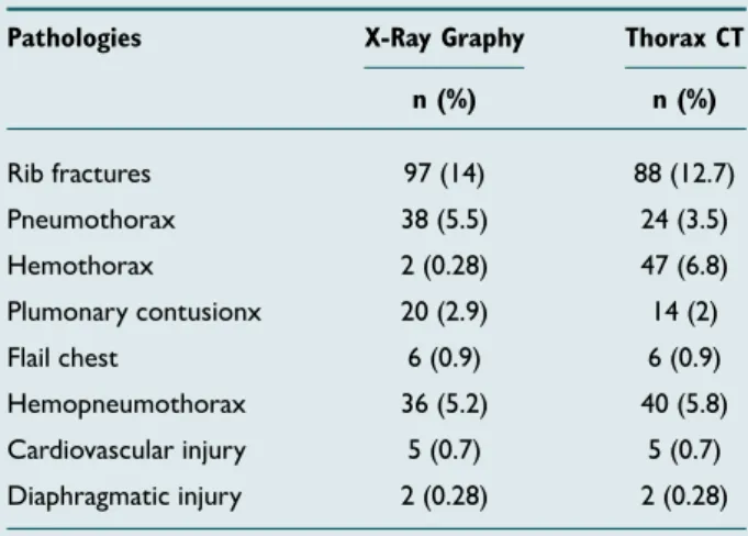
There were no additional injuries in 491 (71.2%) patients with multiple trauma, 52 (7.5%) patients with head, 23 (3.3%) patients with abdominal, 12 (1.7%) patients with pelvis, 11 (1%) 6) extremity and 62 (9%) patients had multiple trauma. 366 (53%) patients had no BT, whereas 324 (47%) patients had BT, although X-ray is normal, 63 (9.1%) patients due to suspected thoracic trauma. 136 (19.7%) patients had BT for detailed examination, 31 (4.5%) patients because of rou-tine in thorax trauma, 95 (13.8%) patients for additional BT even though X-ray is abnormal. According to CT results, 88 (12.5%) patients had rib fractures and 24 (3.5%) patients had pneumothorax (Table 3).
The findings showed that 111 (16.08%) patients with trau-matic thoracic injuries were followed, 56 (8.11%) patients underwent tube thoracostomy and 14 (2%) patients were treated with mechanical ventilation.
When hospitalizations were evaluated, 478 (70.6%) patients were discharged from the emergency department, 159 (23%) patients were admitted to the service, 40 (5.8%) patients were admitted to the intensive care unit and 4 (0.6%) pa-tients were ex.
When Model-1 results were evaluated, it was found that 11.2, 11.6 and 11.7 attributes were very significant for predicting the presence of pathology in X-ray. Sensitivity parameter of Model-1 was calculated as 0.983. In other words, Model-1 accurately estimates 98.3% of patients with pathology on X-ray. The estimated rate of patients without pathology is 37.9%. In addition, in Table 4, there is the complexity matrix of Model-1. According to this matrix, when the weight ratios given in Model-1 are used, 178 patients with pathology were classified as pathology, while three patients were incorrect-ly classified as no pathology. Similarincorrect-ly, 193 patients without pathology were classified as having no pathology, while 316 patients were incorrectly classified as having pathology (Ta-ble 5, 6).
Table 3. Pathologies detected in X-Ray and thorax CT
Pathologies X-Ray Graphy Thorax CT n (%) n (%) Rib fractures 97 (14) 88 (12.7) Pneumothorax 38 (5.5) 24 (3.5) Hemothorax 2 (0.28) 47 (6.8) Plumonary contusionx 20 (2.9) 14 (2) Flail chest 6 (0.9) 6 (0.9) Hemopneumothorax 36 (5.2) 40 (5.8) Cardiovascular injury 5 (0.7) 5 (0.7) Diaphragmatic injury 2 (0.28) 2 (0.28)
X: Flail chest was diagnosed clinically and radiologically. CT: Computed tomography.
Table 4. Attribute weights and performance metrics
AttributeWeigth Sensivity Specificity Model 11.1 11.2 11.3 11.4 11.5 11.6 11.7
Model-1 0 1 0 0 1 0 1 98.3% 37.9%
Model-2 0 0.96 0.56 0.56 1 0 1
Model 1: Pathology on X-Ray, Model 2: Pathology on thorax CT 11.1: Above 60 years of age, 11.2 Sudden deceleration mechanism, 11.3 chest pain, 11.4 chest wall sensitivity, 11.5 intoxication, 11.6 abnormal alertness / The mental state represents 11.7 irritating painful injuries.
Table 5. Model-1 parameters
Property Value
Population size 19
Maximumnumber of the generations 19
P crossover 0.6
Crossover type Shuffle
Selections scheme Roulette wheel
Table 6. Model-1 complexity matrix
True-no True-yes
Prediction X-Ray result no disea-se 193 316
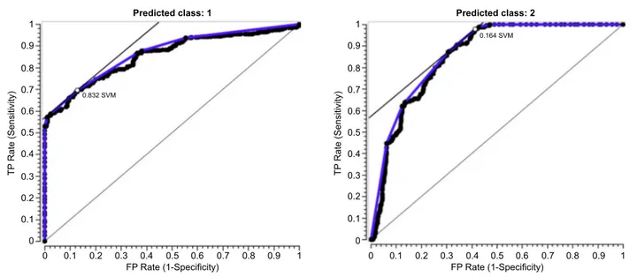
The strength of the diagnostic value for X-Ray if there is any pathology (No (1) and Yes (2) distinguishing power) is ex-pressed by the area under the ROC curve. The diagnostic value increases as the field value approaches 1. At 100% diag-nostic power, the field value is equal to 1. The results of the analysis performed with the Orange program in this study, , the ROC curve graphs obtained for are shown in Figure 1a and b that indicates the presence of pathology and CT results in the X-Ray. Support vector machines (SVM) were used as the classification method in the program.
DISCUSSION
Trauma is the transfer of kinetic energy to the patient (ac-celeration, de(ac-celeration, or blastic) by different mechanisms. Traumatic damage may vary depending on the duration of the trauma, the mechanism of occurrence, the object or the physical structure of the patient. Thoracic traumas have been reported to be more common in young males.[13,14] In our study, the most common cause of thoracic trauma was an in-vehicle traffic accident. Males were the majority and the mean age of the patients was 45 years.
Complaints of chest pain, thoracic tenderness with palpation and sudden deceleration mechanism criteria were requested in the patients and traumatic thoracic pathology was observed in approximately 25% of all images. The most common thoracic trauma was rib fracture. Thorax CT was requested in 45% of the patients for further examination. When the patients were evaluated according to nexus thorax radiography rules, sudden slowdown mechanism, intoxication and painful injury were found to be statistically significant in detecting trauma. The overall sensitivity and specificity of Nexus thorax radiographs were 98% and 38%, which was consistent with the literature.[6,8] According to the literature data, the most decisive criteri-on for blunt thoracic trauma has been reported to be the
mechanism of sudden deceleration.[6,8,15] In our study, it was concluded that one of the most important criteria concern-ing the indication of request for radiological examination and the detection of traumatic pathology is the sudden decel-eration mechanism in accordance with the literature data. In addition, the symptom of chest pain in trauma was found to be as important and determinative as the criteria of sud-den slowdown mechanism. Any condition that disrupts the consciousness of the person or the presence of additional injuries that may neglect the injury in the thorax is also the parameters that should be a serious stimulus for physicians. In our study, we found that 73% of patients underwent X-ray, 47% underwent thorax CT, and 25% of our patients had tho-racic pathology, and the most common pathologies were rib fracture, pneumothorax and hemothorax. Safari et al.[16] stat-ed that they detectstat-ed the rate of radiography around 43% and most commonly seen jeans fracture and hemothorax. In another study, 43% of the patients underwent X-ray, 42% of them underwent CT, and 15% of the patients reported that they detected thoracic pathology and most commonly found rib fracture and pneumothorax.[6] In another study, it was stated that the rate of X-ray use was 50% and the rate of using CT was about 50%, and the rate of thoracic pathol-ogy was 15%.[15] As can be seen from the literature, CT scan results and detected pathologies are compatible with each other, but it will be seen that we take a little more X-ray at the rate of radiography. It may be difficult to determine why more is taken, but the aim of our study is to reduce the rate of radiography, which means that such studies are needed because we already believe that many graphics are required. In this context, we think that this result can be interpreted by us in accordance with the literature.
Because it is considered a forensic case concerning trauma, physicians may be reluctant to evaluate a trauma patient and may require unnecessary radiographs and CTs to secure
Figure 1. Graph of the ROC curve obtained for the X-ray (a) and CT result (b) on the X-Ray after the analysis with the Orange program.
0.3 0.2 0.1 0 0 0.1 0.2 0.3 0.4 FP Rate (1-Specificity) TP Rate (Sensitivity) 0.5 0.6 0.7 0.8 0.9 1 0.3 0.2 0.1 0 0 0.1 0.2 0.3 0.4 FP Rate (1-Specificity) TP Rate (Sensitivity) 0.5 0.6 0.7 0.8 0.9 1
them. At this point, the algorithms or scoring systems that will relieve the physician from this feeling of insecurity seem to be applicable concerning forensic/medical relief. This also applies to the patient. Patients get rid of irradiation due to unnecessary radiographs, and as a result of these sys-tems, which have a sensitivity of almost 100%, they can be examined more safely with less radiation. There are many examples of this situation. For example, Canada Brain CT rules used in patients with head trauma have reduced the rate of brain CT scans by 30%, but the probability of patient skipping has decreased to almost 0%.[13] Of course, there is also a protocol now known as head-to-pelvis CT proto-cols. In this protocol, physicians undergo full-body CT to reduce time loss and minimize skippable injuries in patients with multiple trauma, hemodynamically unstable patients and even recommend this application by predicting less radiation from CTs to be taken separately.[17–19] Although it is very popular in this regard, ATLS instead focuses on system evaluations where physical examination and clinical evaluations are at the forefront.[20] This application of ATLS requires more clinical findings and physical examinations, the physician experience and the subjective evaluations of the physician more. With the scoring rules to be added to these practices, these subjective evaluations can be further secured with objective parameters and will comfort the pa-tient and the physician.
The algorithm or parameters that should be considered while developing such a rule are easy to apply and can be used for tracking purposes. From this perspective, it is evident that the rules of nexus thorax radiography are easy to apply pa-rameters. It has been tried and found to be easy to apply in many studies, including our study.[15,21,22] One of the import-ant parameters here is the omission of a potentially fatal inju-ry is extremely critical and eliminates the implementation of the algorithm. In the previous studies in this context, the fatal injury - the aortic injury as the fatal injury - was not omitted and the reliability of the rules seems to be confirmed.[6] The absence of a fatal pathology missed in our study supports the previous studies and confirms the reliability of the rules. In this context, we think that the necessity of entering these parameters into the algorithms will be on the agenda soon. In conclusion, when the physicians note that the risk of nexus thorax radiography/CT is low risk in detail in the patient’s file, we think that the rules of nexus thorax radiography/CT will protect physicians both legally and conscientiously in pa-tients with blunt thoracic trauma. In this context, we believe that these rules can be applied. However, medicine is an art. Physicians, who are practitioners of this art, should not forget to examine and follow the patient and try to integrate these rules, which we mentioned in the quality and effective exam-ination. As long as this is realized, we think that the diagnosis will be minimal in patients.
This research did not receive any specific grant from funding agencies in the public, commercial, or not-for-profit sectors.
Ethics Committee Approval: Approved by the local
eth-ics committee.
Peer-review: Internally peer-reviewed.
Authorship Contributions: Concept: E.A.; Design: E.A.,
A.D.; Supervision: B.Y., R.M.K., A.Z Resource: A.D., G.Y.; Data: A.D., G.K., Ö.D.A.; Analysis: O.Ö; Literature search: E.A., Ö.D.A.; Writing: E.A.; Critical revision: B.Y., R.M.K., A.Z.
Conflict of Interest: None declared.
Financial Disclosure: The authors declared that this study
has received no financial support.
REFERENCES
1. Ekeke ON, Okonta KE. Trauma: a major cause of death among surgical inpatients of a Nigerian tertiary hospital. Pan Afr Med J 2017;28:6. 2. Osime OC, Ighedosa SU, Oludiran OO, Iribhogbe PE, Ehikhamenor E,
Elusoji SO. Patterns of trauma deaths in an accident and emergency unit. Prehosp Disaster Med 2007;22:75–8. [CrossRef ]
3. Shekhar C, Gupta LN, Premsagar IC, Sinha M, Kishore J. An epidemi-ological study of traumatic brain injury cases in a trauma centre of New Delhi (India). J Emerg Trauma Shock 2015;8:131–9. [CrossRef ]
4. Curran JA, Brehaut J, Patey AM, Osmond M, Stiell I, Grimshaw JM. Un-derstanding the Canadian adult CT head rule trial: use of the theoretical domains framework for process evaluation. Implement Sci 2013;8:25. 5. Mata-Mbemba D, Mugikura S, Nakagawa A, Murata T, Kato Y,
Tatewa-ki Y, et al. Canadian CT head rule and New Orleans Criteria in mild traumatic brain injury: comparison at a tertiary referral hospital in Japan. Springerplus 2016;5:176. [CrossRef ]
6. Rodriguez RM, Anglin D, Langdorf MI, Baumann BM, Hendey GW, Bradley RN, et al. NEXUS chest: validation of a decision instrument for selective chest imaging in blunt trauma. JAMA Surg 2013;148:940–6. 7. Nishijima DK, Simel DL, Wisner DH, Holmes JF. Does this adult
pa-tient have a blunt intra-abdominal injury?. JAMA 2012;307:1517–27. 8. Rodriguez RM, Hendey GW, Mower WR. Selective chest imaging for
blunt trauma patients: The national emergency X-ray utilization studies (NEXUS-chest algorithm). Am J Emerg Med 2017;35:164–70. [CrossRef ]
9. Paul S, Das S. Simultaneous feature selection and weighting – An evo-lutionary multi-objective optimization approach. Pattern Recognition Letters 2015;65:51–9. [CrossRef ]
10. Komosiński M, Krawiec K. Evolutionary weighting of image features for diagnosing of CNS tumors. Artif Intell Med 2000;19:25–38. [CrossRef ]
11. Altman DG, Bland JM. Diagnostic tests. 1: Sensitivity and specificity. BMJ 1994;308:1552. [CrossRef ]
12. Parikh R, Mathai A, Parikh S, Chandra Sekhar G, Thomas R. Under-standing and using sensitivity, specificity and predictive values. Indian J Ophthalmol 2008;56:45–50. [CrossRef ]
13. Wright DW, Merck LH. Head Trauma. In: Tintinalli JE, Stapczynski JS, Ma OJ, Yealy DM, Meckler GD, Cline DM. Tintinalli’s Emergency Medicine A Comprehensive Study Guide. 8th edition. McGraw-Hill Ed-ucation 2016.p.1695–707.
14. Yalçınkaya İ, Sayır F, Kurnaz M, Çobanoğlu U. Chest trauma: analysis of 126 cases. Ulus Travma Acil Cerrahi Derg 2000;6:288–91.
15. Rodriguez RM, Langdorf MI, Nishijima D, Baumann BM, Hen-dey GW, Medak AJ, et al. Derivation and validation of two decision instruments for selective chest CT in blunt trauma: a multicenter prospective observational study (NEXUS Chest CT). PLoS Med 2015;12:e1001883. [CrossRef ]
OLGU SUNUMU
Künt toraks travmasında Nexus X-ray kurallarının değerlendirilmesi
Dr. Ethem Acar,1 Dr. Ahmet Demir,1 Dr. Birdal Yıldırım,1 Dr. Gökhan Kaya,1
Dr. Ömer Doğan Alataş,1 Dr. Rabia Mihriban Kılınç,2 Dr. Arife Zeybek,3 Dr. Osman Özkaraca4 1Muğla Sıtkı Koçman Üniversitesi Tıp Fakültesi, Acil Tıp Anabilim Dalı, Muğla
2Muğla Sıtkı Koçman Üniversitesi Tıp Fakültesi, Radyoloji Anabilim Dalı, Muğla 3Muğla Sıtkı Koçman Üniversitesi Tıp Fakültesi, Göğüs Cerrahisi Anabilim Dalı, Muğla 4Muğla Sıtkı Koçman Üniversitesi Teknoloji Fakültesi, Bilgisayar Donanımı Anabilim Dalı, Muğla
AMAÇ: Künt toraks travmasının değerlendirilmesi için hala kabul edilmiş bir radyografik kural yoktur. Acil hekimleri tecrübelerine ve muayene bulgularına göre radyografi istemektedirler. Bu konuda çeşitli çalışmalar yapılmış ve bu çalışmaların bir kısmında radyografi kurallarının ilk adımlarını destekleyebilecek sonuçlarda tespit edilmiştir. Bunlardan biri Nexus toraks radyografi kuralıdır. Çalışmamızda, Nexus toraks radyografi kurallarının doğruluğunu belirlemeyi amaçlıyoruz.
GEREÇ VE YÖNTEM: Çalışmamız üniversite hastanemiz acil servisinde yapılan ileriye yönelik bir kohort çalışmasıdır. Çalışmada 690 hasta değer-lendirildi.
BULGULAR: Çalışmamız sonucunda göğüs ağrısı, palpasyonla toraksta hassasiyeti, ani yavaşlama mekanizması nedeniyle daha fazla toraks grafisi çe-kildiğini ve tüm görüntülemenin yaklaşık %25’inde patoloji tespit edildiği gözlenmiştir. En sık rastlanan patolojiler kaburga kırığı idi. Hastaların yaklaşık %45’ine toraks bilgisayarlı tomografi (BT) çekildi ve toraks BT en sık olarak detaylı inceleme için istendi. Hastaları Nexus toraks radyografi kuralları-na göre değerlendirdiğimizde ani yavaşlama, zehirlenme ve rahatsız edici ağrılı yaralanma mekanizmasının diğer parametrelerden daha önemli olduğu görülmektedir. Nexus toraks radyografilerinin genel duyarlılığı ve özgüllüğü sırasıyla %98 ve %38 olarak bulundu.
TARTIŞMA: Künt toraks travmasının değerlendirilmesinde, Nexus toraks radyografisi kuralları patolojik tanı açısından faydalı olarak değerlendiril-mektedir.
Anahtar sözcükler: Acil servis; künt toraks travması; Nexus kuralları.
Ulus Travma Acil Cerrahi Derg 2020;26(6):920-926 doi: 10.14744/tjtes.2020.55594