R E S E A R C H
Open Access
Extracellular microRNAs: key players to
explore the outcomes of in vitro
fertilization
Haroon Latif Khan
1, Shahzad Bhatti
1,2,3*, Sana Abbas
1, Celal Kaloglu
4, Ahmed M. Isa
5, Hooria Younas
6,
Rachel Ziders
7, Yousaf Latif Khan
1, Zahira Hassan
8, Bilgün Oztürk Turhan
9, Aysegul Yildiz
10,
Hikmet Hakan Aydin
11and Ender Yalcinkaya Kalyan
12Abstract
Background: MicroRNAs (miRNAs) are small RNA molecules that modulate post-transcriptional gene regulation. They are often used as promising non-invasive biomarkers for the early diagnosis of cancer. However, their roles in assisted reproduction are still unknown.
Methods: This prospective study was designed to evaluate the expression profiles of seven extracellular miRNAs (miR-7-5p, miR-202-5p, miR-378-3p, miR-224, miR-320a, miR-212-3p, and miR-21-5p) in human follicular fluid (FF) to explore the outcomes of in vitro fertilization (IVF). Of 255 women, 145 were without polycystic ovary syndrome (PCOS), and their ovarian assets were normal (NOR), while 110 were with normo-androgenic PCOS.
Results: The combination of six FF miRNAs expression profile discriminated between PCOS and NOR women with a sensitivity of 79.2% and a specificity of 87.32% (AUC = 0.881 [0.61; 0.92],p = 0.001). MiR-202-5p significantly had a lower abundance level, and miR-378-3p had a high abundance level in pooled FF samples from patients treated with human menopausal gonadotropin (hMG) than those treated with recombinant follicle-stimulating hormone (rFSH) (p < 0.001). Our results showed that miRNA-320a was significantly different in top-quality embryos versus non-top-quality embryos on day 3 in NOR patients with a sensitivity of 80% and specificity of 71%, (AUC = [0.753 (0.651; 0.855)],p = 0.001). For clinical pregnancy outcome prediction, FF miRNA-21 exhibited high sensitivity (74.8%) and specificity (83.7%) with the AUC value of 0.774 (0.682; 0.865).
Conclusion: Conclusively, our results provide evidence that miR-7-5p, miR-378-3p, miR-224, miR-212-3p were a differentially high expression in normo-androgenic PCOS patients than NOR patients. While miRNA-320a was significantly different in top-quality embryos versus non-top-quality embryos on day 3 (p = 0.001). The expression level of FF miR-212-3p was significantly related to the probability of embryos to develop into a high-quality blastocyst in patients with normal ovarian reserve.
Keywords: PCOS, miRNA expression, Follicular fluid, IVF, Embryo quality
© The Author(s). 2021 Open Access This article is licensed under a Creative Commons Attribution 4.0 International License, which permits use, sharing, adaptation, distribution and reproduction in any medium or format, as long as you give appropriate credit to the original author(s) and the source, provide a link to the Creative Commons licence, and indicate if changes were made. The images or other third party material in this article are included in the article's Creative Commons licence, unless indicated otherwise in a credit line to the material. If material is not included in the article's Creative Commons licence and your intended use is not permitted by statutory regulation or exceeds the permitted use, you will need to obtain permission directly from the copyright holder. To view a copy of this licence, visithttp://creativecommons.org/licenses/by/4.0/. The Creative Commons Public Domain Dedication waiver (http://creativecommons.org/publicdomain/zero/1.0/) applies to the data made available in this article, unless otherwise stated in a credit line to the data.
* Correspondence:[email protected];[email protected]
1
Lahore Institute of Fertility and Endocrinology, Hameed Latif Hospital, 14 Abu-Bakar Block New Garden Town, 54800 Lahore, Pakistan
2Department of Human Genetics and Molecular biology, University of Health
Sciences, Lahore 54600, Pakistan
Background
Extracellular microRNAs (miRNAs) are among the most abundant yet enigmatic biomolecules. Belonging to a large group of endogenously expressed single-stranded RNA molecules, ranging from 21 to 24 nucleotides in length, they regulate a wide array of biological processes. They do this by silencing genes and typically work as transcrip-tional and post-transcriptranscrip-tional regulators of gene expres-sion. MiRNAs do not encode proteins themselves but possess specific binding sites to messenger RNA (mRNA) targets, promoting their decay and inhibiting translation
[1]. In mammalian reproduction, miRNAs are markedly
involved in controlling several reproductive functions in females, such as folliculogenesis, steroidogenesis, oocyte maturation, early embryonic development, cell prolifera-tion, and apoptosis [2]. They are primarily expressed in all human reproductive tissues and biological fluids, includ-ing follicular fluid (FF) [3]. Extracellular miRNAs can be readily quantified as they are highly conserved and notably stable in a harsh environment with the presence of en-dogenous RNase activity [4]. Seemingly, they are often used as promising non-invasive biomarkers for the early diagnosis of cancer; however, their roles in assisted reproduction treatments remain unknown [5].
Currently, the ovarian reserve assessment is inferred through serum levels of anti-Mullerian hormone (AMH) and basal antral follicle count (AFC) by consultants. These markers are also routinely used to monitor the ovarian response during gonadotrophin therapy. None-theless, it became increasingly distinct that variability be-tween laboratory procedures and lack of a standardized international assay is one of the fundamental limitations of AMH testing in ART [6]. Polycystic ovary syndrome (PCOS) is the most frequent endocrine disorder, which has an intricate inheritance pattern among multifaceted endocrinopathies, affecting women of childbearing age. Abnormal preantral folliculogenesis in PCOS is the prcipal cause of more than 75% of cases of anovulatory in-fertility and menstrual cycle disturbances [7].
Altered miRNAs expression profile has been reported in the serum and FF of patients with PCOS. Therefore, these differential miRNAs might be classified as specific therapeutic targets and diagnostic biomarkers of PCOS. They could also reflect the real status of fertilization po-tential in couples undergoing IVF/intracytoplasmic sperm injection (ICSI) treatment [8].
Over the past decade, numerous articles have ex-plained that several miRNAs are fundamentally con-cerned with the steroidogenic regulation of ovarian follicles, yet the impact of gonadotropin therapy on miRNA expression in the follicular microenvironment remains within the exploratory phase [9,10]. Specifically, human FF is a complex biological fluid with intrinsic regulation of the micro-environment, determining the
oocyte developmental potential and post-fertilization de-velopment of an embryo. Subsequently, metabolic changes in its components have been designated as ideal noninvasive biomarkers to predict oocyte and embryo quality. Additionally, by comparing gene expression pro-files, it was revealed that miRNAs might be key media-tors in specific disease etiology and certain cellular processes; notably, they have an immense contribution as a regulator of oogenesis and embryogenesis [11,12].
Circulating miRNA-21 acts as key oncomiRs, which are ex-tensively overexpressed in numerous cancer forms, and mul-tiple studies have demonstrated that aberrant overexpression endorses proliferation, invasion, and migration through down-regulation of various tumor suppressor genes [13,14]. How-ever, the downregulation of miRNA-21-5p in granulosa cells (GCs) promotes apoptosis by targeting PI3K/AKT and JAK/ STAT3 kinase cascades [15]. It is also linked with RNA-induced silencing complex in GCs and regulates cell prolifera-tion, differentiaprolifera-tion, and survival through TGF-β signaling [16,
17]. Earlier studies exhibited that miR-320a is of the top ten highly expressed miRNAs identified in the FF samples and positively linked with embryo quality [18,19]. Several studies have established that miR-7-5p acts as a key mediator in the feedback loop of the hypothalamus-pituitary-ovary axis by regulating gonadotropins, and knockout leads to infertility due to hypogonadism in the mouse model [20–22]. Previous evi-dence from in vitro study demonstrated that miR-212 has a potential role in regulating gonadotropins, including follicular development and ovulation in equines [23]. The miR-202-5p is the most dominant form of the miRNAs abundantly expressed in GCs and non-fertilized oocytes. The knockout of miR-202-5p compromises the quantity and quality of oocytes [24]. Likewise, the significant upregulation of miR-378-3p in-creases the density of the primordial follicles in the ovary. It also induces an increased autophagy level in response to oxi-dative stress and delays the apoptotic events inside the devel-oping follicles [25]. Li and his fellows highlight the diverse role of miR-224 in ovaries and a promising marker of PCOS. They explained that the overexpression of miR-224 reduces the Ptx3 and Smad4 gene expression responsible for cell expan-sion and oocyte development [26].
This contemporary study was designed with a primary objective to seek out seven extracellular miRNAs (7-5p, miR-202-5p, miR-378-3p, miR-224, miR-320a, miR-212-3p, miR-21-5p) expression profiles in the follicular microenvironment of IVF patients with polycystic ovar-ies and with normal ovarovar-ies. Further, we examined the importance of these miRNAs to predict the success of ARTs in normal responders.
Methods
Subjects
This prospective study was conducted on 255 women undergoing IVF/ICSI at the Lahore Institute of Fertility
and Endocrinology, Hameed Latif Hospital Lahore, be-tween January 2017 to January 2020. Among 255 women, 145 with normal ovarian reserve (NOR), while 110 women had normo-androgenic PCOS, diagnosed based on Rotterdam criteria, i.e., clinical or biochemical hyperandrogenism, oligo-and/or anovulation, and ap-pearance of polycystic ovaries on ultrasound examin-ation. The functional ovarian response was determined by measuring the AMH concentration in serum and AFC through transvaginal sonography on cycle day 3. Patients were informed about the study’s objective, and written informed consent was taken before collecting FF samples. This study was approved by the institutional ethical review board, and research was conducted under the recommended guidelines.
IVF procedure
Of 255 women, 141 received gonadotrophin-releasing hormone (GnRH) agonist (Decapeptyl, Ipsen Pharma), and 114 were undergoing COS (Controlled ovarian stimulation) with antagonist protocol. Two types of go-nadotropins were used in COS: r-FSH and HP-hMG. Serum E2 levels and folliculograms were used to assess the ovarian response. Ovulation was triggered with ex-ogenous human chorionic gonadotropin (hCG) (LG, Life
Sciences 5000–10,000 IU) once 3 follicles reached a
mean diameter of ≥18 mm. After 36 h of hCG
adminis-tration, oocyte retrieval was performed utilizing transva-ginal ultrasound and a single lumen needle (Cook, KOPS-1230-VUB, Limerick, Ireland). Flushing during these retrievals was prevented, and cumulus-oocyte complexes (COC) were removed mechanically. FF sam-ples were stored at − 80 °C for further evaluation. Oo-cytes were cultured singly in 30μl of cleavage medium (Vitrolife) and under mineral oil (Ovoil™) at 37 °C in a humid setting containing O2 5%, CO26%, and N2 89%,
respectively. The fertilization examination was done after 18 h of insemination, and normal fertilization is demon-strated by the appearance of two pronuclei (2PN) and two polar bodies.
Embryo quality was documented on day 3 based on the cleavage-stage-scoring system, including the number of cells, blastomere regularity, degree of fragmentation, and multinucleation. Top-quality embryos on day 3 were considered those with 6–8 regular blastomeres and less than 15% fragmentation [27]. Blastocysts were vitrified
and kept in liquid nitrogen (− 196 °C) until embryo
transfer. Morphological evaluation of blastocysts was done through the Gardner scoring system [28], and only fully expended blastocysts (IV/V grade) with cohesive trophectoderm (A/B quality) were warmed in a closed vitrification system and transferred under the procedure recommended by Irvine Scientific. Clinical pregnancy
was verified by fetal cardiac activity with at least one gestational sac detected on ultrasonography.
Follicular fluid (FF) sample preparation
After egg retrieval, intrafollicular fluid from both groups (110 PCOS and 145 NOR) pooled separately and subject to centrifugation at 4000 rpm for 15 min. However, in NOR patients, 0.5 mL of the FF from each mature fol-licle (n = 280) with ≥18 mm diameter having a volume of 2–3 cm3
were handled separately to associate the miR-NAs profiles with embryo quality or with pregnancy rate. Moreover, in NOR patients, each mature oocyte, related embryo, and FF sample were processed separ-ately in the IVF laboratory. Clear supernatant fluids were separated and filtered, applying 0.45μL syringe filters into sterile 10 ml tubes by excluding dead cells and then stored at − 80 °C. All follicular aspirations were per-formed between 8: 30 h and 10.00 h.
Hormonal analysis
BMI was calculated based on height and weight through standardized equipment. Venous blood was drawn be-tween 8: 00 am to 9: 00 am. Baseline hormones such as follicular stimulating hormone (FSH), luteinizing hor-mone (LH), 17β-estradiol (E2), thyroid-stimulating
hor-mone (TSH), Total Testosterone (TT), sex horhor-mone- hormone-binding globulin (SHBG), and anti-mullerian duct hor-mone (AMH) were assessed on 2nd day of the menstrual cycle through electrochemiluminescence immunoassay, according to the manufacturer’s instructions (Elecsys® Roche Diagnostics, Indianapolis, USA). The antral fol-licle count (AFC) was assessed using transvaginal ultra-sonography (TVS) on the 2nd or 3rd day of the menstrual cycle.
MicroRNAs (miRNA) extraction
MiRNAs extraction was done according to the manufac-turer’s protocol. Specific miRNAs were evaluated from 4 ml of each FF pool using QIAamp Circulating Nucleic Acid Kit (Qiagen). In short, in the lysis step, 3 ml of FF
sample pool with 500μL Proteinase K (20 mg/ml) and
4.5μL of Buffer ACL were mixed that inactivated the
DNases and RNases, resulting in the release of nucleic acids from vesicles. Then the mixture was incubated at 60 °C for 30 min. Next, to improve miRNAs’ binding capability to the membrane, 10 ml ABC buffer was added to the lysate and shifted lysate onto a QIAamp Mini Column after mixing by pulse vertexing. The miR-NAs were incorporated onto a small silica membrane as the mixture is drawn through by vacuum pressure. The residual contaminants were thoroughly washed away after three steps-washing of the membrane, and pure cir-culating nucleic acids were eluted in buffer AVE at room temperature.
Evaluation of miRNAs differential expression profile of human FF through qRT-PCR
The candidate miRNAs assay was determined through qRT-PCR with TaqMan™ MicroRNA assay (Applied Bio-systems), and universal stem-loop primers (USLP) with a low intra-assay coefficient of variation (ICV) were used
to generate complementary DNA (cDNA). The 15μL of
RT reaction mix comprised FF pool sample: 5μL (1–10
ng of total RNA), 100 mM dNTP: 0.15μL, 10X RT
buf-fer: 1.5μL, 50 U/ μL MultiScribe™ RT enzyme: 1 μL, 20
U/ μL RNase inhibitors: 0.19 μL, nuclease-free water:
4.16μL, and TaqMan™ 5X RT primers: 3 μL. The reverse
transcription was performed under standard cycling con-ditions: 16 °C for 30 min, 42 °C for 30 min, 85 °C for 5 min, and hold step at 4 °C. CFX-96® touch RT-PCR de-tection system (Bio-Rad-Life-Sciences, USA) was used for quantitative PCR, carried out in a 384-well plate, and duplicated for each sample with negative control (water).
The 10μL PCR reaction mix contained cDNA: 3 μL,
PCR MasterMix: 5μL (TaqMan®, Applied Biosystems),
and primer: 2μL under the following conditions: initial denaturation step at 95 °C for 10 min, 50 cycles of 95 °C for 15 s, and anneal at 60 °C for 60 s. miRNA-16 was ap-plied as endogenous control [29], and eq. 2-ΔCt was used
to determine the relative expression of 7-5p, miR-202-5p, miR-378-3p, miR-224, miR-320a, miRNA-212-3p, miR-21-5p in each FF pool. These seven miRNAs were se-lected based on their extensive clinical importance in oo-cyte maturation and embryo quality [11, 19,30, 31]. We estimated the fold change by evaluating the relative ex-pression levels between normo-androgenic PCOS patients
and NOR patients by applying the 2-ΔΔCrt method and
normalized to miR-16 expression in FF [32,33].
Pathway analysis and gene ontology (Go)
MiRNAs were uploaded for KEGG pathway enrichment analysis and Gene Ontology (GO). Putative target genes of studied miRNAs were predicted through eight
avail-able bioinformatic algorithms such as miRanda (www.
cbio.mskcc.org/miRNA2003/miranda.html) version 1.9, TargetScan (http://www.targetscan.org/vert_72/) version 7 . 2 , D I A N A - m i c r o T (h t t p : / / d i a n a . i m i s . a t h e n a -innovation.gr/DianaTools/index.php?r=microT_CDS/ index) version 4, miRDB (http://www.mirdb.org/), Pic-Tar (https://pictar.mdc-berlin.de/), miRWalk (http://zmf. umm.uni-heidelberg.de/apps/zmf/mirwalk2/), RNA hy-brid (https://bibiserv.cebitec.uni-bielefeld.de/rnahybrid/),
and target gene prediction at EMBLE (http://www.
russell.embl.de/miRNAs/). However, to minimize the number of false positives targets, we include only those target genes that were predicted by 6 out of 8 programs and proceeded for further use in GO annotation and KEGG (keto Encyclopedia of Genes and Genomes)
path-way enrichment analysis using DAVID (https://david.
ncifcrf.gov/) version 6.8 integrate functional genomic an-notation [34]. Benjamini and Hochberg’s multiple testing
correlation was used to adjust thep-values (−log 10 FDR adjustedp values = < 0.05).
Statistical analysis
Categorical variables are presented as numbers and per-centages, and continuous parametric data are shown as mean ± standard deviation (SD). The Shapiro-Wilk test was used to verify the normality of data, while to com-pare and correlate quantitative variables, we employed the Mann-Whitney and the Spearman Rank tests, re-spectively. Univariate analysis was conducted to relate the relationship with PCOS for clinical parameters and each FF miRNA relative expression. Those miRNAs whose relative expressions were significantly associated with the PCOs (p < 0.05) in univariate analysis were fur-ther subjected to multivariate analysis. Similarly, univari-ate analysis showed that BMI trajectory had significantly associated with PCOS, thus integrated into the multi-variate analysis to calculate the adjusted odds ratios for the six miRNAs. ROC curves were plotted to find the cutoff values regarding FF miRNAs’ ability to discrimin-ate between normo-androgenic PCOs and normal
ovar-ian reserves. We also predicted the blastocyst
developmental potential and clinical pregnancy out-comes by calculating the area under the curve [AUC (95% CI)]. Sensitivity and specificity were calculated for the optimal cutoff values using XLSTAT 2020 software
and SPSS (version 27; SPSS Inc., Chicago, IL.USA).
P-values less than 0.05 were considered statistically significant.
Results
Baseline clinical characteristics of the subjects
The baseline clinical characteristics of all subjects (110
PCOS and 145 NOR women) were given in Table 1. A
significant difference was perceived in terms of FSH, LH, AMH, LH/FSH ratio, AFC, and total dose of gonadotro-phins between women with normo-androgenic PCOS compared with those possessing normal ovarian reserve (NOR) (p < 0.05). However, age, BMI, duration of infer-tility, estradiol levels, total testosterone, prolactin, TSH, and SHBG concentrations were not significantly differ-ent between normo-androgenic PCOS and NOR pa-tients. Furthermore, the number of retrieved oocytes, the number of MII- oocytes, rate of mature oocytes, fertilization rate, and total fragmentation rate were sig-nificantly higher in PCOS than in the NOR group (p < 0.05). The blastulation rate, expanded blastocyst rate, and clinical pregnancy rate per transfer was significantly increased in the NOR group compared to the PCOS group (p < 0.05), as presented in Table2.
Altered miRNAs expression in human FF among PCOS and normal responders
Comparative expression analysis revealed significant lower abundance levels of miRNA-202-5p and
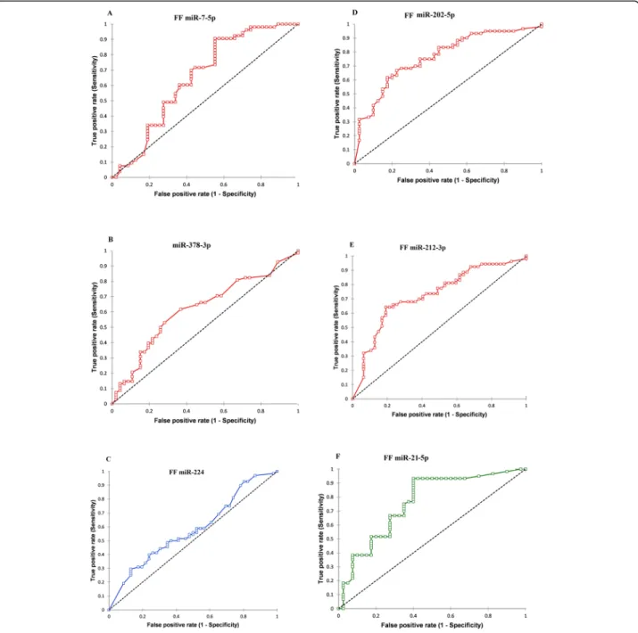
miR-21-5p (p = 0.001), and higher abundance levels of miR-7-miR-21-5p, miR-378-3p, miR-224, miR-212-3p (p = 0.001) in pooled samples of normo-androgenic PCOS (n = 110) compared to women without PCOS (n = 145) as shown in Fig.1a-f.
Table 1 Baseline clinical characteristics of all participants with normo-androgenic PCOS and without PCOS
Total No. of Participants
(n = 255) Women with NOR (n =145)
Women with normo-androgenic PCOS (n = 110)
Mann-Whitney test P-value
Variables Means ± SD N (%) Means ± SD N (%) Means ± SD N (%)
Age (years) 34.8 ± 1.4 – 34.6 ± 1.1 – 35.6 ± 1.2 – 0.31
BMI (kg/m2) 22.1 ± 1.5 – 21.3 ± 0.7 23.7 ± 1.0 – 0.12
No. of IVF attempts 2.3 ± 1.3 – 2.1 ± 1.5 – 2.2 ± 1.4 – 0.65
Age at menarche (years) 12.9 ± 1.1 – 12.8 ± 1.9 – 12.7 ± 1.7 – 0.17
Infertility status
Primary infertility – 136 (53.3) – 88 (60.6) – 48 (43.6) –
Secondary Infertility – 119 (46.6) 57 (39.3) – 62 (56.3) –
Duration of infertility (years) 3.6 ± 3.3 – 3.4 ± 1.1 – 3.5 ± 1.2 – 0.17
Infertility diagnosis
Male factor – 2 (0.78) – 0 – 2 (1.8) –
Female factor 146 (57.2) – 48 (33.1) – 98 (89) –
Mixed factors – 10 (3.92) – 0 – 10 (9.0)) –
Unexplained factors – 97 (38) – 97 (66.8) – 0 –
Baseline serum hormonal evaluation on 2nd day of Menstrual cycle
FSH (IU/I) 6.5 ± 0.98 – 6.6 ± 0.42 – 5.87 ± 0.53 – 0.002 LH (IU/I) 6.8 ± 0.74 – 6.1 ± 0.55 – 8.3 ± 0.44 – 0.001 LH/FSH 1.02 ± 0.14 – 0.98 ± 0.07 – 1.84 ± 0.49 – 0.001 E2 (pg/ml)) 44.01 ± 0.01 – 44.8 ± 1.13 – 43.2 ± 1.58 – 0.71 AMH (ng/ml) 5.8 ± 1.03 – 3.21 ± 1.14 – 8.13 ± 1.52 – 0.001 Prolactin (ng/ml) 15.6 ± 1.30 – 17.01 ± 1.17 – 14.77 ± 1.97 – 0.53 TSH (μIU/ml) 2.03 ± 0.98 – 2.17 ± 0.11 – 1.99 ± 0.17 – 0.18 Total T (nmol/L) 1.02 ± 0.89 – 0.98 ± 0.71 – 1.21 ± 0.19 – 0.05 Free T (nmol/L) 0.031 ± 0.11 – 0.02 ± 0.002 – 0.04 ± 0.002 – 0.35 SHBG (nmol/L) 57.49 ± 1.03 – 59.09 ± 1.21 – 55.74 ± 1.11 – 0.24 AFC 23.1 ± 1.63 – 15.94 ± 0.63 – 28.1 ± 0.48 – 0.001 Stimulation protocol – – – Agonist – – 72 (49.6) – 69 (62.7) – – Antagonist – – 73 (50.3) – 41 (37.2) – –
Total days of stimulation 10.2 ± 1.20 – 9.99 ± 1.4 – 10.1 ± 1.5 – 0.81
Total dose of gonadotrophins (IU/I) 2187 ± 198.74 – 2288.21 ± 101.32 – 1974.36 ± 106.3 – 0.001
rFSH – – 75 (51.72) – 89 (80.9) – –
HP-hMG – – 70 (48.2) – 21 (19.0) – –
Serum endocrine level on hCG decision day
E2 (pg/ml)) 1810.12 ± 111.89 – 1858.36 ± 211.89 – 1982.41 ± 332.13 – 0.52
P (ng/ml) 0.89 ± 0.31 – 0.85 ± 0.01 – 0.93 ± 0.02 – 0.14
LH (IU/I) 1.99 ± 0.44 – 1.98 ± 0.98 – 2.01 ± 1.02 – 0.74
Endometrium at oocyte retrieval (mm) 10.25 ± 1.02 10.42 ± 1.05 0.63
BMI body mass index, IVF in-vitro fertilization, FSH follicle-stimulating hormone, LH luteinizing hormone, E2 estradiol, AMH anti-Mullerian hormone, TSH thyroid-stimulating hormone, T testosterone, SHBG sex hormone-binding globulin, AFC antral follicle count, rFSH recombinant follicle-stimulating hormone, HP-hMG highly purified human menopausal gonadotropin, P progesterone. The values are presented as mean ± SD. P-value (Mann-Whitney test) < 0.05 considered statistically significant
The fold change analysis of FF miRNAs at oocyte retrieval day among PCOS and NOR patients was listed in TableS1, showing no significant change in the fold change expression of miRNA-320a in the follicular fluid of both groups.
The multivariate regression analysis exhibited that the six-miRNA signature was independent prognostic factor associated with PCOS after adjusting for body mass index (BMI) (adjusted OR 5.21 [1.79; 12.75], p = 0.001; 0.6 [0.27; 0.97], p = 0.001; 5.01 [1.75; 12.03], p = 0.001; 5.27 [1.89; 12.99], p = 0.005; 5.18 [1.99; 13.56]; p = 0.004; 0.98 [0.78; 0.99], p = 0.003
re-spectively as shown in Table 3. Receiver Operating
Characteristics (ROC) curve analysis was used to evaluate the sensitivity, specificity, and area under the curve (AUC). The ROC plays a central role in determining the relationship between the differential expression of miRNAs (7-5p, 202-5p, miR-378-3p, miR-224) and PCOS in the FF samples. For the discrimination of PCOS, the AUC values of the
differential expression profile of miR-7-5p was (0.711
[0.63; 0.89], p = 0.001, cut-off point ≥0.931 with a
sensitivity of 63.4% and a specificity of 87.1%;
miR-378-3p, AUC 0.620 [0.51; 0.72], p = 0.001, cut-off
point ≥25.1with a sensitivity of 68.9% and a
specifi-city of 88.7; miR-224p, AUC 0.581 [0.47; 0.68], p =
0.001, cut-off point ≥0.987 with a sensitivity of
64.8% and a specificity of 86.4%; miR-202-5p, AUC 0.798 [0.59; 0.85], p = 0.003, cut-off point ≤15.8 with a sensitivity of 71.5% and specificity of 88.5%;
miR-212-3p, AUC 0.726 [0.58; 0.89], p = 0.00, cut-off
point ≥35.14 with a sensitivity of 78.8% and a
speci-ficity of 87.2%; miR-21-5p, AUC 0.643 [0.53; 0.76], p = 0.003, cut-off point ≤41.21 with a sensitivity of
61.5% and a specificity of 81.74% respectively
(Table 4, Fig. 2a-f). However, the AUC value of
miR-NAs was increased to 0.881 [0.61; 0.92], p = 0.001
when combined in multivariate analysis. While the
combination of these six miRNAs raised the
Table 2 Reproductive outcomes of all patients (n = 255) and of each group: women with normo-androgenic PCOS (n = 110) and without PCOS having normal ovarian reserve (NOR) (n = 145)
Reproductive outcomes Total No. of
Participants (n = 255)
Women with NOR (n = 145)
Women with normo-androgenic PCOS (n = 110)
Mann-Whitney test P-value Ovarian outcome parameters
No. of Follicles aspirated [median (95% population limit)]
16.20 (11.2; 20.6) 13 (10.3; 18.2) 17 (15.4; 25.6) 0.03
No. of oocytes Retrieved [median (95% population limit)]
13.02 (10.32; 18.3) 11 (8.2; 13.5) 14.6 (11.3; 20.8) 0.001
No. of MII- oocytes [median (95% population limit)] 9.81 (8.01; 13.8) 9.4 (7.9; 12.3) 10.5 (8.7; 15.8) 0.04
Rate of mature oocytes (%) (0–100) 80 84 76 0.001
No. of Fertilized oocytes [median (95% population limit)]
9.10 (7.2; 11.98) 8.91 (6.4; 11.3) 9.86 (7.7; 13.54) 0.04
Fertilization rate (mean ± SD) 6.01 ± 1.68 4.92 ± 1.96 8.65 ± 1.33 0.001
Embryological outcome parameters
No. of Normal Fertilized oocytes (2PN) [median (95% population limit)]
8.21 (6.01; 10.2) 8.41 (5.9; 10.6) 8.03 (6.7; 10.55) 0.09
No. of Early cleaved zygotes [median (95% population limit)]
8.00 (5.01; 8.51) 8.01 (5.2; 9.97) 7.99 (4.5; 9.69) 0.71
No. of blastomeres (6–8 cells) with regular symmetry at day 3 median (95% population limit)]
7.01 (4.89; 8.74) 7.22 (4.87; 8.14) 7.19 (4.96; 9.01) 0.66
No. of High-Quality day 3 embryos [median (95% population limit)]
2.00 (1.05; 3.01) 2.10 (1.98; 3.21) 2.20 (2.01; 2.99) 0.73
Total fragmentation rate (%) 15 13 18 0.001
Blastulation rate (%) at day 5 (0–100) 43 48 36 0.001
Expended blastocyst rate (%) at day 5 (0–100) 46 49 37 0.001
Clinical outcomes
Clinical pregnancy rate per transfer (%) (0–100) 35 39 26 0.001
The values are presented as median (95% population limit) and percentage (%) point scale (0–100). P-value (Mann-Whitney test) < 0.05 considered statistically significant. Blastocyst rate was based on the number of blastocysts obtained / the number of zygotes cleaved. The number of fertilized oocytes means the total number of oocytes that were fertilized. A number of normally fertilized oocytes mean total fertilized oocytes obtained / abnormally fertilized oocytes. The fertilization rate is based on the number of oocytes obtained / the number of zygotes cleaved
sensitivity to 79.2% with a specificity of 87.32% (see Table 4).
Our results revealed that expression of six miRNAs (miR-7-5p, miR-202-5p, miR-378-3p, miR-224,
miR-21-5p, miR-212-3p) between patients with
normo-androgenic PCOS and without PCOS give the significant AUC value with greater sensitivity and specificity to-gether. Based on these findings, we proposed that these miRNAs signatures might be a potential noninvasive biomarker for the discrimination between normo-androgenic PCOS and non-PCOS patients.
MiRNAs expression profile in follicular fluid (FF) samples in response to the exogenous gonadotropin therapy
The differential miRNA expression profile in FF sam-ples was evaluated in response to the exogenous go-nadotropin administration in NOR patients (n = 145). The miR-202-5p and miR-378-3p exhibiting modest expression differences according to the gonadotropin treatment. Remarkably, significantly lower abundance level of FF miR-202-5p and higher abundance levels of FF miR-378-3p was reported in pooled samples
from patients treated with human menopausal
Fig. 1 Relative miRNAs expression profile in follicular fluid (FF) pools of NOR patients (n = 145) compared to PCOS women (n = 110). a FF miR-7-5p b FF miR-378-3p c FF miR-224 d FF miR-202-miR-7-5p e FF 212-3p f FF miR-21-miR-7-5p.P < 0.05 is considered statistically significant (Mann-Whitney test)
gonadotropin (hMG) for those treated with recombin-ant follicle-stimulating hormone (rFSH) (p < 0.001)
re-spectively, as presented in Fig. 3a. Furthermore,
without concerning the type of treatment, the expres-sion of miR-202-5p was significantly increased in FF pool samples from those patients with higher doses regimen of gonadotropin (≥ 3000 IU/l) than those who received < 3000 IU/I (P = 0.001), as displayed in Fig. 3b.
Similarly, the correlation coefficient exhibited that miR-202-5p has a significant but positive correlation
with the overall dosage of gonadotropins (R2
= 0.669, p = 0.001) (see Fig. 3c). Patients with a low propor-tion of mature oocytes (MII) retrieved (≤ 3) showed a significantly lower concentration of FF miR-378-3p and miR-224 levels than those with more than 3 (p = 0.001) oocytes retrieved after 36 h post hCG
ad-ministration (Fig. 3d). Spearman correlation
coeffi-cient showed that the relative expression of miR-378-3p and miR-224 levels was significantly
associ-ated with the number of mature oocytes (R2
= 0.760,
p = 0.003, R2
= 0.927, p = 0.001), respectively (Fig. 3 e-f).
FF miR-320a and day 3 embryo quality
Our study expressed a significant association between FF miRNAs and day 3 high-quality embryos in NOR pa-tients; we found that miRNA-320a was significantly dif-ferent in top-quality embryos versus non-top-quality embryos on day 3 (p = 0.001) as depicted in Fig. 3g. Ac-cordingly, miRNA-320a had an AUC of [0.753 (0.651; 0.855),p = 0.001], cutoff value ≥1.35 with a sensitivity of 80% and specificity of 71% respectively (Fig.3h).
FF miR-212-3p expression level and blastulation
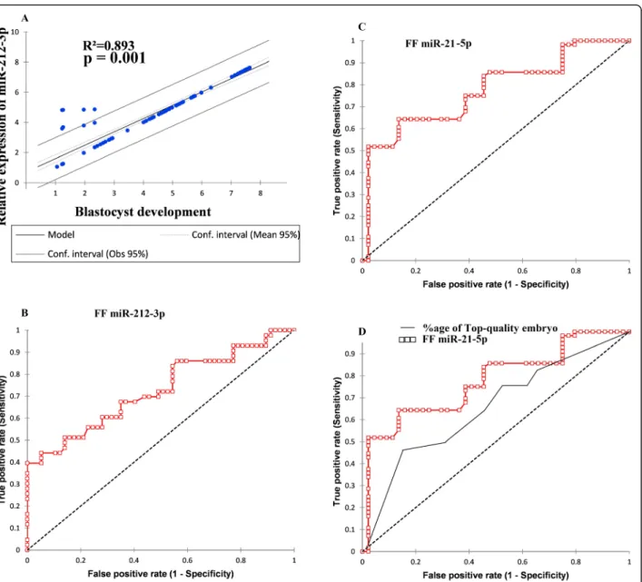
We observed an a significant positive relationship be-tween miR-212-3p expression level and high-quality blastocyst development (R2
= 0.893,p = 0.001) by analyz-ing the group of women without PCOS (n = 145) as shown in Fig. 4a. In fact, a high abundance of FF
miR-212-3p was significantly associated with
normo-androgenic PCOS. In our study, we noticed that the
Table 3 Multivariate logistic regression analysis exhibited the relationship between FF miRNAs relative expression with patients having polycystic ovaries
FF miRNAs relative expression related to normo-androgenic PCOS
Univariate logistic regression Analysis Multivariate logistic regression Analysis
FF miRNAs β SE Wald Crude Odd ratio
[95% Cl] P-value β SE Wald BMI adjusted Oddratio [95% Cl] P-value
miR-7-5p 0.81 0.038 141.36 5.01 [2.01; 11.02] 0.001 1.02 0.052 126.31 5.21 [1.79; 12.75] 0.001 miR-202-5p 0.97 0.041 132.7 0.74 [0.29; 0.89] 0.001 0.89 0.041 96.32 0.64 [0.27; 0.97] 0.001 miR-378-3p 0.89 0.032 48.62 4.93 [1.88; 10.36] 0.001 0.64 0.034 87.01 5.01 [1.75; 12.03] 0.001 miR-224 0.64 0.541 35.87 5.13 [1.91; 11.35] 0.004 0.53 0.059 96.32 5.27 [1.89; 12.99] 0.005 miR-212-3p 0.84 0.453 256.4 4.87 [1.68; 9.97] 0.003 0.99 0.034 52.14 5.18 [1.99; 13.56] 0.004 miR-21-5p 0.91 0.521 41.02 0.91 [0.89; 1.03] 0.002 1.23 0.061 75.97 0.98 [0.78; 0.99] 0.003
FF follicular fluid, Cl confidence interval,β beta coffecient, SE standard error
Table 4 Predictive values of sensitivity and specificity evaluation for the probability of PCOS
FF miRNAs Prediction for normo-androgenic PCOS
AUC [(95% population limit)] S.E.M *P-Value **Cut-off point Sensitivity (%) Specificity (%) miR-7-5p 0.711 [0.63; 0.89] 0.061 0.001 ≥ 0.931 63.4 87.1 miR-378-3p 0.620 [0.51; 0.72] 0.053 0.001 ≥ 25.1 68.9 88.7 miR-224 0.581 [0.47; 0.68] 0.044 0.001 ≥ 0.987 64.8 86.4 miR-202-5p 0.798 [0.59; 0.85] 0.054 0.002 ≤ 15.8 71.5 88.5 miR-212-3p 0.726 [0.58; 0.89] 0.064 0.001 ≥ 35.14 77.8 87.2 miR-21-5p 0.643 [0.53; 0.76] 0.057 0.003 ≤ 41.21 61.5 81.74 Combination of FF miR-7-5p, 202-5p, 378-3p, 224, 212-3p, 21-5p 0.881 [0.61; 0.92] 0.040 0.001 – 79.2 87.32
S. M. E means ± standard error of the mean, *The null hypothesis was true area = 0.5, after adjusting a number of attempts and the number of embryos. P-values in bold letters are considered statistically significant p < 0.05. ** Estimated cut points that maximize sensitivity and specificity for observed range predictors
expression level of FF miR-212-3p was significantly re-lated to the probability of embryos to develop into a high-quality blastocyst in patients with normal ovarian reserve (crude odds ratio = 1.32 [0.98; 2.01], p = 0.03 while the AUC value of FF miR-212-3p to predict blas-tulation was 0.744 (0.648; 0.841) with 79% sensitivity and 69% specificity at the cutoff of ≤20.1 (see Fig. 4b). Similarly, in NOR patients, the intrafollicular expression level of miR-212-3p was significantly correlated with the
likelihood of achieving an expanded blastocyst (crude odds ratio = 1.31 [0.98; 2.01],p = 0.01 with an AUC value of 0.726 (0.623; 0.829 having 71% sensitivity and 88% specificity at the cutoff point≤15.43).
FF miR-21-5p expression level is associated with clinical pregnancy outcome
In NOR patients (n = 145), the level of FF miRNA-21 was associated with clinical pregnancy outcome
Fig. 2 Receiver operating characteristic (ROC) analysis is used to find the power of discrimination of upregulated and downregulated FF miRNAs relative expression profile for PCOS diagnosis. a FF miR-7-5p b FF miR-378-3p c FF miR-224 d FF miR-202-5p e FF miR-212-3p f FF miR-21
Fig. 3 a Comparative expression profile of FF miR-202-5p and FF miR-378-3p in NOR patients relative to the gonadotropin type. rFSH (n = 80) (recombinant follicle-stimulating hormone), HP-hMG (n = 65) (highly purified human menopausal gonadotropin (Mann-Whitney test, p = 0.001). b Expression profile of FF miR-202-5p in NOR patients relative to the total dose of gonadotropin (≥ 3000, n = 90, ≤ 3000, n = 55) Mann-Whitney test, p = 0.001. c The relative expression of FF miR-202-5p has a significant but positive correlation with the overall dosage of gonadotropins (R2=
0.669,p = 0.001). d Differential expression of FF miR-378-3p and FF miR-224 in NOR patients according to the number of retrieved oocytes on day 3 (≥ 3, n = 125, ≤ 3, n = 20) Mann-Whitney test, p = 0.001. e Spearman correlation coefficient depicted a significant positive association between FF miR-378-3p and the number of mature oocytes in NOR patients (n = 145). f The relative expression of FF miR-224 has a significant positive correlation with the number of mature oocytes in NOR patients. g Relative expression profile of FF miR-320a in NOR patients between top-quality embryos and non-top-quality embryos (Mann-Whitney test, p = 0.001). h The receiver operating characteristic (ROC) curve showing the
[crude odds ratio = 1.99 [0.99; 3.97], p = 0.03 as given
in Table S2. Furthermore, ROC curve analysis showed
that the possible AUC value of FF miRNA-21 to
pre-dict clinical outcome was 0.774 (0.682; 0.865), p =
0.001 with a sensitivity of 74.8% and specificity of
83.7% at the cutoff point of ≥63.68 (Fig. 4c).
Add-itionally, when we compared the discrimination power of FF miRNA-21 and the percentage of good quality embryos for predicting clinical pregnancy, we found
that AUC value of about 0.78 (0.589; 8.712), p =
0.001, which was much greater than that for the
percentage of good quality day 3 embryos (6–8 cells) (Fig. 4d).
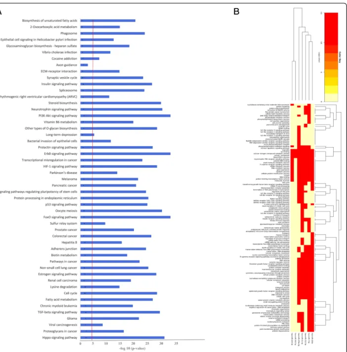
Predicted pathway analysis
MiRNAs were uploaded for KEGG pathway enrich-ment analysis and Gene Ontology (GO). The genes common in six out of eight programs (miRanda, Tar-getScan, DIANA-microT, miRDB, PicTar, miRWalk, RNA hybrid, target gene prediction at EMBLE) were selected to predict targets. We identified significant enriched biological processes based on differential
Fig. 4 a The relative expression of FF miR-212-3p is significantly positively associated with blastocyst development in women with the normal ovarian reserve (n = 145). b Receiver operating characteristic (ROC) analysis revealed that FF miR-212-3p expression is a promising biomarker to predict blastocyst formation in NOR patients (n = 145). c The receiver operating characteristic (ROC) curve illustrated the discriminative ability of FF miR-21-5p to predict the clinical pregnancy outcome in NOR patients (n = 145). d Comparative ROC curve exhibiting discrimination between the relative expression profile of FF miR-21-5p and top-quality embryo proportion for clinical pregnancy outcome in NOR patients (n = 145)
expression of studied miRNAs between normo-androgenic PCOS and NOR patients; the 51 topmost
enriched biological processes were shown in Fig. 5a.
The most significant biological events involving the targets of our miRNAs were related to cell prolifera-tion, regulation of cellular processes, as well as devel-opmental processes. These biological processes were
further validated by available literature data [10, 35,
36]. It was established that TGF beta signaling path-way, Hippo signaling pathpath-way, p53 signaling pathpath-way, cell cycle regulation, oocyte meiosis, HF-1 signaling pathway, FoxO signaling pathway, MAPKK signaling pathway, Notch signaling pathway, insulin receptor signaling pathway, and EGF signaling pathway were
Fig. 5 a Functional pathways Analysis- Top GO biological processes were identified using DAVID blue bars denoted pathways enriched among miRNAs detected in all FF samples of patients based on differential expression of studied miRNAs between normo-androgenic PCOS and NOR patients. Small numbers in each blue bar presented a number of gene count in each enriched term are associated with those miRNAs. The red line exhibited a statistically significant FDR thresh-hold. Benjamini and Hochberg multiple testing correlation were used to adjust thep-values (−log 10 FDR adjusted p values = < 0.05). b Predicted pathways; Heatmap analysis of miRNAs red color shows lower p values, and high expression, yellow indicates intermediate expression levels
all actively involved in ovarian pathophysiology (Fig.
5b). Our results indicate that the differential expres-sion of studied miRNAs in FF was potentially
associ-ated with cell growth, differentiation, and
developmental processes during folliculogenesis and oocyte maturation.
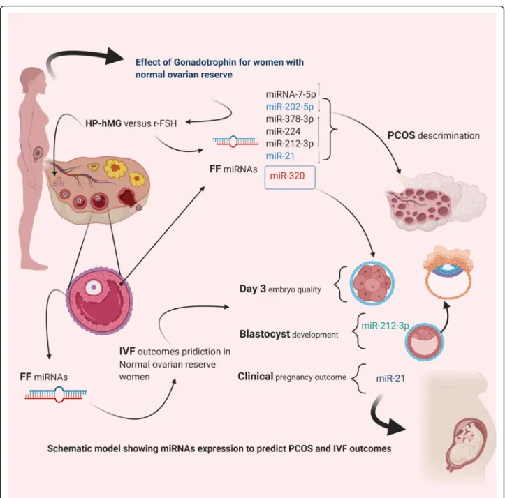
Discussion
The present study explored seven circulating miRNAs (miR-7-5p, miR-202-5p, miR-378-3p, miR-224, miR-320a, miRNA-212-3p, and miR-21-5p) within the FF pool of
normo-androgenic PCOS and NOR women undergoing treatment by IVF/ICSI. In particular, our study revealed that the expression levels of these miRNAs were signifi-cantly varied when compared to the patient’s ovarian
re-serve status, ovarian response to gonadotropin
administration, and IVF outcomes (see Fig.6).
Our data revealed that expression of miR-378-3p was significantly upregulated, and miR-202-5p was signifi-cantly lower abundance levels in FF pools from patients administered with human menopausal gonadotropin
(hMG) in comparison with those treated with
recombinant follicle-stimulating hormone (rFSH). This corresponds with the previous time course study con-ducted on a mouse model system, which demonstrated a biphase regulation of miRNAs through FSH in GCs. The study also observed that 202-5p and 14 other miR-NAs were progressively downregulated by FSH exposure during the first 12 h but did not meet the significance level [37]. Inconsistent with our findings, the downregu-lation of miR-202-5p in GCs disturbs the oocyte-somatic cell interactions and severely interrupts the onset of primordial follicles’ development [38, 39]. It was subse-quently noticed that downregulation of miR-202-5p re-sults in reduced expression of one of the family members of KELCH, which has potential participation in the cross-talk between the oocyte and its companion somatic cell, disrupts the normal ovarian function exclu-sively in drosophila [39]. Furthermore, this hypothesis is strengthened by the SLIT-ROBO signaling pathway’s dysregulation due to reduced miR-202-5p levels in ovar-ian cancer, ultimately leading to an interruption in cellu-lar communication across the ovarian tissues [38]. These findings clearly illustrate that the potential targets of miR-202-5p perform integrated regulatory functions and actively participate in follicular recruitment and growth. Recently, this was made clear by Gay et al. that knockout of the miR-202-5p gene in medaka ovaries reduces the expression of Gdf and Foxl2b genes that result in either no oogenesis or reduced oocyte production, which were later unable to mature and fertilize [24]. In mammals, FF miR-202-5p targeting the PI3K and TGFβR2 and posi-tively associated with fertilization potential and oocytes maturation [40–43]. On these grounds, we suggest that the altered expression of miR-202-5p in human ovarian follicles might play a significant biological role in follicu-lar development [40,44].
PCOS is responsible for excessive follicle formation, signifying that the normal cross-talk between GCs and oocytes in mature preovulatory follicles might be badly mismanaged in the initial phase of development [45]. Our data of PCOS patients revealed an upregulation of miR-378-3p than in NOR patients. In mice ovaries, the upregulation of miR-378-3p leads to an increased dens-ity of primordial follicles and decreased apoptosis induc-tion both in vivo and in vitro cultures [25]. Another study’s key finding suggested that the overexpression of miR-378-3p is responsible for an accelerated transition of follicles from primordial to the primary stages due to increased GC proliferation in anovulatory females [46]. Keeping all facts in mind, it is worth mentioning that ab-normal preantral folliculogenesis due to overexpression of miR-378-3p might play a fundamental role in ovarian pathogenesis. It may be responsible for the relatively high density of the primary population of follicles in PCOS patients.
The significance of miRNAs in the early stages of em-bryonic development has been extensively discussed in
several mammalian species [29]. Our results
demon-strated that FF miR-320a was significantly linked to the embryonic developmental potential in NOR patients. Moreover, our findings showed that FF miR-320a was differentially expressed in top-quality embryos than im-paired quality embryos. The recent publication using the mouse knockdown model for miR-320a revealed that the proportion of blastocyst-stage embryos was affected con-siderably by attenuating Wnt signaling components in early embryonic development both in vivo and in vitro [19,47,48]. Considering all these facts, it is inferred that miR-320 might be regarded as a potential contributor in regulating the embryo quality in patients undergoing IVF procedures [36].
Accumulative shreds of evidence indicate that miR-212-3p was preferentially enriched in FF and robustly expressed in preovulatory GCs and cumulus-oocyte complex (COC) of mammalian ovaries [10, 48, 49]. We found that FF miR-212-3p levels were significantly asso-ciated with embryo developmental potential in NOR pa-tients. Therefore, the significant dysregulation of miR-212-3p expression in FF samples probably reflects the abnormal endocrine environment, impairing oocyte mat-uration. Comparably, the downregulation of miR-212-3p induces decreased implantation potential in cryopre-served vitrified mouse blastocysts due to the pervasive aberrant expression of a panel of genes necessary for embryo implantation [50]. Previous studies have shown that miR-212-3p also has a regulatory contribution to follicular development and steroidogenesis [51–53]. Spe-cifically, experiments with the bovine model revealed that FF miR-212-3p may also play a decisive role in regulating FIGLA, an oocyte-specific transcriptional fac-tor belonging to a basic family Helix-loop-Helix dimeric protein that is essential for regulating primordial folli-cles’ formation [54,55]. In an in vitro study, FIGLA was known to bind promoter elements (E-Box) in the oocyte-zona-pellucida glycoprotein genes (Zp1, Zp2, Zp3), regulate their expression profile, and can effect-ively influence the blastocyst development [56]. Hence, the FF miR-212-3p expression evaluation could define the best embryo culture strategies for blastocyst develop-ment and IVF outcomes in patients with normal ovarian reserve.
Our study found higher abundance levels of FF miR-224 in PCOS patients than in NOR patients. Analysis of the previous literature has shown that overexpression of miR-224 of cumulus cells led to the cell expansion-dependent impairment and downregulation of gene ex-pression associated with the oocyte maturation, most specifically by targeting Ptx3 [26]. Moreover, our results
decreased in those NOR patients who have a small num-ber of mature oocytes (≤ 3) than those with a more sig-nificant number of (> 3) oocytes retrieved (p = 0.001). In situ hybridization through locked-nucleic-acid-modified probes revealed that miR-224 is mainly expressed in GCs of follicles at varying degrees of development and suggestively increased the proliferation potential of GCs. Moreover, transfected GCs by miR-224 inhibitors halt their proliferation potential predominantly during follicle differentiation by altering the steroidogenic function of GCs [31].
As alluded to earlier, the phenomenon of embryo im-plantation is a crucial phase in early pregnancy establish-ment in mammals, but endometrium receptivity remains a limiting step in the overall success of IVF in humans. Our results showed that FF miR-21-5p differential ex-pression significantly predicts the NOR group’s clinical pregnancy outcome with much higher sensitivity than the percentage of good quality embryos. Previous work demonstrated that FF miR-21-5p has a dynamic role in preimplantation embryo development by regulating apoptotic proteins [57, 58]. Moreover, downregulation of miR-21-5p in PCOS contributes to cell apoptosis by tar-geting PI3K/AKY and JAK/STAT3 signaling pathways
[52]. It has been demonstrated experimentally that
SMAD7 is a principal mediator and regulator of the TGF-beta signaling, which is one of the critical pathways involv-ing in folliculogenesis, directly targeted, and inhibited by downregulation of miR-21-5p in PCOS [54].
Gene-GO terms demonstrated that the differential ex-pression of miRNAs (up and down-regulation) might be responsible for the massive alterations in the harmony of the biological system. KEGG enrichment analysis high-lights the presumptive targets of the studied miRNAs into major interactive signal transduction pathways. While pathway enrichment analysis revealed that the TGF-βeta signaling pathway, AKT pathway, and Hippo signaling pathway regulate normal ovarian function and folliculogenesis [17]. Mainly, one critical downstream mediator of the PI3K/AKT pathway, such as active FoxO family members, performs a crucial role during ovarian folliculogenesis by inhibiting primordial follicles’ activa-tion. However, inactivation of FoxO3 leads to premature stimulation of primordial follicles and depletion of ma-ture follicles [59]. A brief overview of the in-silico com-putational strategy demonstrated that FoxO3 is a promising target of miR-21-5p, which might be pro-jected as an intricate network in PCOS, contributing through androgenic excess [35]. Notably, regulation of these miRNAs by various intrinsic and extrinsic factors in conjunction with their underlying mechanism of gene targeting generates complex networks of regulatory ele-ments that have a substantial role in disease etiology, such as PCOS. One of this study’s strengths was the
same age-matched large groups of NOR, and PCOS women, who were all undergoing IVF procedures with the attention of subsequent studied based on FF.
This study’s limitation is that the analysis was from different COS protocols that might affect miRNA ex-pression. A more precise assessment of miRNA’s role requires using the same COS protocol in all patients. A more precise assessment of the role of miRNA in predicting individual embryo outcome would require the analysis of individual FF and tracking that through to the individual blastocyst stage.
Conclusion
Conclusively, our results provide evidence that miR-7-5p, miR-378-3p, miR-224, miR-212-3p were a differen-tially high expression in normo-androgenic PCOS pa-tients than NOR papa-tients. While 202-5p and miR-21-5p were found significantly high levels in NOR pa-tients versus PCOS papa-tients. Similarly, the expression level of FF miR-212-3p was significantly related to the probability of embryos to develop into a high-quality blastocyst in patients with normal ovarian reserve. In the future, these miRNAs in FF could improve personalized IVF treatment strategies while treating infertility. More informatic considerations are required to highlight the detailed biological input of these miRNAs in the FF microenvironment to predict embryo quality, their re-sponse to gonadotropin therapy, and identifying their direct contribution to female infertility.
Abbreviations
IVF:In vitro fertilization; AFC: Antral follicle count; AMH: Anti-Müllerian hormone; PCOS: Polycystic Ovarian Syndrome; NOR: Normal ovarian reserve; FF: Follicular fluid; miRNA: MicroRNA; ROC: Receiving operating
characteristics; E2: 17β-estradiol; BMI: Body mass index; FSH: Follicular stimulating hormone; LH: Luteinizing hormone (LH); TSH: Thyroid-stimulating hormone; TVS: Transvaginal ultrasonography; COS: Controlled ovarian stimulation; ICSI: Intracytoplasmic sperm injection; CI: Confidence interval
Supplementary Information
The online version contains supplementary material available athttps://doi. org/10.1186/s12958-021-00754-9.
Additional file 1 : Table S1. Fold change in the FF miRNAs at oocyte retravel day between women with normo-androgenic PCOS (n = 110) ver-sus NOR women (n = 145). Table S2. Univariate analysis exhibiting asso-ciation of specific FF miRNAs with blastocyst formation and pregnancy outcome.
Acknowledgments
We acknowledge the research initiative and gratefully thank Professor Dr. RLK, Professor of Emeritus in Obstetrics and Gynecology, in manuscript editing. The study is supported by the infertility research center of HLA hospital. Moreover, The authors would like to express their appreciation to the College of Medicine Research Center, the Deanship of Scientific Research, King Saud University for their technical support.
Authors’ contributions
HLK, YLK Reviewing; SB Conceptualization, Methodology, Software, Data curation, Project administration, performing experimental work,
Writing-Original draft preparation. Supervision, Writing- Reviewing and Editing; SA Formal analysis, Validation, Methodology, performing experimental work, Investigation, Writing- Reviewing and Editing; CK Writing- Reviewing and Editing; Data curation, Writing- Original draft preparation; ZH, NOT, AY, and EYK Reviewing and Editing, AMI reviewing, Software, HY Reviewing and Editing, RZ Reviewing and Editing; HHY Software, Validation, Methodology. The author(s) read and approved the final manuscript.
Funding
Lahore institute of Fertility and Endocrinology, Hameed Latif Hospital, Lahore, Pakistan.
Availability of data and materials
The data set used and analyzed during the current study is available from the corresponding author on reasonable request.
Declarations
Ethics approval and consent to participate
The study was approved by our Institutional Ethical Committee (IEC). Informed consent was obtained from all subjects before the research and publishing of the results of the investigation.
Consent for publication Not applicable.
Competing interests
The authors declared that they have no conflict of interest.
Author details
1Lahore Institute of Fertility and Endocrinology, Hameed Latif Hospital, 14
Abu-Bakar Block New Garden Town, 54800 Lahore, Pakistan.2Department of
Human Genetics and Molecular biology, University of Health Sciences, Lahore 54600, Pakistan.3Department of Medical Education, Rashid Latif
Medical College, Lahore, Pakistan.4Department of Histology and
Embryology, Cumhuriyet University Faculty of Medicine, 58140 Sivas, Turkey.
5
Assisted Conception Unit, Obstetrics and Gynecology Department, College of Medicine, King Saud University, Riyadh, Saudi Arabia.6Department of
Biochemistry, Kinnaird College Lahore, Lahore, Pakistan.7Your Family Fertility,
1408 Sweet Home Road Suite 9, Amherst NY 14228, USA.8Department of
Cellular Pathology, Royal Free Hospital, London NW3 2QG, UK.9Department of Obstetrics and Gynecology, Nisdetiye Maha,İstanbul, Turkey.
10Department of Molecular Biology and Genetics, Faculty of Science, Mugla
Sitki Kocman University, Kotekli, 48000 Mugla, Turkey.11Department of
Medical Biochemistry, Ege University School of Medicine, Bornova, Izmir, Turkey.12Department of IVF unit, Private Adatip Hospital, Yenisehir mahallesi
Kardelen sokak 2, Pendik, 34912 Istanbul, Turkey.
Received: 1 February 2021 Accepted: 27 April 2021
References
1. Catalanotto C, Cogoni C, Zardo G. MicroRNA in control of gene expression: an overview of nuclear functions. Int J Mol Sci. 2016;17(10):1712.https://doi. org/10.3390/ijms17101712.
2. Eisenberg I, Kotaja N, Goldman-Wohl D, Imbar T. MicroRNA in human reproduction. In: microRNA: Medical Evidence. Switzerland: Springer International Publishing; 2015. p. 353–87.
3. Tesfaye D, Gebremedhn S, Salilew-Wondim D, Hailay T, Hoelker M, Grosse-Brinkhaus C, et al. MicroRNAs: tiny molecules with a significant role in mammalian follicular and oocyte development. Reproduction. 2018;155(3): R121–35.https://doi.org/10.1530/REP-17-0428.
4. Sohel MH. Extracellular/circulating microRNAs: release mechanisms, functions and challenges. Achiev Life Sci. 2016;10(2):175–86.https://doi. org/10.1016/j.als.2016.11.007.
5. Mishra PJ. MicroRNAs as promising biomarkers in cancer diagnostics. Biomarker Res. 2014;2(1):19.https://doi.org/10.1186/2050-7771-2-19. 6. Ferguson JM, Pépin D, Duru C, Matejtschuk P, Donahoe PK, Burns CJ.
Towards international standardization of immunoassays for Müllerian inhibiting substance/anti-Müllerian hormone. Reprod BioMed Online. 2018; 37(5):631–40.https://doi.org/10.1016/j.rbmo.2018.08.012.
7. March WA, Moore VM, Willson KJ, Phillips DI, Norman RJ, Davies MJ. The prevalence of polycystic ovary syndrome in a community sample assessed under contrasting diagnostic criteria. Hum Reprod. 2010;25(2):544–51.
https://doi.org/10.1093/humrep/dep399.
8. Long W, Zhao C, Ji C, Ding H, Cui Y, Guo X, et al. Characterization of serum microRNAs profile of PCOS and identification of novel non-invasive biomarkers. Cell Physiol Biochem. 2014;33(5):1304–15.https://doi.org/10.11 59/000358698.
9. Sang Q, Yao Z, Wang H, Feng R, Wang H, Zhao X, et al. Identification of microRNAs in human follicular fluid: characterization of microRNAs that govern steroidogenesis in vitro and are associated with polycystic ovary syndrome in vivo. J Clin Endocrinol Metabolism. 2013;98(7):3068–79.https:// doi.org/10.1210/jc.2013-1715.
10. Santonocito M, Vento M, Guglielmino MR, Battaglia R, Wahlgren J, Ragusa M, et al. Molecular characterization of exosomes and their microRNA cargo in human follicular fluid: bioinformatic analysis reveals that exosomal microRNAs control pathways involved in follicular maturation. Fertil Steril. 2014;102(6):1751–1761. e1751.
11. Gross N, Kropp J, Khatib H. MicroRNA signaling in embryo development. Biology. 2017;6(3):34.https://doi.org/10.3390/biology6030034. 12. Andronico F, Battaglia R, Ragusa M, Barbagallo D, Purrello M, Di Pietro C.
Extracellular vesicles in human oogenesis and implantation. Int J Mol Sci. 2019;20(9):2162.https://doi.org/10.3390/ijms20092162.
13. Mase Y, Ishibashi O, Ishikawa T, Takizawa T, Kiguchi K, Ohba T, et al. MiR-21 is enriched in the RNA-induced silencing complex and targets COL4A1 in human granulosa cell lines. Reprod Sci. 2012;19(10):1030–40.https://doi. org/10.1177/1933719112442245.
14. Li X, Xin S, He Z, Che X, Wang J, Xiao X, et al. MicroRNA-21 (miR-21) post-transcriptionally downregulates tumor suppressor PDCD4 and promotes cell transformation, proliferation, and metastasis in renal cell carcinoma. Cell Physiol Biochem. 2014;33(6):1631–42.https://doi.org/10.1159/000362946. 15. Ma L, Tang X, Guo S, Liang M, Zhang B, Jiang Z. miRNA-21–3p targeting of
FGF2 suppresses autophagy of bovine ovarian granulosa cells through AKT/ mTOR pathway. Theriogenology. 2020;157:226–37.https://doi.org/10.1016/j. theriogenology.2020.06.021.
16. Han X, Xue R, Yuan H-J, Wang T-Y, Lin J, Zhang J, et al. MicroRNA-21 plays a pivotal role in the oocyte-secreted factor-induced suppression of cumulus cell apoptosis. Biol Reprod. 2017;96(6):1167–80.https://doi.org/10.1093/biolre/iox044. 17. Raja-Khan N, Urbanek M, Rodgers RJ, Legro RS. The role of TGF-β in
polycystic ovary syndrome. Reprod Sci. 2014;21(1):20–31.https://doi.org/1 0.1177/1933719113485294.
18. Wang X-L, Wang K, Zhao S, Wu Y, Gao H, Zeng S-M. Oocyte-secreted growth differentiation factor 9 inhibits BCL-2-interacting mediator of cell death-extra long expression in porcine cumulus cell. Biol Reprod. 2013;89(3): 56, 51-59.https://doi.org/10.1095/biolreprod.113.108365.
19. Feng R, Sang Q, Zhu Y, Fu W, Liu M, Xu Y, et al. MiRNA-320 in the human follicular fluid is associated with embryo quality in vivo and affects mouse embryonic development in vitro. Sci Rep. 2015;5(1):8689.https://doi.org/10.1038/srep08689. 20. Ahmed K, LaPierre MP, Gasser E, Denzler R, Yang Y, Rülicke T, et al. Loss of
microRNA-7a2 induces hypogonadotropic hypogonadism and infertility. J Clin Invest. 2017;127(3):1061–74.https://doi.org/10.1172/JCI90031. 21. He J, Xu S, Ji Z, Sun Y, Cai B, Zhang S, et al. The role of miR-7 as a potential
switch in the mouse hypothalamus-pituitary-ovary axis through regulation of gonadotropins. Mol Cell Endocrinol. 2020;518:110969.https://doi.org/10.1 016/j.mce.2020.110969.
22. Chakrabarti M, Ray SK. Experimental procedures for demonstration of microRNA mediated enhancement of functional neuroprotective effects of estrogen receptor agonists. In: Estrogen receptors. New York: Humana Press; 2016. p. 359–72.
23. Schauer S, Sontakke S, Watson E, Esteves C, Donadeu F. Involvement of miRNAs in equine follicle development. Reproduction. 2013;146(3):273–82.
https://doi.org/10.1530/REP-13-0107.
24. Gay S, Bugeon J, Bouchareb A, Henry L, Delahaye C, Legeai F, et al. MiR-202 controls female fecundity by regulating medaka oogenesis. PLoS Genet. 2018;14(9):e1007593.https://doi.org/10.1371/journal.pgen.1007593. 25. Sun X, Klinger FG, Liu J, De Felici M, Shen W, Sun X. miR-378-3p maintains
the size of mouse primordial follicle pool by regulating cell autophagy and apoptosis. Cell Death Dis. 2020;11(9):1–14.
26. Li X, Wang H, Sheng Y, Wang Z. MicroRNA-224 delays oocyte maturation through targeting Ptx3 in cumulus cells. Mech Dev. 2017;143:20–5.https:// doi.org/10.1016/j.mod.2016.12.004.
27. The Istanbul consensus workshop on embryo assessment: proceedings of an expert meeting. Hum Reprod. 2011;26(6):1270–83.
28. Gardner DK, Lane M, Stevens J, Schlenker T, Schoolcraft WB. Blastocyst score affects implantation and pregnancy outcome: towards a single blastocyst transfer. Fertil Steril. 2000;73(6):1155–8.https://doi.org/10.1016/S0015-0282(00)00518-5. 29. Schmittgen TD, Livak KJ. Analyzing real-time PCR data by the comparative C T
method. Nat Protoc. 2008;3(6):1101–8.https://doi.org/10.1038/nprot.2008.73. 30. Pan B, Toms D, Shen W, Li J. MicroRNA-378 regulates oocyte maturation via the
suppression of aromatase in porcine cumulus cells. Am J Physiol-Endocrinol Metabol. 2015;308(6):E525–34.https://doi.org/10.1152/ajpendo.00480.2014. 31. Yao G, Yin M, Lian J, Tian H, Liu L, Li X, et al. MicroRNA-224 is involved in
transforming growth factor-β-mediated mouse granulosa cell proliferation and granulosa cell function by targeting Smad4. Mol Endocrinol. 2010;24(3): 540–51.https://doi.org/10.1210/me.2009-0432.
32. Bustin SA, Benes V, Garson JA, Hellemans J, Huggett J, Kubista M, Mueller R, Nolan T, Pfaffl MW, Shipley GL. The MIQE guidelines: M inimum I nformation for publication of Q uantitative real-time PCR E xperiments. UK: Oxford University Press; 2009.
33. Yang L-h, Wang S-l, L-l T, Liu B, Wang L-l, Wang Z-y, et al. Universal stem-loop primer method for screening and quantification of microRNA. PLoS One. 2014;9(12):e115293.https://doi.org/10.1371/journal.pone.0115293. 34. Dennis G, Sherman BT, Hosack DA, Yang J, Gao W, Lane HC, et al. DAVID:
database for annotation, visualization, and integrated discovery. Genome Biol. 2003;4(9):1–11.
35. Naji M, Aleyasin A, Nekoonam S, Arefian E, Mahdian R, Amidi F. Differential expression of miR-93 and miR-21 in granulosa cells and follicular fluid of polycystic ovary syndrome associating with different phenotypes. Sci Rep. 2017;7(1):1–14.
36. Ivey KN, Srivastava D. microRNAs as developmental regulators. Cold Spring Harb Perspect Biol. 2015;7(7):a008144.https://doi.org/10.1101/cshperspect.a 008144.
37. Yao N, Yang B-Q, Liu Y, Tan X-Y, Lu C-L, Yuan X-H, et al. Follicle-stimulating hormone regulation of microRNA expression on progesterone production in cultured rat granulosa cells. Endocrine. 2010;38(2):158–66.https://doi. org/10.1007/s12020-010-9345-1.
38. Kidder GM, Vanderhyden BC. Bidirectional communication between oocytes and follicle cells: ensuring oocyte developmental competence. Can J Physiol Pharmacol. 2010;88(4):399–413.https://doi.org/10.1139/Y10-009.
39. Charlier C, Montfort J, Chabrol O, Brisard D, Nguyen T, Le Cam A, et al. Oocyte-somatic cells interactions, lessons from evolution. BMC Genomics. 2012;13(1):1–18.
40. Moustakas A, Heldin C-H. The regulation of TGFβ signal transduction. Development. 2009;136(22):3699–714.https://doi.org/10.1242/dev.030338. 41. Machtinger R, Rodosthenous RS, Adir M, Mansour A, Racowsky C, Baccarelli
AA, et al. Extracellular microRNAs in follicular fluid and their potential association with oocyte fertilization and embryo quality: an exploratory study. J Assist Reprod Genet. 2017;34(4):525–33.https://doi.org/10.1007/s1 0815-017-0876-8.
42. Yao K, Lau S-W, Ge W. Differential regulation of kit ligand a expression in the ovary by IGF-I via different pathways. Mol Endocrinol. 2014;28(1):138–50.
https://doi.org/10.1210/me.2013-1186.
43. Liu T, Guo J, Zhang X. MiR-202-5p/PTEN mediates doxorubicin-resistance of breast cancer cells via PI3K/Akt signaling pathway. Cancer Biol Ther. 2019; 20(7):989–98.https://doi.org/10.1080/15384047.2019.1591674.
44. Jakimiuk AJ, Weitsman SR, Yen H-W, Bogusiewicz M, Magoffin DA. Estrogen receptorα and β expression in theca and granulosa cells from women with polycystic ovary syndrome. J Clin Endocrinol Metabol. 2002;87(12):5532–8.
https://doi.org/10.1210/jc.2002-020323.
45. Butler AE, Ramachandran V, Hayat S, Dargham SR, Cunningham TK, Benurwar M, et al. Expression of microRNA in follicular fluid in women with and without PCOS. Sci Rep. 2019;9(1):16306. https://doi.org/10.1038/s41598-019-52856-5.
46. Stubbs SA, Stark J, Dilworth SM, Franks S, Hardy K. Abnormal preantral folliculogenesis in polycystic ovaries is associated with increased granulosa cell division. J Clin Endocrinol Metabol. 2007;92(11):4418–26.https://doi. org/10.1210/jc.2007-0729.
47. Harwood BN, Cross SK, Radford EE, Haac BE, De Vries WN. Members of the WNT signaling pathways are widely expressed in mouse ovaries, oocytes, and cleavage stage embryos. Dev Dyn. 2008;237(4):1099–111.https://doi. org/10.1002/dvdy.21491.
48. Salas-Huetos A, James ER, Aston KI, Jenkins TG, Carrell DT, Yeste M. The expression of miRNAs in human ovaries, oocytes, extracellular vesicles, and early embryos: a systematic review. Cells. 2019;8(12):1564.https://doi.org/1 0.3390/cells8121564.
49. Panzan MQ, Mattar R, Maganhin CC, dos Santos SR, Rossi AGZ, da Motta ELA, et al. Evaluation of FAS and caspase-3 in the endometrial tissue of patients with idiopathic infertility and recurrent pregnancy loss. Eur J Obstet Gynecol Reprod Biol. 2013;167(1):47–52.https://doi.org/10.1016/j.ejogrb.2 012.10.021.
50. Zhao X, Hao H, Du W, Zhu H. Effect of vitrification on the microRNA transcriptome in mouse blastocysts. PLoS One. 2015;10(4):e0123451.https:// doi.org/10.1371/journal.pone.0123451.
51. Fiedler SD, Carletti MZ, Hong X, Christenson LK. Hormonal regulation of MicroRNA expression in periovulatory mouse mural granulosa cells. Biol Reprod. 2008;79(6):1030–7.https://doi.org/10.1095/biolreprod.108.069690. 52. Battaglia R, Vento ME, Borzì P, Ragusa M, Barbagallo D, Arena D, et al.
Non-coding rnas in the ovarian follicle. Front Genet. 2017;8:57.https://doi.org/1 0.3389/fgene.2017.00057.
53. Gebremedhn S, Pandey H, Salilew-Wondim D, Hoelker M, Schellander K, Tesfaye D. Dynamics and role of MicroRNAs during mammalian follicular development. Anim Reprod (AR). 2018;13(3):257–63.
54. Webber L, Stubbs S, Stark J, Trew G, Margara R, Hardy K, et al. Formation and early development of follicles in the polycystic ovary. Lancet. 2003; 362(9389):1017–21.https://doi.org/10.1016/S0140-6736(03)14410-8. 55. Liu L, He D, Wang Y, Sheng M. Integrated analysis of DNA methylation and
transcriptome profiling of polycystic ovary syndrome. Mol Med Rep. 2020; 21(5):2138–50.https://doi.org/10.3892/mmr.2020.11005.
56. Soyal SM, Amleh A, Dean J. FIGalpha, a germ cell-specific transcription factor required for ovarian follicle formation. Development. 2000;127(21): 4645–54.https://doi.org/10.1242/dev.127.21.4645.
57. Zhang C, Shi Y-R, Liu X-R, Cao Y-C, Zhen D, Jia Z-Y, et al. The anti-apoptotic role of berberine in preimplantation embryo in vitro development through regulation of miRNA-21. PLoS One. 2015;10(6):e0129527.https://doi.org/1 0.1371/journal.pone.0129527.
58. Bartolucci AF, Uliasz T, Peluso JJ. MicroRNA-21 as a regulator of human cumulus cell viability and its potential influence on the developmental potential of the oocyte. Biol Reprod. 2020;103(1):94–103.https://doi.org/10.1 093/biolre/ioaa058.
59. Yang J-L, Zhang C-P, Li L, Huang L, Ji S-Y, Lu C-L, et al. Testosterone induces redistribution of forkhead box-3a and down-regulation of growth and differentiation factor 9 messenger ribonucleic acid expression at early stage of mouse folliculogenesis. Endocrinology. 2010;151(2):774–82.https://doi. org/10.1210/en.2009-0751.
Publisher’s Note
Springer Nature remains neutral with regard to jurisdictional claims in published maps and institutional affiliations.