https://doi.org/10.1007/s00403-019-02032-6
ORIGINAL PAPER
Epicardial fat thickness is ıncreased and associated with disease
severity in hidradenitis suppurativa
Emine Tugba Alatas1 · Murat Biteker2 · Omer Dogan Alatas3
Received: 15 November 2019 / Revised: 12 December 2019 / Accepted: 19 December 2019 / Published online: 1 January 2020 © Springer-Verlag GmbH Germany, part of Springer Nature 2020
Abstract
Previous studies showed an association between various dermatological diseases and epicardial fat thickness (EFT). How-ever, EFT, which has been accepted as a cardiometabolic risk factor in recent years, has not been studied in the context of hidradenitis suppurativa (HS). Therefore, we aimed to determine whether EFT is increased in HS patients, and whether EFT is associated with disease severity. Forty adult patients with HS and 100 age- and sex-matched controls were included. Patients with diabetes mellitus, cardiovascular diseases, chronic renal or hepatic diseases, or other inflammatory condi-tions were excluded. The EFT was measured by transthoracic echocardiography. Disease severity was assessed by Hurley stage, and stage III patients have been described as having severe disease. High-sensitivity C-reactive protein (hs-CRP) and EFT were significantly higher in HS patients compared to controls. There were positive correlations between EFT and the duration of the disease, hs-CRP, and Hurley stage. The EFT was proportionally increased in HS patients with increasing disease severity; the largest EFT was found in Hurley stage 3 patients (7.34 ± 2.30 mm), followed by Hurley stage 2 patients (6.12 ± 1.82 mm), and Hurley stage 1 patients (4.83 ± 0.98 mm). Multivariate analysis showed that male gender, hs-CRP, body mass index, and EFT ≥ 5.9 mm were independent predictors of severe disease. The EFT is increased in HS, and it is an independent predictor of disease severity in adult patients with HS.
Keywords Epicardial fat thickness · Hidradenitis suppurativa
Introduction
Previous studies have suggested that hidradenitis suppura-tiva (HS) is not only a cutaneous disease, as the patients with HS have a higher risk of stroke and cardiovascular adverse events compared with controls, but also a systemic disease [1–3]. The pathophysiology of HS has not been well elucidated, but current evidence supports the association between HS and obesity [4], insulin resistance [5], hyper-lipidemia [6], and metabolic syndrome [7]. Several studies also showed that obese HS patients have more severe disease
and more joint problems than patients with lower body mass indexes [8].
Epicardial adipose tissue is a fat depot on the surface of the myocardium surrounding the coronary arteries [9]. However, it is not only fat tissue, but also produces several inflammatory and atherogenic cytokines [10]. Epicardial fat thickness (EFT) has been shown to be associated with coronary artery disease, insulin resistance, and metabolic syndrome [11]. These results motivated the physicians to investigate the association between EFT and chronic inflam-matory skin diseases. Although EFT has been investigated in patients with lichen planus [12], psoriasis [13], and rosa-cea [14], there has been no study investigating EFT in HS patients. Therefore, we aimed to examine the association between EFT and HS, and also aimed to identify the relation between the severity of the disease and EFT in HS.
* Emine Tugba Alatas [email protected]
1 Department of Dermatology, Faculty of Medicine, Muğla
Sıtkı Kocman University, Muğla, Turkey
2 Department of Cardiology, Faculty of Medicine, Muğla Sıtkı
Kocman University, Muğla, Turkey
Methods
Study patients
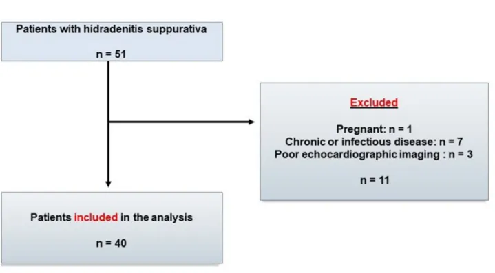
A total of 40 patients with HS and 100 age- and gen-der-matched controls were enrolled in the present study from June to September 2019. All consecutive adult (aged ≥ 18 years) HS patients admitted to our dermatol-ogy outpatient and emergency clinic were prospectively included during the study period. However, patients with any acute illness, diabetes mellitus, and with a history of chonic renal or hepatic insufficiency, cardiovascular, coro-nary, cerebrovascular, chronic inflammatory or connec-tive tissue diseases, pregnant women, and patients with poor echocardiographic imaging were not included in the patient or control groups in our study. The control group consisted of age- and gender-matched subjects admitted to the dermatology outpatient clinic during the study period due to skin disorders other than HS. Our study protocol was approved by the Muğla University ethics committee, and informed consent was obtained from all subjects. The study population included 40 consecutive patients with HS and 100 subjects as controls.
Data collection
The demographic characteristics and clinical features, including age, smoking habits, gender, height, weight, and body mass index, were recorded. Routine biochemical measurements including fasting blood glucose, albumin, electrolytes, creatinine, high-sensitivity C-reactive protein (hs-CRP), and lipid profile were obtained at admission.
Information about disease-specific characteristics such as duration of the disease and the number of involved areas was also obtained from HS patients. The severity of HS was assessed by the Hurley staging [15], and stage III patients were defined as having severe HS.
Epicardial fat thickness measurement
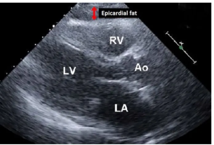
A single experienced cardiologist who was blinded to the subjects’ data performed transthoracic echocardiography in all HS patients and in control group using a Philips EPIQ system (Koninklijke Philips N.V.; Best, The Neth-erlands). The EFT was identified as the echo-free space between the epicardial layers and it was measured on the free wall of the right ventricle from the parasternal long-axis and short-long-axis views at the enddiastole [16] (Fig. 1).
Statistical analysis
All analyses were performed using SPSS Statistics ver-sion 23.0 for Windows (SPSS Inc., Chicago, IL, USA). Quantitative variables were compared by the Student t test or Mann–Whitney U tests, and qualitative variables were compared by the Pearson 2 test or Fisher exact tests. Pear-son and Spearman’s analyses were used to find out the cor-relation of EFT with other factors in patients with HS. To identify the independent predictors of ‘’severe disease’’, univariate and multivariate linear regression analyses were performed. An optimal cutoff value of EFT for the detec-tion of ‘’severe disease’’ was determined by receiver oper-ating characteristics (ROC) analysis.
Results
Forty patients with HS (mean age 41.7 ± 10.6 years; 47.5% female) and 100 controls (mean age 42.2 ± 11.3 years; 47% female) were enrolled in the study (Fig. 2). Comparison of demographic, clinical, laboratory characteristics, and EFTs of patients with HS and control subjects is presented in Table 1. No differences were found between HS and age- and gender- matched controls in terms of body mass index, lipid profile, or serum glucose levels. However, the hs-CRP (2.12 ± 1.86 vs. 1.32 ± 0.85 mg/L, respectively;
p < 0.001) and EFT (6.8 ± 1.2 vs. 4.9 ± 0.7 mm,
respec-tively; p = 0.001) were significantly increased in patients with HS compared with the controls.
The disease-specific characteristics of HS patients are presented in Table 1. The mean duration of HS was 12.1 ± 8.3 years, and mean age of onset was
Fig. 1 Echocardiography in the parasternal long-axis view of a 36-year-old male with HS showed an EFT of 7.1 mm. Ao aorta; LA left atrium; LV left ventricle; RV right ventricle
21.6 ± 9.4 years. The mean number of affected sites was 4.1 ± 1.9, and the most commonly involved sites were groin and axilla. Of the study population, the proportion of the patients with severe disease was 27.5% according to the Hurley staging.
Correlation between EFT and other parameters
There were no significant correlations between EFT and age, smoking, or lipid profile. However, there was a positive cor-relation between EFT and Hurley stage (corcor-relation coeffi-cient (r) = 0.432, p = 0.001), hs-CRP (r = 0.471, p = 0.001), and disease duration (r = 0.365, p = 0.032) (Table 2).
Association of EFT with disease severity
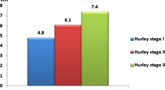
The EFT increased significantly with increase in dis-ease severity; the largest EFT was found in Hurley stage 3 patients (7.34 ± 2.30 mm), followed by Hurley stage 2 patients (6.12 ± 1.82 mm), and Hurley stage 1 patients (4.83 ± 0.98 mm) (Fig. 3).
Predictors of severe disease
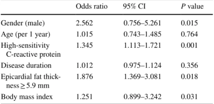
Multivariate and ROC analyses were performed to identify the predictors of Hurley stage III HS patients (Table 3). The
ROC analysis revealed that the optimal cutoff EFT value to predict severe HS was 5.9 mm (AUC = 0.877; 95% CI 0.815–0.923; p < 0.001). Multivariate analysis showed that male gender (OR 2.562; 95 CI 0.756–5.261; p = 0.015), hs-CRP (OR 1.345; 95 CI 1.113–1.721–5.261; p = 0.001), body mass index (OR 1.251; 95 CI 0.899–3.242; p = 0.031), and EFT ≥ 5.9 mm (OR 1.876; 95 CI 1.369–3.01; p = 0.018) were independent predictors of severe disease (Table 3).
Dıscussıon
The main findings of this preliminary study are as follows: [1] HS patients had a greater EFT than those without HS [2]. Increased EFT positively correlated with the duration of the disease and Hurley stage, and [3] EFT ≥ 5.9 mm indepen-dently predicted a higher severity of HS.
Increased risk for cardiovascular diseases has been dem-onstrated in various dermatological diseases in recent dec-ades [17]. Although the underlying mechanism remains not completely understood, chronic inflammation is thought to provide a connection between these two distinct entities [17]. However, the linkage and association between der-matological and cardiovascular diseases are often investi-gated in patients with psoriasis [18]. On the other hand, HS patients are known to have a high prevalence of overlapping
conditions with cardiovascular diseases such as hyperlipi-demia, insulin resistance, diabetes, and metabolic syndrome, making these patients more susceptible to atherosclerosis [2]. Moreover, Egeberg and colleagues demonstrated that the risk of cardiovascular mortality was higher in patients with HS compared with the risk in those with psoriasis [19]. Therefore, the early prediction of severe disease and optimal management of high-risk patients in HS may prevent late-onset cardiovascular complications.
Obesity is one of the most important modifiable risk factors for cardiovascular diseases such as coronary artery disease, heart failure, and atrial fibrillation. Although body mass index, waist, circumference, and other measures are useful indications of general adiposity, recent interest has focused on EFT as it plays a significant role in the development of cardiovascular diseases through secret-ing several inflammatory cytokines [20]. Different stud-ies conducted during the last few years have demon-strated that EFT is also associated with the presence and severity of different chronic inflammatory skin diseases [12–14]. Ertem et al. enrolled 54 patients with lichen planus, and 50 control subjects [12]. Like the results we found in patients with HS, they found positive correlations between EFT and duration of lichen planus, and hs-CRP levels. Bacaksız and colleagues included 115 adult patients with psoriasis and 60 healthy individuals [13]. They found that EFT and hs-CRP were significantly higher in the pso-riasis group than in the control group. The EFT was cor-related with body mass index, waist circumference, hs-CRP levels, and psoriasis severity index [13]. Belli et al. enrolled 40 patients with rosacea and 40 control subjects to investigate EFT in rosacea [14]. Rosacea patients had sig-nificantly higher EFT than controls, and EFT was indepen-dently associated with another marker of atherosclerosis;
Table 1 Comparison of hidradenitis suppurativa (HS) patients and control subjects
Values are presented as number (%), or mean ± standard deviation HS hidradenitis suppurativa, Hs-CRP high-sensitivity C-reactive pro-tein, HDL high-density lipoprotein; LDL: Low-density lipoprotein
HS (n = 40) Control (n = 100) P value
Female 19 (47.5) 47 (47) 0.769
Age (years) 41.7 ± 10.6 42.2 ± 11.3 0.425
Weight, m 1.67 ± 0.2 1.66 ± 0.1 0.965
Height, kg 80.5 ± 16.9 78.8 ± 15.9 0.096
Body mass index, kg/m2 27.3 ± 6.2 27.8 ± 5.9 0.125
Current smoking 14 (35.0) 34 (34.0) 0.844 Laboratory findings Creatinine (mg/dL) 0.90 ± 0.32 0.91 ± 0.30 0.741 Hs-CRP (mg/L) 2.12 ± 1.86 1.32 ± 0.85 < 0.001 Hemoglobin (g/dL) 12.8 ± 3.6 12.7 ± 2.9 0.562 LDL cholesterol (mg/ dL) 129.2 ± 32.4 127.2 ± 27.9 0.254 HDL cholesterol (mg/ dL) 47.2 ± 11.9 48.3 ± 12.5 0.366 Triglycerides (mg/dL) 129.3 ± 80.3 125.9 ± 81.2 0.101 Total cholesterol (mg/ dL) 182.4 ± 35.4 179.9 ± 35.6 0.069 Fasting glucose (mg/dL) 90.2 ± 10.6 89.7 ± 9.9 0.112 Epicardial fat thickness
(mm) 6.8 ± 1.2 4.9 ± 0.7 0.001
Age at onset 21.6 ± 9.4
Duration of the disease
(years) 12.1 ± 8.3
Hurley stage
I 4 (10)
II 25 (62.5)
III 11 (27.5)
Number of affected sites 4.1 ± 1.9
Fig. 3 Changes in epicardial EFT according to the severity of the disease. (p = 0.013 for Hur-ley stage I vs. stage II, p = 0.001 for Hurley stage II vs. stage III, and p < 0.001 for Hurley stage I vs. stage III)
carotid intima–media thickness [14]. The results of these three studies were in line with our findings that the EFT is increased and associated with the severity of the chronic inflammatory skin diseases. Although our and others stud-ies could not determine the causality due to the case–con-trol, and cross-sectional design of the studies, we postu-late that persistent inflammation may be the reason for the increased EFT in HS. The association between duration of the disease and EFT supports our hypothesis, as the longer duration of HS causes higher cumulative effect of inflammation. Whatever the reason, our study showed that EFT was increased and associated with the duration and severity of the disease in HS.
The EFT can be easily and non-invasively measured by echocardiography, and it may have the potential to be a marker of subclinical cardiovascular diseases and a predic-tor of severity of the disease in patients with HS. Therefore, increased EFT may guide the patient’s cardiovascular risk in HS. It remains, however, unknown whether a reduction in the amount of EFT could reduce the risk of cardiovascular diseases in patients with HS or treatment of HS reduces the thickness of epicardial adipose tissue.
Limitations
The results of our study may not be applicable to all patients due to strict inclusion criteria and the small sample size of the study. This is a cross-sectional study, and the prognostic value of serial changes in EFT could not be assessed. We did not use magnetic resonance imaging, which is the gold standard method in assessing EFT.
Conclusions
Although the pathological background of increased EFT in HS is not known, this study is the first to demonstrate that EFT is increased in HS patients, and it is associated with the severity and the duration of the disease. Further large volume prospective studies are warranted to examine the EFT in HS and to investigate whether increased EFT has prognostic importance in HS.
References
1. Napolitano M, Megna M, Timoshchuk EA, Patruno C, Balato N, Fabbrocini G, Monfrecola G (2017) Hidradenitis suppurativa: from pathogenesis to diagnosis and treatment. Clin Cosmet Inves-tig Dermatol 10:105–115. https ://doi.org/10.2147/CCID.S1110 19 2. Crowley JJ, Mekkes JR, Zouboulis CC, Scheinfeld N, Kimball A,
Sundaram M, Gu Y, Okun MM, Kerdel F (2014) Association of hidradenitis suppurativa disease severity with increased risk for systemic comorbidities. Br J Dermatol 171(6):1561–1565. https ://doi.org/10.1111/bjd.13122
3. Kimball AB, Sundaram M, Gauthier G, Guérin A, Pivneva I, Singh R, Ganguli A (2018) The comorbidity burden of hidrad-enitis suppurativa in the United States: a claims data analysis. Dermatol Ther (Heidelb) 8(4):557–569. https ://doi.org/10.1007/ s1355 5-018-0264-z
4. Kromann CB, Ibler KS, Kristiansen VB, Jemec GB (2014) The influence of body weight on the prevalence and severity of hidrad-enitis suppurativa. Acta Derm Venereol 94(5):553–557. https :// doi.org/10.2340/00015 555-1800
5. Vilanova I, Hernández JL, Mata C, Durán C, García-Unzueta MT, Portilla V, Fuentevilla P, Corrales A, González-Vela MC, González-Gay MA, Blanco R, González-López MA (2018) Insulin resistance in hidradenitis suppurativa: a case–control study. J Eur Acad Dermatol Venereol 32(5):820–824. https ://doi. org/10.1111/jdv.14894
6. Lee JH, Kwon HS, Jung HM, Kim GM, Bae JM (2018) Prevalence and comorbidities associated with hidradenitis suppurativa in Korea: a nationwide population-based study. J Eur Acad Dermatol Venereol 32(10):1784–1790. https ://doi.org/10.1111/jdv.15071 7. Phan K, Charlton O, Smith SD (2019) Hidradenitis
suppura-tiva and metabolic syndrome—systematic review and adjusted meta-analysis. Int J Dermatol 58(10):1112–1117. https ://doi. org/10.1111/ijd.14500
8. Theut Riis P, Saunte DM, Benhadou F, Del Marmol V, Guillem P, El-Domyati M, Abdel-Wahab H, Antoniou C, Dessinioti C, Gürer MA, Beksaç B, Szepietowski JC, Matusiak L, Emtestam L, Lapins J, Riad H, Doss N, Massa AF, Hamzavi I, Nicholson C, Dolenc-Voljc M, Kim KH, Ohn J, Zouboulis CC, Karagiannidis I, Mokos
Table 2 Pearson correlation analysis between epicardial fat thickness and other confounding variables in patients with hidradenitis suppu-rativa
Hs-CRP high-sensitivity C-reactive protein
Correlation coefficient (r) p value
Smoking 0.098 0.201 Age 0.080 0.358 Creatinine (mg/dL) 0.096 0.420 Hs-CRP (mg/dL) 0.471 0.001 Disease duration 0.365 0.032 Hurley stage 0.432 0.001 Total cholesterol 0.086 0.562
Table 3 Multivariate regression analysis for the prediction of severe disease (Hurley stage III)
Hs-CRP high-sensitivity C-reactive protein
Odds ratio 95% CI P value
Gender (male) 2.562 0.756–5.261 0.015
Age (per 1 year) 1.015 0.743–1.485 0.764
High-sensitivity
C-reactive protein 1.345 1.113–1.721 0.001
Disease duration 1.012 0.975–1.124 0.356
Epicardial fat
thick-ness ≥ 5.9 mm 1.876 1.369–3.081 0.018
ZB, Durinec P, Jemec GBE (2018) Low and high body mass index in hidradenitis suppurativa patients—different subtypes? J Eur Acad Dermatol Venereol 32(2):307–312. https ://doi.org/10.1111/ jdv.14599
9. Berg G, Miksztowicz V, Morales C, Barchuk M (2019) Epicar-dial adipose tissue in cardiovascular disease. Adv Exp Med Biol 127:131–143. https ://doi.org/10.1007/978-3-030-11488 -6_9 10. Alexopoulos N, Raggi P (2018) Epicardial adipose tissue:
another tassel in the complex fabric of atherosclerosis. Car-diovasc Hematol Disord Drug Targets 18(1):17–26. https ://doi. org/10.2174/18715 29X17 66617 01251 03555
11. Patel VB, Shah S, Verma S, Oudit GY (2017) Epicardial adipose tissue as a metabolic transducer: role in heart failure and coro-nary artery disease. Heart Fail Rev. 22(6):889–902. https ://doi. org/10.1007/s1074 1-017-9644-1
12. Ertem AG, Erdogan M, Koseoglu C, Akoglu G, Ozdemir E, Koseoglu G, Sivri S, Keles T, Durmaz T, Aktas A, Bozkurt E (2016) Epicardial fat tissue thickness is increased in patients with lichen planus and is linked to inflammation and dyslipidemia. Rev Port Cardiol 35(10):525–530. https ://doi.org/10.1016/j. repc.2016.04.010
13. Bacaksız A, Tasal A, Sevgili E, Erdoğan E, Onsun N, Sönmez O, Topukcu B, Asoğlu E, Göktekin O (2014) Epicardial fat thick-ness in patients with psoriasis vulgaris. Turk Kardiyol Dern Ars 42(1):47–54. https ://doi.org/10.5543/tkda.2014.78949
14. Belli AA, Altun I, Altun I (2017) Thickness of carotid intima and epicardial fat in rosacea: a cross-sectional study. An Bras Derma-tol 92(6):820–825. https ://doi.org/10.1590/abd18 06-4841.20176 832
15. Hurley H (1989) Axillary hyperhidrosis, apocrine bromhidro-sis, hidradenitis suppurativa and familial benign pemphigus:
surgical approach. In: Roenigk RK, Roenigk HH (eds) Roenigk and Roenigk’s dermatologic surgery principles and practice. Mar-cel Dekker Inc, New York, pp 623–645
16. Iacobellis G, Assael F, Zappaterreno RMC, A, Alessi G, Di Mario U, Leonetti F, (2003) Epicardial fat from echocardiography: a new method for visceral adipose tissue prediction. Obes Res 11(2):304–310. https ://doi.org/10.1038/oby.2003.45
17. Gijón E, Meseguer-Yebra C, Palacio-Aller L, Godoy-Rocati DV, Lahoz-Rallo C (2016) New populations at increased cardiovascular risk: Cardiovascular disease in dermatological diseases. Clin Investig Arterioscler 28:143–153. https ://doi. org/10.1016/j.arter i.2015.08.001
18. Jung KJ, Kim TG, Lee JW, Lee M, Oh J, Lee SE, Chang HJ, Jee SH, Lee MG (2019) Increased risk of atherosclerotic car-diovascular disease among patients with psoriasis in Korea: a 15-year nationwide population-based cohort study. J Dermatol 46(10):859–866. https ://doi.org/10.1111/1346-8138.150 19. Egeberg A, Gislason GH, Hansen PR (2016) Risk of major
adverse cardiovascular events and all-cause mortality in patients with hidradenitis suppurativa. JAMA Dermatol 152:429–434. https ://doi.org/10.1001/jamad ermat ol.2015.6264
20. Le Jemtel TH, Samson R, Ayinapudi K, Singh T, Oparil S (2019) Epicardial adipose tissue and cardiovascular disease. Curr Hyper-tens Rep. https ://doi.org/10.1007/s1190
6-019-0939-Publisher’s Note Springer Nature remains neutral with regard to