BIOINSPIRED MATERIALS FOR REGENERATIVE MEDICINE
AND DRUG DELIVERY APPLICATIONS
A THESIS SUBMITTED TO
THE GRADUATE SCHOOL OF ENGINEERING AND SCIENCE OF BILKENT UNIVERSITY
IN PARTIAL FULFILLMENT OF THE REQUIREMENTS FOR THE DEGREE OF
MASTER OF SCIENCE IN
MATERIALS SCIENCE AND NANOTECHNOLOGY
By
SEREN HAMSICI October 2016
ii
BIOINSPIRED MATERIALS FOR REGENERATIVE MEDICINE AND DRUG DELIVERY APPLICATIONS
By Seren Hamsici September 2016
We certify that we have read this thesis and that in our opinion it is fully adequate, in scope and in quality, as a thesis for the degree of Master of Science.
Ayşe Begüm Tekinay (Advisor)
Mustafa Özgür Güler (Co-advisor)
Semra İde
Aykutlu Dana
Approved for the Graduate School of Engineering and Science
Ezhan Karaşan
iii
ABSTRACT
BIOINSPIRED MATERIALS FOR REGENERATIVE MEDICINE AND DRUG DELIVERY APPLICATIONS
Seren Hamsici
MSc in Materials Science and Nanotechnology Advisor: Ayşe Begüm Tekinay
Co-advisor: Mustafa Özgür Güler October, 2016
The structural organization and functional capabilities of natural materials have inspired many technological and scientific developments. Biological systems are under constant pressure for innovation due to the constraints imposed by natural selection, which has allowed various organisms to surmount engineering challenges in ways that can scarcely be matched by modern science. Biomimetics or bioinspiration is a field that focuses on the adaptation of engineering principles observed in biological models to fabricate materials capable of circumventing longstanding problems in fields such as energy and medicine. This transition from biological systems has facilitated the design of effective materials, structures or processes within the range of nature’s adaptations and strategies.
In the first study of this thesis, I describe the development of a bioactive scaffold composed of adamantyl-conjugated, laminin-derived bioactive IKVAV peptide molecules enmeshed in electrospun cyclodextrin nanofiber (CDNFs). Accordingly, host-guest interactions between adamantyl groups on peptide termini and
iv
cyclodextrin molecules on electrospun nanofiber surfaces were utilized to produce a composite material for the treatment of neurodegenerative disorders. Electrospun CDNFs provided a 3-dimensional environment conductive for the growth of PC12 cells and expressed functionalized bioactive epitopes on their surfaces to enhance the differentiation of neural progenitors. In addition, CDNFs further supported neural growth through their highly aligned mesh structure. Neural bIII tubulin and synaptophysin I gene expression levels significantly increased when PC12 cells were cultured on aligned and IKVAV-functionalized CDNFs. Neurite extension of PC12 cells also increased significantly when cultured on aligned and IKVAV-functionalized CDNFs when compared to random and unIKVAV-functionalized electrospun CDNFs. As such, these nanofibers are able to effectively induce the neural differentiation of PC-12 cells through the physical and biochemical signals provided by their structure and bioactive sequence.
The second part of the present thesis focuses on the local delivery of gemcitabine, a cytotoxic cancer drug that is rapidly degraded in plasma and cannot be encapsulated in conventional delivery vesicles due to its highly hydrophobic nature. In order to overcome these limitations, gemcitabine was coupled with Fmoc-Gly and integrated into a peptide-based nanocarier system in order to control drug concentration within the therapeutic range and minimize the adverse effects. Two oppositely-charged amyloid inspired peptides (Fmoc-AIPs) were chosen as drug carrier systems. These molecules self assemble into nanofiber structures at physiological conditions through non-covalent interactions.
Overall, the present thesis demonstrates the significance of peptide-based materials for the purpose of designing functional bioinspired/biomimetic materials for various
v
cellular applications such as tissue engineering and drug delivery. The complexity of nature necessitates the design of biomaterials that can mimic the cellular microenvironment for the treatment of diseases, and further insight into natural processes will no doubt enhance our ability to overcome the engineering challenges presented by modern medicine.
Keywords: neurite outgrowth, drug delivery, host-guest interaction, regenerative medicine, biomaterials, amyloid inspired peptides
vi
ÖZET
BİYOESİNLENİLMİŞ MALZEMELERİN REJENERATİF TIP VE İLAÇ TAŞINIMI ALANLARINDA UYGULAMALARI
Seren Hamsici
Malzeme Bilimi ve Nanoteknoloji, Yüksek Lisans Tez Danışmanı: Ayşe Begüm Tekinay Tez Eşdanışmanı: Mustafa Özgür Güler
Ekim, 2016
Doğanın düzeni ve kabiliyeti teknolojik gelişmeler açısından pek çok bilim alanına ilham vermiştir. Evrim esnasında doğada birçok zorluklar yaşanmış buna bağlı olarak gösterilen insanların yaptıklarına kıyasla çoğu alanda üstün olmuştur. Biyobenzetim veya biyoesinlenme, seçilen biyolojik modellerden fikir almayı içeren ve bu modelleri sağlık sorunlarının giderilmesi veya yeni aygıtların veya malzemelerin geliştirilmesi amacıyla kullanmayı hedefleyen bir alandır. Biyolojik sistemlerdeki bu geçiş etkili malzemelerin dizaynında ve doğanın adaptasyon stratejileri çerçevesinde farklı yapılar ve malzemeler tasarlamayı olanak kılar.
Bu tezin ilk çalışmasında, nörodejeneratif hastalıkların tedavisi için kullanılabilinir yapı iskeleleri oluşturmak amacıyla, adamantil konjuge edilen ve laminin proteininden türetilmiş IKVAV peptit epitopu ile elektroeğirme yöntemi ile üretilmiş siklodekstrin nanofiberleri (SDNF) arasında konakçı- konuk etkileşiminden faydalanılmıştır. Elektroeğirme ile üretilmiş SDNF üç boyutlu bir biyo-uyumlu ortam sağlamasının yanısıra yüzeyinin biyoaktif epitop ile işlevselleştirilmiş olması
vii
PC-12 hücrelerinin nöral farklılaşmasını yönlendirmesini sağlar. SDNF nöral farklılaşma için fiziksel olarak belli bir yöne yönelme ile üretilmiştir. PC12 hücreleri hizalanmış ve IKVAV ile işlevselleştirilmiş SDNF üzerinde kültürlendiğinde nöral βIII tubulin ve sinaptofizin gen ifade seviyeleri önemli ölçüde artmıştır. Aynı zamanda rastgele olan ve herhangi bir peptit ile fonksiyonalize edilmemiş SDNF'lere göre hizalanmış ve IKVAV ile işlevselleştirilmiş SDNF'lerde kültürlenen PC-12 hücrelerinde nörit uzaması önemli ölçüde artmıştır. Çalışmadaki tüm sonuçlar hizalanma ve biyoaktif peptit epitopunun PC-12 hücrelerindeki nöral farklılaşmanın önemini vurgulamaktadır.
İkinci çalışmada ise gemsitabin ilacının konrollü salınımı için peptit bazlı yeni bir system geliştirilmiştir. Gemsitabinin hızlı plazma bozulması, ilacın terapötik etkinliğinin azalmasına sebep olur. Aynı zamanda gemsitabinin hidrofilik yapısı enkapsülasyonunu ve etkili bir şekilde taşınması için zorluklar yaratarak düzgün olmayan bir salım profiline neden olur. Bu sınırlamaların üstesinden gelmek amacıyla, gemsitabin Fmoc-Gly'ye konjuge edilmiş ve sonrasında peptit bazlı nanotaşıyıcı sisteme entegre edilerek hem ilaç konsantrasyonu terapötik aralık içinde tutulmuş hem de yan etkileri en aza indirgenmiştir. İlaç taşıyıcı sistem için iki farklı zıt yüklü amiloidden esinlenilmiş peptit (Fmoc-AIP) kullanılmıştır.
Bu tez doku mühendisliği ve ilaç salınımı gibi farklı hücresel uygulamalar için ve fonksiyonel biyolojik tabanlı biyobenzetim veya biyoesinlenilme yolu ile dizayn edilmiş peptit bazlı malzemelerin önemini vurgulamaktadır.
Anahtar kelimeler: Nörit uzaması, ilaç taşınımı, konakçı- konuk ilişkisi, rejeneratif tıp, biyomalzemeler
viii
ACKNOWLEDGEMENT
I would like to thank Prof. Mustafa Özgür Güler and Prof. Ayşe Begüm Tekinay for their support and guidance throughout my graduate studies.
I would like to express my special thanks to Dr. Göksu Çınar, Dr. Melis Şardan, Gökhan Günay, Egemen Deniz Eren, Melike Sever and Ayşegül Dede for their contribution to my studies and most importantly for their friendship and making me feel the part of a big family. Without their support, love and encouragement I could not achieve any of these studies.
I would like to acknowledge my old and present lab mates Dr. Aslı Çelebioğlu, Çağla Eren, Özüm Şehnaz Günel, M. Aref Khalily, Dr. Ruslan Garifullah, Begüm Dikeçoglu, Meryem Hatip, Oya İlke Şentürk, Hatice Kübra Kara, Dr. Özlem Erol, Dr. Gözde Uzunallı, Elif Arslan, Berna Şentürk, Alper Özkan, Zeynep Orhan, Dr. Ayşe Özdemir, Merve Şen, Canelif Yılmaz and Idil Uyan.
I would also like to acknowledge The Scientific and Technological Research Council of Turkey (TÜBİTAK BIDEB-2210 C and 114Z562) for funding my MSc research and financially supporting me.
I would like to indebted to my dearest and sincere friends M. Dilan Cindioğlu, Melis Kaplan and Başak Mecit for their encouragement, enthusiasm and not only during my thesis but also during my undergraduate years. I would never forget all beautiful moments even in stressful and difficult times. Finally my deepest gratitude goes to my family for their unconditional support, endless love throughout my life. My grandmother, no matter where you are I always feel like you are with me.
ix
CONTENTS
ABSTRACT ... iii ÖZET ... vii ACKNOWLEDGEMENT ... viii CONTENTS ... ixLIST OF FIGURES ... xii
LIST OF TABLES ... xv
Abbreviations ... xvii
Chapter 1 ... 1
INTRODUCTION ... 1
1.1 Design Considerations of Bioinspired Materials ... 2
1.1.1 Peptide Based Materials ... 3
1.1.2 Hybrid Materials ... 6
1.2 Bioinspired Materials for Biomedical Applications ... 8
1.2.1 Regenerative Medicine...8
1.2.2 Drug Delivery...10
Chapter 2 ... 13
BIOACTIVE PEPTIDE FUNCTIONALIZED ALIGNED CYCLODEXTRIN NANOFIBERS FOR NEURITE OUTGROWTH ... 13
2.1 INTRODUCTION ... 14
2.2 Experimental Section ... 17
x
2.2.2 Synthesis and Characterization of Peptides ... 17
2.2.3 Electrospinning of Cyclodextrin Nanofibers...….19
2.2.4 Critical Aggregation Concentration (CAC) Determination ... 19
2.2.5 Functionalization of CDNFs with peptides ... 20
2.2.6 Scanning Electron Microscopy (SEM) ... 20
2.2.7 X-Ray Photoelectron Spectroscopy (XPS) ... 20
2.2.8 FT-IR Analysis ... 20
2.2.9 Elemental Analysis ... 21
2.2.10 Isothermal Titration Calorimetry (ITC) ... 21
2.2.11 Contact Angle Measurements ... 21
2.2.12 Fluorescent Imaging ... 21
2.2.13 Viability Assay ... 22
2.2.14 Adhesion Assay ... 23
2.2.15 Immunocytochemistry ... 23
2.2.16 Quantitative Reverse Transcription Polymerase Chain Reaction (qRT-PCR) ... 24
2.2.17 Statistical Analysis ... 25
2.3. RESULTS AND DISCUSSION ... 25
2.4 CONCLUSION ... 39
Chapter 3 ... 40
GEMCITABINE INTEGRATED NANOPRODRUG CARRIER SYSTEM FOR BREAST CANCER THERAPY ... 40
xi
3.2 EXPERIMENTALSECTION....…..………....…………43
3.2.1 Materials ... 43
3.2.2 Synthesis of Gemcitabine Conjugated amino acid, Fmoc-G-Gem ... 44
3.2.3 Synthesis and Characterization of Fmoc-AIPs ... 44
3.2.4 Preparation of Fmoc-G-Gem integrated Fmoc-AIPs Carrier System .. 45
3.2.5 Critical Aggregation Concentration (CAC) Determination ... 45
3.2.6 FT-IR Spectroscopy ... 46
3.2.7 Circular Dichroism (CD) ... 46
3.2.8 X-Ray Diffraction (XRD) ... 46
3.2.9 STEM Imaging ... 47
3.2.10 Congo Red Staining ... 47
3.2.11 Controlled Release Behavior ... 47
3.2.12 Cell Culturing/Maintenance and Dose Determination ... 48
3.2.13 Cellular Viability ... 48
3.3 RESULTS and DISCUSSION ... 49
3.4 CONCLUSION ... 59
Chapter 4 ... 60
CONCLUSION AND FUTURE PERSPECTIVES ... 60
xii
List of Figures
Figure 1.1 Top-down and bottom-up approaches for material construction ... 2 Figure 1.2 Schematic illustrations of different types of self-assembled peptides ... 4 Figure 2.1 Chemical representations of a) Adamantane-6-aminohexanoic acid-GGKIKVAV-Am (IKVAV) b)Adamantane-6-aminohexanoic acid-GGKK-Am (KK) c) Schematic representations of electrospun and aligned CDNFs and host-guest interaction with bioactive peptide epitope ... 26 Figure 2.2 Liquid chromatography-mass spectroscopy (LC-MS) analyses of the synthesized adamantane conjugated peptides. ... 27 Figure 2.3 Blue shift of nile red in different concentrations of guest molecules ... 27 Figure 2.4 Isothermal titration calorimetry...28 Figure 2.5 Scanning electron microscopy (SEM) images of random and aligned CDNFs with peptide functionalized forms...29 Figure 2.6 Fiber diameters of electrospun random and aligned nanofibers ... 29 Figure 2.7 Aligned (a) and random (b) CDNFs functionalized with fluorescent-labeled IKVAV peptide. Red arrow shows the alignment direction c. XPS spectra of functionalized and aligned CDNFs. Inset shows the N1s (red line) and the presence of the peptide on the surface d. FTIR spectrum of IKVAV, CDNFs and CDNFs/IKVAV.. ... 30 Figure 2.8 XPS analyses of random (a-c) and aligned (d-f) CDNFs. a. Random only CDNFs, b. Functionalized with Ada-KK, c. Ada-IKVAV, d. Aligned only CDNFs, e. Functionalized with Ada-KK. ... 31 Figure 2.9 FT-IR spectrum of random organized CDNF only, CDNF/KK, CDNF/IKVAV and only bioactive and non-bioactive peptide molecules. ... 32
xiii
Figure 2.10 Contact angle analyses of a)random b)aligned CDNFs show wettability
behavior. ... 34
Figure 2.11 a) The relative viability of PC-12 cells cultured on random and aligned CDNFs with peptide functionalized system compared to control group treated with Poly-D-Lysine (PDL) b) The adhesion profiles of PC-12 cells after 2 h incubation in the presence of BSA and cyclohexamide.. ... 35
Figure 2.12 a) Relative viability on CDNFs after 48 h of incubation b) Cellular adhesion on CDNFs after 4 h of incubation ... 35
Figure 2.13 a) Synaptophysin expression of PC-12 cells cultured on random and aligned CDNFs with bioactive and non-bioactive functionalized peptide groups. Scale bars: 40 µm b) Neurite length and percentage of neurite bearing cells quantified by Image J at day 7 after induction reveals the aligned CDNFs functionalized with IKVAV is the most suitable scaffold for neurite outgrowth... ... 37
Figure 2.14 Expression of a) SYN1 and b) βIII tubulin normalized to control gene GAPDH.. ... 38
Figure 3.1 Synthesis scheme of Fmoc-G-Gem. ... 50
Figure 3.2 Mass Spectrum of Fmoc-G-Gem ... 50
Figure 3.3 Liquid chromatograms and mass spectra (LC-MS) of peptides ... 51
Figure 3.4 a) Chemical representations of Fmoc-G-Gem, Fmoc-AIP1 (Fmoc-EFFAAE) and Fmoc-AIP2 (Fmoc-KFFAAK), b) Schematic representations of coassembly of Fmoc-AIPs and Fmoc-G-Gem c) STEM image of Fmoc-AIPs and Fmoc-G-Gem ...51
xiv
Figure 3.5 Liquid chromatograms and mass spectra (LC-MS) of peptides ... 52 Figure 3.6 Blueshift of Nile Red against concentration of a) AIP1, Fmoc-AIP2, Fmoc-G-Gem, b) Fmoc-AIPs, Fmoc-AIPs w/ Fmoc-G-Gem, the blueshift was calculated by subtracting maximum emission of samples from Nile Red (654 nm).. 52 Figure 3.7 Secondary structure analysis of AIPs, G-Gem and Fmoc-AIPs w/ Fmoc-G-Gem conducted by a) Circular Dichroism (CD), b) Congo Red Assay, c) FT-IR analysis at physiological conditions ... 53 Figure 3.8 XRD patterns of Fmoc-AIPs and Fmoc-AIPs w/ Fmoc-G-Gem ... 54 Figure 3.9 The release behavior of Fmoc-G-Gem through Fmoc-AIPs for 480 h (20 days) at pH 7.4 and 5.5. ... 54 Figure 3.10 Cell viability of 4T1 cells for a) 24 h and b) 48 h by using alamar blue assay ... 55 Figure 3.11 Cell Viability of 4T1 cells treated with different concentrations of Fmoc-G-Gem encapsulated Fmoc-AIPs for 24 h and 48 h...56
Figure 3.12 a) Representative images of cellular viability of 4T1 cells treated with Fmoc-AIPs, Fmoc-AIPs w/ Fmoc-G-Gem, Fmoc-G-Gem and Gem Only groups (green: live, red: dead), b) The percentages of cellular viability for 3 days... 58 Figure 3.13 a) Representative images of live (stained green) and dead (stained red) assay of 4T1 cells treated with AIPs, Gem Only, G-Gem and Fmoc-AIPs w/ Fmoc-G-Gem. The percentage of cellular viability for 96 h, and one-day interval during 4 days (b, c respectively). ... 59
xv
List of Tables
Table 1.1 Recently published self-assembled peptide based hybrid materials ... 6 Table 2.1 Atom weight percentages of random and aligned CDNFs and functionalized forms ... 33 Table 2.2 Peptide amount in 100 g of functionalized random and aligned CDNFs……… ... ...33
xvi
Abbreviations
ANOVA Analysis of variance
CD Circular dichroism
CDNF Cyclodextrin nanofibers
CAC Critical aggregation concentration
DCM Dichloromethane
DIEA N,N-diisopropylethylamine
DMEM Dulbecco's modified Eagle's medium
DMF N,N-dimethylformamide
DMSO Dimethyl sulfoxide
ECM Extracellular matrix
FITC Fluorescein isothiocyanate
Fmoc 9-Fluorenylmethoxycarbonyl
FTIR Fourier transform infrared spectroscopy
HBTU N,N,N′,N′-Tetramethyl-O-(1H-benzotriazole-1-yl) uronium
hexafluorophosphate
HPLC High pressure liquid chromatography
ICC Immunocytochemistry
ITC Isothermal titration calorimetry
LC-MS Liquid chromatography-mass spectroscopy
NMR Nuclear magnetic resonance
PA Peptide amphiphile
PBS Phosphate buffered saline
xvii
QTOF Quadrapole time of flight
RT Room temperature
SEM Standard error of mean
SEM Scanning electron microscopy
SPPS Solid phase peptide synthesis
STEM Scanning transmission electron microscopy
TCP Tissue culture plate
TFA Trifluoroacetic acid
THF Tetrahydrofuran
XPS X-ray photoelectron spectroscopy
1
Chapter 1
1. Introduction
Although mankind is often proud of its accomplishments, human engineering can scarcely match the complexity and functionality of natural designs. This is hardly surprising, as evolution has far stricter demands for perfection and a billion-year head-start on intelligence! As such, natural systems are far superior to their artificial counterparts in many respects, and the prospect of repurposing these designs for human use is attractive for modern scientists and engineers alike [1]. Indeed, the field of biomimetics has been developed around this idea, and involves the adaptation of ideas found in nature for the development of novel materials, structures or processes [2]. This allows one to circumvent the material optimization process, which has already been performed through natural selection, and directly utilize designs that are known to be effective [3].
Biomimetic or bioinspired materials are at the frontier between biological and material sciences, chemistry, physics and biotechnology [4], [5]. The combination of design principles inherent to these fields allows the development of materials capable of rapidly diagnosing emerging diseases, directly releasing a drug to its target organ, or providing bioactive signals for the development of cells, tissues and organs [6]. Recent advances in the synthesis of biomaterials, polymers, and natural and synthetic biomacromolecules have greatly expanded the scope of these efforts, allowing the production of multifunctional materials that elicit a desired set of biological effects through their size, shape, surface chemistry and attached ligands [7], [8].
2
1.1 Design Considerations of Bioinspired Materials
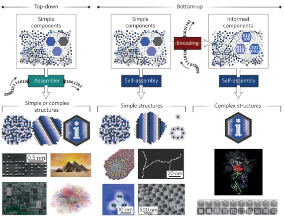
The low-cost, high-yield, impurity-free synthesis of biomaterials is crucial for a wide range of biomedical applications, and a broad range of top-down and bottom-up approaches exist for this purpose [9] (Figure 1.1). Top-down approaches create nanoscaled structures by scaling down the bulk material into its component parts by lithography techniques, while bottom-up approaches involve the assembly of supramolecular structures from basic components such as atoms and molecules [10]. Biological systems are especially adept at utilizing bottom-up processes to create incredibly complex biomolecules, such as proteins and nucleic acids [11].
Figure 1.1 Top-down and bottom-up approaches for material construction. (Adapted from Ref. 11 with permission from Nature Materials)
3
1.1.1 Peptide-Based Materials
Biomaterials for medical use must be compatible with the tissue they are interacting with, and must provide the necessary combination of mechanical integrity, chemical composition and biological signals that are required to elicit the desired physiological response [12]. Proteins and peptides are especially popular in nanobiotechnological research due to their ability to satisfy these conditions. Peptides are composed of amino acids, which in turn consist of an alpha carbon atom, an amino group (NH2), a
hydrogen atom (H), a carboxy group (COOH) and a side chain. Side chains are especially important for peptide design, as they are largely responsible for determining the structures, physicochemical properties, and biological functions of any given amino acid sequence [13]. Amino acids can be divided into four groups based on their side chains: charged (positive and negative), polar, non-polar and aliphatic. The side chains of peptides define their structures via non-covalent and covalent intreractions such as hydrophobic interactions, aromatic stacking, hydrogen bonding, disulfide bridges and electrostatic interactions. Even though these interactions are individually weak, their coordination allows the production of stable secondary structures through self-assembly [14].
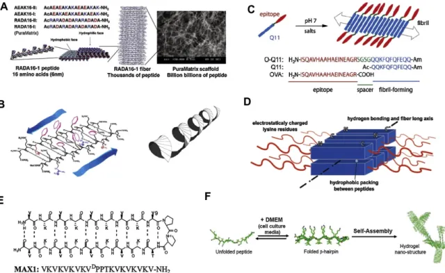
Peptide molecules often incorporate alternating hydrophobic and hydrophilic amino acids to facilitate their assembly into β-sheets (Figure 1.2).The (RADA)4 peptide, for
example, is an ionically complementary peptide that consists of repeating hydrophobic (alanine) and hydrophilic (arginine and aspartic acid) groups, which form stable β-sheet structures and allow the peptide to self-assemble into rigid hydrogels [15], [16]. RADA (Ac-RADARADARADARADA-Am) peptides are sold
4
commercially as Puramatrix® and used in cell culture to promote the differentiation of progenitor cells [17]. Frech and Morgan modified Puramatrix® with peptides and and investigated that this design enhances the differentiation capacity of human neural progenitor cells (hNPCs) into neuronal cells [18]. Furthermore, Dissanayaka
et al. used Puramatrix® for the aim of encapsulation of two differenct cell lines to
improve dental pulp regeneration by obtaining a combination of mineralization and angiogenesis[19].
Figure 1.2 Schematic illustrations of different types of β-sheet forming self-assembled peptides (Adapted from Ref.12 with permission from Acta Materialia)
Hartgerink et. al. designed another peptide motif capable of self-assembling into β-sheet through the presence of the ABA amino acid motif, where A consists of positively-charged residues and B domain composes of hydrophilic and hydrophobic amino acids [20] to maintain hydrophobic packing and antiparallel β-sheets [21].
5
They modified this sytem by integrating cell adhesion epitopes to the peptide backbone and developed a series of possible formulations for tissue remodeling [22].
Another example of a β-sheet forming motif is provided by insoluble amyloid aggregates, which cause Alzheimer’s disease as a result of their deposition in the nervous system [23]. The assembly of short peptides containing diphenylalanine, which is the core motif of amyloid fibrils, gained considerable interest in recent years due to their aromatic stacking arrangements that result in stable self-assembled systems [24].
Hydrophobic interactions are one of the most important driving forces for biological self-assembly, especially for structures such as phospholipid cell membranes. Peptide amphiphile (PA) molecules contain hydrophobic fatty acids, such as palmitic and lauric acids, which facilitates hydrophobic collapse and allows the hydrophilic peptide region to interact with water to form micellar structures [25]. These micelles are usually ordered into high-aspect-ratio nanosystems (i.e. nanofibers) due to the hydrogen bonding interactions among peptide molecules. The peptide-based composition of these nanostructures enhance their biocompatibility, biodegradability and non-immunogenicity, making them ideal for biomedical applications [26].
Peptide amphiphiles are often produced using solid-phase synthesis methods, which provide precise control over the peptide sequence, this method can be used for the mass-production of high-purity peptide samples without the need of harsh chemical reaction conditions [27]. Synthesis procedure begins with the conjugation of first amino acid to the solid support from its C- terminal, followed by the covalent linkage of subsequent attachment of amino acids to the N- terminal end of the alpha amino
6
group [28]. Functional side chains of the amino acids are protected in order to prevent side reactions [28]. The resulting peptide is organized into ordered structures by noncovalent interactions such as hydrogen bonding between amide-containing peptide groups, electrostatic interactions formed by charged side chains, and hydrophobic interactions [29] that drive the self-assembly mechanism.
1.1.2 Hybrid Materials
Hybrid biomaterials contain at least two distinct classes of molecules that are conjugated either covalently or non-covalently. Different conjugation designs enable the fabrication of novel materials with properties superior to these of individual molecules (Table 1.1). Conjugation of peptide domains to polymers, nucleobases, saccharides, aromatic groups or halogenic elements allows their self-assembly into different structural organizations, including nanoribbons, nanofibrils, nanotubes or nanospheres, through the synergistic combination of nanostructure components.
Table 1.1 Self-assembled peptide-based hybrid materials
Hybrid system Structure Ref.
Fmoc-FF, Fmoc-FG, Fmoc-GG, Fmoc-GF Nanoribbon/fiber/sheet [30] Fmoc-n-X-Phe
(n=2, 3 or 4 and X= F, Cl or Br) Fibril [31]
Fmoc-3-F-Phe-X and Fmoc-F5-Phe-X (X=OH,
NH2 or OMe) Fibril [32]
Naphthalene-FF-taurine Nanotube [33]
Pyrenebutyryl-ɛ-Ahx-VVAGH-Am Nanofiber [34]
Boc-FF Nanosphere [35]
Terthiophene-XE (X=G,V,I and L) Nanosheet/nanotube [36]
mPEO7-F4-OEt Nanotube [37]
PEG-Pep-fluorophore-Pep-PEG Micelle [38]
CREKA-PEG2000-DSPE Micelle [39]
7
Introducing aromatic moieties into the peptide backbone is another approach to drive the self-assembly process. While the self-assembly of peptide amphiphiles is driven by their linear hydrophobic tails, supramolecular assembly can also be provided by planar aromatic moieties with preferred π-stacking arrengements [41]. Fmoc (9-fluorenylmethoxycarbonyl) is a well-known molecule that promotes hydrophobic and π-stacking interactions through its fluorenyl rings. A combination of Fmoc and aromatic side-chains such as phenylalanine (F), tyrosine (Y) and tryptophan (W) allows the synergic formation of π–π interactions between Fmoc groups and side-chain phenyl rings, thus inducing faster self-assembly with higher stability and elacticity in the resulting hydrogels [42].
The conjugation of a peptide with a polymer yields a hybrid material that combines the versatility of the bioactive amino acid sequence with the prolonged lifetime, thermal stability and stimulus-responsiveness inherent to polymers [43]. There are two main types of peptide-polymer conjugates: either a hydrophobic peptide is integrated into a hydrophilic polymer, or a hydrophilic peptide is combined with a hydrophobic peptide [38]. The well-known physical and chemical properties and biocompatible behavior of the hydrophilic polymer polyethylene glycol (PEG) makes a popular choice for drug delivery and tissue scaffolding applications. Hamley et al. designed PEG-conjugated amyloid-forming peptides (FFKLVFFF, KLVFF, and AAKLVFFF) and showed that the hybrid material had a higher tendency for fibrillization with additional hydrophobic units [44]. The same group also investigated the effect of PEG chain length (either 5, 11 or 27) on nanostructure formation by the FFKLVFF sequence. While the shortest PEG chain resulted in twisted ribbons, the longest one formed uniform fibrils [45], indicating the influence
8
of PEG chain length on strand morphology.
1.2 Bioinspired Materials for Biomedical Applications
1.2.1 Regenerative Medicine
Regenerative medicine is one of the main areas in healthcare that stands to benefit from advances in nanobiotechnology. In order to mimic extracellular matrix (ECM), bioinspired materials aim to replace the degenerated tissues of the human body by enabling scaffolds functionalized with cells and biologically active molecules [46]. Within tissues, cells are surrounded by an ECM consisting of a complex, three-dimensional (3D), fibrous and hierarchically organized network that presents a wide range of biochemical and physical signals [47]. Furthermore, the ECM provides cell support, directs cell behavior, activates cascades of intracellular signaling pathways and regulates cell-cell and cell-soluble factors [48]. Therefore, mimicking the same level of complexity and functionality of ECM with engineered biomaterials is pivotal for regenerative medicine [49]. As such, tissue-engineered materials should not only serve as structural components that physically support cells, but also provide suitable environments for cell functionality and show key properties such as enhancing cell attachment, proliferation, differentiation and new tissue formation [50]. Recent advances in the fabrication of biomaterials have allowed the design of biomaterials capable of exhibiting many of these functions.
PA nanofiber networks are inherently similar to the ECM due to their high water content and porous network [51]. Synthetic PA nanofiber scaffolds stimulate cells to adhere, proliferate, migrate and differentiate to specific cellular fates [52],[20]. Laminin is one of the most abundant proteins in the ECM and plays an important role
9
in cell adhesion [53]. The laminin-derived Ile-Lys-Val-Ala-Val (IKVAV) peptide epitope is able to mimic the adhesive behavior of this molecule and stimulates the extension of neurites, which are crucial for the recovery of spinal cord injuries in cell-based approaches [54]. Stupp et al. showed that the use of IKVAV-PA nanofibers allowed reduced scar formation and increased axon regeneration in spinal cord injury models [55]. This group has also demonstrated the importance of alignment for guiding neurites along the nanofiber matrix, reporting that aligned nanofibers, when exposed to a CaCl2–containing solution, enhanced neurite
outgrowth and facilitated the differentiation of encapsulated neural progenitor cells into neurons showing electrical activity and synaptic connections [56].
Arg-Gly-Asp (RGD) is another peptide motif derived from fibronectin and facilitates cell adhesion by interacting with cell surface integrin receptors. It can be incorporated into the PAs to impart that functionality [57]. Koda et al. designed a palmitoyl-GGGHGPLGLARK-Am peptide, where PLG is the cleavage site that is recognized by the cancer-associated enzyme matrix metalloproteinase-7 (MMP-7) [58]. After enzyme treatment, the PA molecule changed its morphological characteristics and turned into long fibers from spherical micelles. Maruyama et al. used the same cleavage site in order to facilitate the selective entry of a cytotoxic peptide into cancer cells but not healthy cells [59]. Hamley et al. investigated the self-assembly and cytotoxicity of six alanine residues attached to the RGD tripeptide sequence, and showed that both factors are altered based on the concentration of the peptide [60]. Lastly, Mata et al. showed that self-assembled peptide nanostructured gels could be used for bone regeneration [61] by combining fibronectin-derived
10
epitope palmitoyl-AAALLLKKRGDS (RGDS-PA) and a phosphorylated serine residue-bearing palmitoyl-AAALLLEE(P)SG(S(P)-PA) [61].
In bone regeneration studies, bioactive peptide molecules derived from osteogenic growth peptides (OGPs) have been shown to enhance the differentiation of mesenchymal stem cells into osteoblasts [62]. Zhang et al. demonstrated that the functionalization of RADA with OGP at the C-terminus greatly improves its osteoinductive properties [63]. The level of differentiation was determined through the evaluation of ALP activity, and cellular proliferation and differentiation were observed to be significantly altered in the presence of this peptide system [63].
As a conclusion, PA systems present a versatile platform for a broad range of applications, as the peptide sequence can be designed and developed to answer a variety of biological challenges due to their self-supported and extracellular matrix mimicking properties.
1.2.2 Drug Delivery
A wide variety of nanostructured drug carriers have been proposed in order to enhance therapeutic indices, increase chemical and biological stability, reduce systemic toxicity, provide controllable drug delivery within the therapeutic window, and overcome both under-dosing and over-dosing issues [64]. These nanocarriers are composed of organic materials (such as polymers, liposomes, vesicles and peptides) or inorganic materials (such as gold nanoparticles and quantum dots) to serve as possible drug vehicles for cancer treatment [65].
11
sustained pace, and peptide hydrogels are highly suitable for this purpose. These gels maintain plasma rates of their drug cargo by adjusting their rate of release [26]. Recently, Gazit et al. demonstrated that short peptide sequences can form hydrogels that, due to their low molecular weight, are not bioaccumulated in in vivo applications [26]. However, in some conditions, cytotoxicity would be challenge in these systems. Xu et al. investigated dipeptides that capable of forming hydrogels through their exposed N-terminal residues and inhibit the growth of cancer cells [66]. Hartley et al. studied hydrophobic tripeptide (D
Leu-Phe-Phe) and used it to encapsulate ciprofloxacin, an antibacterial drug that is effective against several types of bacteria [67]. The peptide hydrogel could accumulate up to 30% of its weight in ciprofloxacin, and release kinetics experiments showed that the drug could be released in a sustained equilibrium 2-3 days after encapsulation. Xu group designed small peptides with D-amino acids and used them for the delivery of naproxen (Npx), a nonsteroidal anti-inflammatory drug. The self-assembly mechanism of diphenylalanine enable to conjugate with Npx in order to improve the stability of the supramolecular structure. However, these methods nonetheless face certain limitations such as low encapsulation efficiency, drug leakage and disruption of gel formation by drug molecules during the self-assembly process [68]. More recently, the use of pro-drug based nanostructures has emerged as a new approach for drug delivery. The advantage of this technique is a substantial increase in drug loading, since each molecule in the gel carries its own drug molecule [69]. The existence of enzymes in the intestine is not allowed drugs to take orally. Therefore, the chosen way for drug-peptide conjugates is administration via the parenteral route [70]. Special targeting peptide epitopes can be used for selective cell surface binding and
12
increase the drug’s circulation times within the body, allowing it to reach its target cells and become internalized through peptide-receptor interactions. Ephrin type-A receptor 2 (EphA2) is overexpressed in various cancer types such as metastatic breast, lung, and colon carcinomas [71,72]. Wang et al. investigated that the conjugation of EphA2-targeting peptide YSA (YSAYPDSVPMMS) with the anticancer drug paclitaxel (PTX) effectively prevented tumor growth in prostate cancer models [73].
13
CHAPTER
2
BIOACTIVE PEPTIDE FUNCTIONALIZED ALIGNED
CYCLODEXTRIN NANOFIBERS FOR NEURITE OUTGROWTH
14
2.1 INTRODUCTION
Neurotrauma, including spinal cord and peripheral nerve injury, is a significant health problem across the world and causes severe disabilities with more than a hundred thousand new injuries each year [74]. Nerve autografts constitute the gold standard for the treatment of peripheral nerve injuries, however, only half of the patients get full recovery after treatment because of donor shortage, mismatch between donor nerve and injury site, immunological problems and the need of second surgery [75,76]. These problems have led to the development of alternative medical therapies, focused on polymeric biomaterials, which are able to guide and direct neural cells to promote recovery of motor or sensory functions [77,78]. Biomaterials are also used for the treatment of neurodegenerative diseases by repairing and regenerating damaged tissue through mimicking the native environment of the neuronal cells [79,80]. Extracellular matrix (ECM) acts as a structural support and includes a wide range of polysaccharide chains and fibrous proteins offering the biochemical and biomechanical cues for cells to proliferate, differentiate and migrate [81]. Therefore, design of bioactive scaffolds mimicking ECM can facilitate attachment of cells, enable nutrient transport with its porous structure and provide relevant signals to cells [82].
Electrospinning is a widely used fabrication technique for producing fibers with a range from nanometers to micrometers. The interest behind this technique is mainly due to the ease of control on structural, mechanical and biological properties of the fabricated materials by simple changes such as functionalization with different kinds of compounds or agents [83–85]. The most frequently used polymers in electrospinning process for nerve tissue-engineered grafts are poly(l-lactic acid)
15
(PLLA), poly(lactic-co-glycolic-acid) (PLGA) and poly(ε-caprolactone) (PCL) due to their biodegradability and biocompatibility [86]. On the other hand, non-bioactive and hydrophobic nature of these polymers limit the cell-materials interactions, which cascade the essential cellular bioprocesses for neural differentiation [87]. In order to improve hydrophilicity and bioactivity on the surface of electrospun polymeric materials, additional approaches such as chemical covalent binding [88], air plasma treatment [89] and blending during electrospinning [90] were previously reported. Furthermore, electrospun nanofibers were chemically functionalized with laminin or laminin derived epitopes to promote neural cell affinity, recovery and neurite extension [91–93]. Becker et al. functionalized PLLA fibers with laminin derived epitope by click chemistry and showed the improved effect of the peptide conjugated fibers on neural cells in terms of neurite extension [92]. In another approach, Lee et al. applied air plasma method using ammonia gas to develop electrospun PLGA scaffolds presenting amino rich and hydrophilic surface characteristics for cellular adhesion and proliferation [94]. However, adjusting the surface homogeneity and controlling the exposure time are challenging issues since the air plasma affects only limited depth of electrospun scaffolds [95]. In addition to the chemical conjugation approaches, Ramakrishna et al. prepared PCL nanofibers which were either blended with laminin or encapsulated laminin in the core of PCL nanofibers via electrospinning process [96]. However, the natural proteins could be denatured during the electrospinning process, because of the harsh conditions of organic solvents. Besides functionalization, the orientation of the electrospun fibers also constitutes an important feature for designing biomaterials for neural tissue engineering[97,98]. Uniaxially aligned fibers guide regenerating axons and induce
16
extension of the neurites along the direction of scaffold, which is able to repair the nerve defects even in the presence of long peripheral nerve gaps [99=101].
Supramolecular host-guest chemistry is a novel surface functionalization method due to specificity of recognition motifs via non-covalent interactions [102]. Cyclodextrins (CDs) are composed of glucopyranose subunits, and they are one of the most intriguing oligosaccharide types that form noncovalent host-guest interaction complexes due to their cyclic and truncated cone shape structures [103,104]. One of the most well-known noncovalent host-guest interactions is observed between CD and adamantane molecules where CD cavity serves as a host while adamantane is a hydrophobic guest molecule [105]. Stupp et al. used this approach to obtain biofunctionality through adamantane conjugated bioactive peptide epitope on the surfaces containing CD moieties in order to enhance focal adhesions of cells [106].
Here we report a versatile and effective approach for design of aligned electrospun CD nanofibers (CDNFs) capable of being functionalized by adamantane conjugated laminin derived peptide epitope. In this system, the pentapeptide epitope including Ile-Lys-Val-Ala-Val (IKVAV), a laminin derived sequence promoting neurite outgrowth [107,108], was utilized as the bioactive epitope. In this study, aligned cyclodextrin nanofibers (CDNFs) produced in environmentally friendly conditions and functionalized with adamantane conjugated IKVAV epitope through host-guest interaction were demonstrated for the first time. We observed that biofunctionalized aligned electrospun CDNFs direct adhesion and neurite outgrowth of neuronal progenitor PC12 cells.
17
2.2 EXPERIMENTAL SECTION
2.2.1 Materials
All protected amino acids, Fmoc-6-aminohexanoic acid, Rink amide MBHA resin, 1-adamantaneaceticacid, fluorescein isothiocyanate isomer I (FITC), N,N,N′,N′-tetramethyl-O-(1H-benzotriazole-1-yl) uronium hexafluorophosphate (HBTU) and diisopropylethylamine (DIEA) were purchased from Novabiochem, ABCR or Sigma-Aldrich. The hydroxypropyl-β-cyclodextrin (HPβCD) (degree of substitution: ~0.6, CavasolÒW7 HP Pharma) was kindly donated by Wacker Chemie AG (Germany). 1,2,3,4-butanetetracarboxylic acid (BTCA, 99%) and sodium hypophosphite hydrate were commercially obtained from Sigma-Aldrich. All other chemicals and materials used in this study were analytical grade and purchased from Invitrogen, Fisher, Merck, Alfa Aesar, and/or Sigma-Aldrich.
2.2.2 Synthesis and characterizations of peptides
Fmoc solid phase peptide synthesis method was used in order to synthesize adamantane-6-aminohexanoicacid-Gly-Gly-Lys-Ile-Lys-Val-Ala-Val-Am (IKVAV), aminohexanoicacid-Gly-Gly-Lys-Lys-Am (KK) and adamantane-6-aminohexanoicacid-Gly-Gly-Lys-Lys(FITC)-Ile-Lys-Val-Ala-Val-Am (FITC-IKVAV). Rink amide MBHA resin (Novabiochem) served as a solid support for the synthesis all peptide molecules. In order to activate carboxylate group of 2 moles equivalents of amino acid, 3 moles equivalents of diisopropylethylamine (DIEA) and 1.95 mole equivalents of N,N,N′,N′-tetramethyl-O-(1H-benzotriazole-1-yl) uronium hexafluorophosphate (HBTU) were used for 1 mole equivalent of solid resin. For each coupling step, 10% acetic anhydride-DMF solution was used to acetylate the
18
unreacted amine groups and Fmoc protecting groups were removed with 20% piperidine/dimethylformamide (DMF) for 20 min. The coupling time for the amino acids and Fmoc-6-aminohexanoic acid was 2 h for each cycle. The coupling mechanism of 1-adamantaneacetic acid was similar to amino acid coupling steps. Between all these steps, resins were washed three times using DMF, DCM and DMF, respectively. For FITC conjugation to IKVAV peptide, 4- methyltrityl (Mtt) protecting group of –Lys was removed using 60 mL cleavage solution containing 2.85 mL TFA, 0.075 mL distilled water, 0.075 mL triisopropylsilane and 57 mL DCM. The resins were shaken with 10 mL of this solution 6 times for 5 min each, then washed with DCM until the yellow color in washing DCM disappeared. Finally, the resins were washed with 5% (v/v) DIEA in DCM prior to FITC coupling. The FITC coupling solution containing 389.4 mg FITC and 256.8 µL DIEA was prepared in 3 mL DMF and added on the resins. The reaction vessel was covered with aluminum folio in order to prevent the bleaching of FITC. For studies utilizing FITC for visualization, it was combined with IKVAV at a ratio of 5-mol %. After the completion of the synthesis, peptides were removed from the solid supports, and the cleavage solution containing 95% trifluoroacetic acid (TFA), 2.5% distilled water, 2.5% triisopropylsilane was added. The cleavage reaction was continued for 3 h by shaking the resins with the cleavage solution. Then, the resins were washed with DCM to collect the cleaved peptides into the flask. Excess TFA and DCM were removed by rotary evaporator, and peptides were precipitated in ice-cold diethyl ether overnight. To remove ether, centrifugation was performed at 8000 rpm for 20 min. The precipitated peptides were dissolved in distilled water and kept at -80 °C overnight. The lyophilization of the frozen peptide solutions was carried out for 3
19
days in order to obtain dried peptide powders. The purity of the peptides was determined by Agilent 6530 quadruple time of flight (Q-TOF) mass spectrometry with electrospray ionization (ESI) source. Further purification was performed with a preparative HPLC system (Agilent 1200 series).
2.2.3 Electrospinning of cyclodextrin nanofibers (CDNFs)
Crosslinked water-insoluble CDNF was obtained by electrospinning of HPβCD
aqueous solution and BTCA was used as a cross-linker. The electrospinning parameters were adjusted as follows; 0.5-2 mL/h flow rate, 10-20 kV applied voltage and 10-20 cm collector distance. While randomly oriented CDNF were deposited on grounded fixed metal collector, rotating drum at speed a speed of 2000 rpm was used to obtain aligned CDNF.
2.2.4 Critical aggregation concentration (CAC) determination
In order to determine the transition phase between peptide assemblies and also control the morphologies, hydrophobic Nile red (9-diethylamino-5-benzo[α]phenoxazinone) fluorescent probe assay was applied to the peptide solutions (ranging from 1 mM to 0.244 µM) prepared in the distilled water at around pH 7. 1.25 mM stock solution of Nile red was prepared in ethanol (1 mL) and then diluted to 78.12 µM by using ethanol. The peptide solutions prepared at the different concentrations were mixed with the same amount of the dye solution; and incubated overnight at room temperature. Final Nile red concentration in the mixtures was 250 nM; and 0.31% (v/v) ethanol was present in the solutions. Cary Eclipse Spectrophotometer was used to collect emission spectra between 580 nm and 750 nm with an excitation of 550 nm.
20
2.2.5 Functionalization of CDNFs with peptides
Electrospun CDNFs were washed with ethanol and water, respectively. Then, they were immersed in the peptide solutions (IKVAV or KK) and incubated for 24 h in order to allow host-guest interaction at 1:1 molar ratio.
2.2.6 Scanning electron microscopy
Morphology of CDNFs and peptide-functionalized CDNFs were analyzed with FEI Quanta 200 FEG scanning electron microscope. Prior to imaging, all samples were coated with 8 nm Au–Pt. The diameters of electrospun fibers were quantified by ImageJ software.
2.2.7 X-ray photoelectron spectroscopy (XPS)
XPS analyses of samples were conducted by using Thermo K- alpha monochromated high performance X-ray photoelectron spectrometer. CDNFs and peptide-immobilized system were analyzed after washing the systems. The survey analyzes were conducted by 5 scans. A high-resolution spectrum for carbon and oxygen were recorded for 10 scans whereas nitrogen was scanned at 15 scans.
2.2.8 FT-IR spectroscopy
The Fourier Transform Infrared Spectrometer (FT-IR) (Bruker-Tensor 37) was used for the collection of IR spectra of the samples. Only CDNFs and the peptide functionalized CDNFs prepared at both aligned and random electrospun fiber organizations were mixed with potassium bromide (KBr) to prepare the pellets by pressing. The IR spectra were recorded between 4000 and 400 cm-1 at a resolution of 4 cm-1 with 64 scans per sample.
21
2.2.9 Elemental analysis
After host-guest functionalization, peptide content in the electrospun CDNFs was analyzed with Thermo Scientific FLASH 2000 series CHNS-O analyzer. Both random and aligned CDNFs and their peptide-functionalized forms were analyzed. As a standard, 2,5-(bis(5-tert-butyl-2-benzo-oxazol-2-yl)) thiophene was used. For each analysis, vanadium pentoxide was used as a catalyst for complete oxidation.
2.2.10 Isothermal titration calorimetry
Stoichiometry and association constants between CD units and guest peptides (IKVAV and KK) were determined using ITC (Microcal ITC200). 40 µL of 5 mM CD solution was injected to the cell containing 280 µL 350 µM IKVAV or 700 µM KK solution, respectively with 1.5 µL increments. During the measurements, the cell temperature was 25 oC, reference power was 3.5 mcal s-1, and stirring speed was 1000 rpm.
2.2.11 Contact angle measurements
The wettability of the electrospun CDNFs was measured using an optical contact angle measurement system (Dataphysis OCA30). Ultra-pure water was used as the testing liquid.
2.2.12 Fluorescent imaging
The functionalization of CDNFs with the peptide molecules through host-guest interactions were also analyzed by incubating FITC conjugated IKVAV peptide with the electrospun CDNFs overnight. Electrospun CDNFs were washed with ethanol and water prior to the imaging. The fluorescence images were acquired using Zeiss LSM510 microscope with a 20X objective. FITC-IKVAV containing peptide
22
solution used for the fluorescent imaging was prepared by mixing non-labeled IKVAV peptide at 1:19 molar ratio, respectively.
2.2.13 Viability assay
PC-12 cellular viability and adhesion analysis were performed in a 48-well plate. Before the cell culture experiments, 9 mm cover slips coated with parafilm were immersed in 96% (v/v) ethanol solution for 30 min; and then sterilized with UV for 15 min. The electrospun nanofibers prepared cut to cover the coverslips were treated with 70% (v/v) ethanol for 30 min for sterilization. Then, the scaffolds were put into cell culture medium at 37 °C for 6 h. PC-12 cells (5.0x10⁴ per well) were seeded on
the electrospun scaffolds containing Roswell Park Memorial Institute medium (RPMI) with 10% horse serum (HS), 5% fetal bovine serum (FBS), 2 mM L-glutamine and 1% penicillin/streptomycin (P/S) incubated at 37°C in 5% (v/v) CO2
for 24 h and 48 h. MTT (3-(4,5-dimethylthiazol-2-yl)-2,5-diphenyltetrazolium bromide) method was used to evaluate the activity of cells on the electrospun fibers. 270 µL of DMEM (Dulbecco’s Modified Eagle Medium) without phenol red and 30 µL of MTT was added into each well and cultivated for 4 h under standard cell culture conditions. All nutrient solution was removed, 300 µL of MTT solubilization solution was added into each well and pipetted at constant temperature in order to solubilize the crystals. The purple solution was transferred into a 96-well culture plate (200 µL/well), and its absorbance was measured at 570 nm using a micro plate reader (M5, Molecular Device)
23
2.2.14 Adhesion assay
The cell adhesion assay was performed as described [109]. Briefly, the cells were incubated with 4 mg/mL BSA and 50 µg/mL cyclohexamide in serum free medium for 2 h and 4 h at 37 oC with 5% CO2. Then, 500 µL of PC12 cells (1 x 10⁵ cells/mL)
were seeded with medium containing 4 mg/mL BSA and 50 µg/mL cyclohexamide for 2 h. After the incubation, unattached cells were washed with PBS twice. The attached cells were fixed with 4% formaldehyde in PBS for 15 min and then stained with 0.5% crystal violet for 1 h. After that point, experiment was conducted under dark conditions. The plates were gently washed with distilled water five times and cellular membrane was distrupted with 2% SDS for 10 min. Absorbance was measured at 570 nm with a M5 microplate reader.
2.2.15 Immunocytochemistry (ICC)
Prior to neural induction, PC-12 cells (5 x 10⁴ per well) were cultured on the materials for 24 h at the standard cell culture conditions. Then the medium was replaced with freshly prepared neural induction medium containing MEM with 2% HS, 1% FBS, 2 mM L-glutamine and 1% P/S along with 20 ng/mL NGF. The cells were cultured on the materials for seven days and fixed with 500µL/well pre-warmed 4% paraformaldehyde/PBS for 15 min at room temperature. After discarding paraformaldehyde, the cells were washed with 0.3% triton-X100/PBS for 15 min at room temperature on shaker. Then, the blocking was carried out with 10% normal goat serum (NGS) and 1% bovine serum albumin (BSA) in PBS with 0.3% Triton-X for 1 h at room temperature on shaker. After aspirating the blocking solution, the cells were treated with primary antibodies against Synaptophysin at a 1: 400 dilution.
24
The cell culture plates were sealed with parafilm and incubated overnight at 4 °C on nutator shaker. Next day, they were incubated for an hour at room temperature. Then, cells were washed with 0.3% PBS/Triton- X100 for 5 min on rocking shaker and twice with PBS for 5 min on rocking shaker. After that point, the experiment was continued at dark conditions. Secondary antibody (GAR AF488) was added with 10% BSA and incubated for 1 h. Washing steps were again applied with PBS and finally distilled water on the shaker. A drop of Prolong Gold Mounting Medium was added onto slides of each coverslip. Then, the coverslips were taken out from each well, inverted and put on mounting solution on slides. 8 images were taken from each well with Zeiss LSM510 microscope with 20x objective. The neurite extension of PC-12 cells was quantified by Image J, and the average neurite lengths were obtained from three independent replicates.
2.2.16 Quantitative reverse transcription polymerase chain reaction (qRT-PCR)
Synaptophysin and βIII tubulin gene expression profiles were examined by qRT-PCR. RNA of PC-12 cells were isolated on day 7 by using TRIzol reagent (Invitrogen). The yield and purity of the isolated RNA were quantified by Nanodrop 2000 (Thermo Scientific). Primer sequences were designed using NCBI database.. Temperature cycling for the reaction was determined as 50 °C for 3 min, 95 °C for 5 min, 40 cycles of 95 °C for 15 s, Tm (58.0 °C for Synaptophysin and βIII tubulin) for
30 s, and 40 °C for 1 min, respectively. Amplification was analyzed by determining the binding of SYBR I to double stranded DNA. The gene expressions were normalized to GAPDH as the internal control gene.
25
2.2.17 Statistical Analysis
For each experimental group, experiments were repeated twice with at least three replicated for each group. Standard error of means (sem) were used for statistical significancy by using one-way or two-way analysis of variance (ANOVA), whichever applicable.
2.3 RESULTS AND DISCUSSION
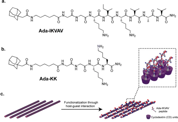
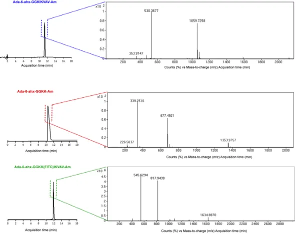
Adamantane (Ada), as a small guest hydrophobic molecule, is able to form host-guest inclusion complex with CD due to its complementary size and high affinity to hydrophobic CD cavity [110,111]. In Figure 2.1, 1-adamantaneacetic acid unit was covalently conjugated to the peptide backbone either presenting laminin mimetic epitope (IKVAV) inducing differentiation of neural progenitor cells into neurons (Figure 2.1a) or non-epitope bearing peptide (KK) through 6-aminohexanoic acid molecule as a spacer (Figure 2.1b). Two glycine residues were introduced between hydrophilic amino acid residues and hydrophobic tail in order to enhance displaying of the bioactive cues at the surface. The functionalization of the non-bioactive polymer surface was achieved via host-guest interaction by incubation of electrospun CDNFs in the peptide solution (Figure 2.1c). The peptide molecules were synthesized by using solid phase peptide synthesis method, purified with HPLC and characterized with LC-MS (Figure 2.2).
26
Figure 2.1 Chemical representations of a) Adamantane-6-aminohexanoic acid-GGKIKVAV-Am (IKVAV), b) Adamantane-6-aminohexanoic acid-GGKK-Am (KK). c) Schematic representation of electrospun and aligned CDNFs and host-guest interaction with bioactive peptide epitope.
The peptide concentrations within the incubation solutions were determined by Nile Red assay [112] (Figure 2.3) in order to prevent peptide aggregation and promote free integration of soluble peptide molecules into CD cavities. Critical aggregation concentration (CAC) of IKVAV and KK peptides were determined as 60 µM and 125 µM, respectively (Figure 2.3). Therefore, CDNFs were allowed for incubation with 50 µM IKVAV and KK peptides for 24 h. The binding affinity of adamantane conjugated IKVAV and KK peptides to soluble CD was determined by ITC measurements (Figure 2.4).
27
Figure 2.2 Liquid chromatography-mass spectrometry (LC-MS) analyses of the synthesized adamantane conjugated peptides.
Figure 2.3 Blue shift of Nile Red in different concentrations of guest molecules. Formation of aggregation takes place for Ada-IKVAV and Ada-KK at concentration around 60 and 125 µM respectively.
28
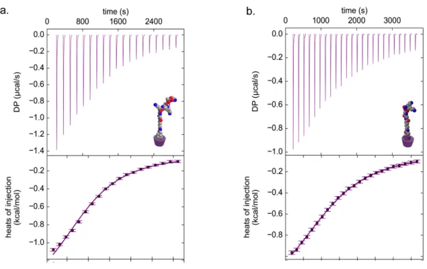
Figure 2.4 Isothermal Titration Calorimetry (ITC) traces of CD titrated into solutions of a) Ada-IKVAV, b) Ada-KK. The purple markers represent each injection, whereas the purple line fitted by software.
The inclusion complex formed at 1:1 molar ratio. In addition, the association constants of both of the IKVAV and KK peptides were found as log KaIKVAV = 4.11
and log KaKK =4.08 respectively, which are comparable with the association
constants presented previously [113]. The structural properties of only CDNFs and the peptide-functionalized materials were also analyzed with SEM (Figure 2.5). The images revealed that the diameters of CDNFs were within the range of hundred nanometers to several microns (Figure 2.6). Furthermore, noncovalent functionalization of CDNFs with peptide molecules did not cause any significant change on the morphology of the CDNFs (Figure 2.5 and Figure 2.6). The presentation of the peptide molecules on the electrospun CDNFs through host-guest
29
functionalization were also investigated using fluorescently labeled IKVAV peptide (FITC-IKVAV).
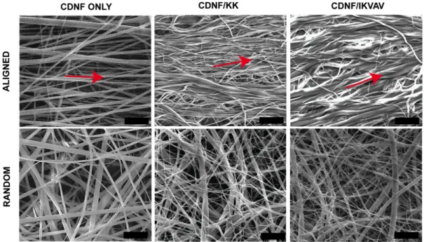
Figure 2.5 Scanning electron microscopy (SEM) images of random and aligned CDNFs with peptide functionalized forms (scale bars: 10 µm). Red arrow shows the alignment direction. Random Aligned 0.0 0.5 1.0 1.5 Fi be r di am et er ( μm ) CDNF only CDNF/KK CDNF/IKVAV
Figure 2.6 Fiber diameters of electrospun random and aligned CDNFs with their peptide functionalized forms. There is not any significant difference between groups in terms of fiber diameter.
30
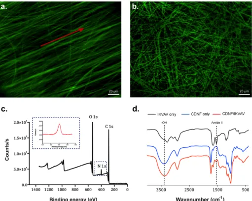
Figure 2.7 Aligned (a) and random (b) CDNFs functionalized with fluorescent-labeled IKVAV peptide. Red arrow shows the alignment direction c. XPS spectra of functionalized and aligned CDNFs. Inset shows the N1s (red line) and the presence of the peptide on the surface d. FTIR spectrum of IKVAV, CDNFs and CDNFs/IKVAV.
After incubation of both aligned and random CDNFs within the peptide solution for 24 h, the confocal images of the samples were acquired to reveal the existence of the peptides on the CDNFs. As shown in Figure 2.7, the fluorescently labeled bioactive peptide molecules were homogenously distributed on both aligned and random electrospun CDNFs (Figure 2.7a). In addition to the fluorescence imaging, the surface characterizations of the samples were performed by XPS. Both random and aligned CDNFs showed only carbon and oxygen atoms (Figure 2.8a-d). On the other hand, functionalization of random and aligned CDNFs with KK and IKVAV (Figure 2.8b-c) demonstrated an additional nitrogen peak, which indicates the existence of the peptides on the surfaces of both random and aligned CDNFs (Figure 2.7c)
31
Figure 2.8 XPS analyses of random (a-c) and aligned (d-f) CDNFs. a. Random only CDNFs, b. Functionalized with Ada-KK, c. Ada-IKVAV, d. Aligned only CDNFs, e. Functionalized with Ada-KK.
32
To further investigate the formation of inclusion complexes between CDNFs and the peptide moieties, FT-IR spectra of the samples were obtained for both aligned and random morphologies (Figure 2.7d and Figure 2.8)
Figure 2.9 FT-IR spectrum of random organized CDNF only, CDNF/KK, CDNF/IKVAV and only bioactive and non-bioactive peptide molecules.
In the case of bare CDNFs, a wide IR absorption band was present at 3390 cm−1 due
to bending vibration of -OH group. However, the peptide functionalization resulted in the red shift on the peak position 3390 cm−1 to 3346 cm−1 due to the interactions between the adamantane-peptide conjugates and hydroxyl groups of CDNFs. In addition, the IR spectra of peptide only group showed typical Amide II peak around 1533 cm−1 due to the N-H bending [114]. Together with host-guest interactions, the
Amide II characteristic band was also shown on the IR spectrum of peptide functionalized CDNFs due to the presence of peptide in the system. Peptide content in the system was quantitatively determined by elemental analysis.
33
Table 2.1 Atom weight percentages of random and aligned CDNFs and functionalized forms wt %C wt %H wt %N RANDO M CDNF 33.32 4.67 - CDNF/KK 35.0 4.76 1.55 CDNF/IKVAV 34.46 4.77 2.37 AL IG NE D CDNF 33.62 4.37 - CDNF/KK 34.31 4.75 1.45 CDNF/IKVAV 35.14 4.86 2.40
Table 2.2 Peptide amount in 100 g of functionalized random and aligned CDNFs. Peptide Amount (g) Random CDNFs (g) Aligned CDNFs (g)
KK 32.90 30.95
IKVAV 30.62 30.81
The carbon, hydrogen, and nitrogen weight percentages within the samples are shown in Table 2.1. The nitrogen cannot be detected on the random and aligned unfunctionalized CDNFs. In addition, KK and IKVAV peptide amounts were calculated as 32.90% (w/w) and 30.62% (w/w) within random CDNFs; and 30.95% (w/w) and 30.81% (w/w) within the aligned CDNFs, respectively (Table 2.2). Surface wettability is an important material characteristic to enhance cellular viability, adhesion and differentiation sustaining cell membrane-surface interactions [115]. Therefore, the surface hydrophilicity of both random and aligned CDNFs was investigated through contact angle measurements (Figure 2.9). Both electrospun CDNFs with random and aligned fiber orientation have highly hydrophilic nature without the need of any additional treatments such as air plasma treatment, immobilization of proteins and chemical modifications [116].
34
Figure 2.10 Contact angle analyses of a. random b. aligned CDNFs show wettability behavior.
Hence, the surface hydrophilicity of the electrospun CDNFs can support the cellular interactions with the materials for further cellular differentiation studies. PC-12 cells have differentiation capacity into neural cells in the presence of the required physical and chemical factors. We used PC-12 cells to study neurite outgrowth and neural differentiation capacities on the presented scaffolds since both physical and biochemical properties of the materials are important factors in order to promote neural differentiation and axonal growth. Figure 2.10 shows biocompatibility and adhesion behaviors of the electrospun CDNFs at random and aligned fiber orientation presenting either IKVAV or KK peptides. We observed that the cellular viabilities on the scaffolds were comparable with poly-D-lysine (PDL) coated surface prepared as positive control (Figure 2.10a and Figure 2.11a). Adhesion profiles of PC-12 cells cultured on CDNFs were investigated because PC-12 cells are suspension cells and need to adhere to the scaffolds for differentiation. The adhesion assay results revealed the well-attached adherent cell profile on the electrospun CDNFs when compared to PDL coated surface (Figure 2.10b and Figure 2.11b).
35
Figure 2.11. a) The relative viability of PC-12 cells cultured on random and aligned CDNFs with peptide functionalized system compared to control group treated with Poly-D-Lysine (PDL) b) The adhesion profiles of PC-12 cells after 2 h incubation in the presence of BSA and cyclohexamide.
Figure 2.12 a. Relative viability on CDNFs after 48 h of incubation b. Cellular adhesion on CDNFs after 4 h of incubation
36
The promoted cellular adhesion behavior on the scaffolds could be due to the affinity of cholesterol molecules located on the cell membrane to the hydrophobic cavity of CDNFs[117, 118]. In Figure 2.123 neural differentiation of the PC-12 cells cultured on CDNFs was studied by immunostaining of the cells using Synaptophysin I antibody, which is the most abundant protein at pre-synaptic axon terminals[119]. Expression of synaptophysin indicated that neurite outgrowth was directed by the alignment of CDNFs (Figure 2.13a). The role of CDNFs in directing neurite outgrowth was investigated by measuring lengths of neurites of PC-12 cells in order to gain quantitative information about neural differentiation. The neurite extension length on aligned and IKVAV functionalized CDNFs were nearly two times longer than non-functionalized and KK functionalized random CDNFs (Figure 2.13b). Among the aligned CDNFs, IKVAV functionalized CDNFs induced significantly longer neurites, which highlighted the importance of bioactive epitope. In addition, the importance of fiber morphology was evident when the results were compared between random and aligned IKVAV functionalized CDNFs (Figure 2.13b). Biofunctionalization and alignment also had influence on the number of cells bearing neurites (Figure 2.13b). Within each group highest number of cells with neurites was observed on the biofunctionalized scaffolds, on the other hand, together with the alignment of CDNFs, number of cells with neurites significantly increased. These results indicated that both fiber alignment and biofunctionalization significantly enhanced the neural differentiation of PC-12 cells on the scaffolds.