DOI 10.1007/s00401-016-1572-y ORIGINAL PAPER
α‑Synuclein‑induced myelination deficit defines a novel
interventional target for multiple system atrophy
Benjamin Ettle1 · Bilal E. Kerman2,8 · Elvira Valera3 · Clarissa Gillmann4 ·
Johannes C. M. Schlachetzki1,9 · Simone Reiprich5 · Christian Büttner6 ·
Arif B. Ekici6 · André Reis6 · Michael Wegner5 · Tobias Bäuerle4 ·
Markus J. Riemenschneider7 · Eliezer Masliah3 · Fred H. Gage2 · Jürgen Winkler1
Received: 23 January 2016 / Revised: 18 March 2016 / Accepted: 24 March 2016 / Published online: 8 April 2016 © Springer-Verlag Berlin Heidelberg 2016
post-mortem MSA brains or transgenic mouse forebrains, respectively, indicating an oligodendrocytic dysfunc-tion in myelin formadysfunc-tion. Corroborating this observadysfunc-tion, overexpression of α-synuclein in primary and stem cell-derived oligodendrocytes severely impaired myelin for-mation, defining a novel α-synuclein-linked pathomecha-nism in MSA. We used the pro-myelinating activity of the muscarinic acetylcholine receptor antagonist benztropine to analyze the reversibility of the myelination deficit. Transcriptome profiling of primary pre-myelinating oli-godendrocytes demonstrated that benztropine readjusts myelination-related processes such as cholesterol and membrane biogenesis, being compromised by oligoden-drocytic α-synuclein. Additionally, benztropine restored the α-synuclein-induced myelination deficit of stem cell-derived oligodendrocytes. Strikingly, benztropine also ameliorated the myelin deficit in transgenic MSA mice, resulting in a prevention of neuronal cell loss. In conclu-sion, this study defines the α-synuclein-induced myeli-nation deficit as a novel and crucial pathomechanism in
Abstract Multiple system atrophy (MSA) is a rare
atypical parkinsonian disorder characterized by a rap-idly progressing clinical course and at present without any efficient therapy. Neuropathologically, myelin loss and neurodegeneration are associated with α-synuclein accumulation in oligodendrocytes, but underlying patho-mechanisms are poorly understood. Here, we analyzed the impact of oligodendrocytic α-synuclein on the forma-tion of myelin sheaths to define a potential intervenforma-tional target for MSA. Post-mortem analyses of MSA patients and controls were performed to quantify myelin and oli-godendrocyte numbers. As pre-clinical models, we used transgenic MSA mice, a myelinating stem cell-derived oligodendrocyte-neuron co-culture, and primary oligo-dendrocytes to determine functional consequences of oligodendrocytic α-synuclein overexpression on myeli-nation. We detected myelin loss accompanied by pre-served or even increased numbers of oligodendrocytes in Electronic supplementary material The online version of this
article (doi:10.1007/s00401-016-1572-y) contains supplementary material, which is available to authorized users.
* Jürgen Winkler
1 Department of Molecular Neurology, University Hospital
Erlangen, Friedrich-Alexander-Universität Erlangen-Nürnberg, Schwabachanlage 6, 91054 Erlangen, Germany
2 Laboratory of Genetics, The Salk Institute for Biological
Studies, La Jolla, CA, USA
3 Department of Neurosciences, University of California San
Diego, La Jolla, CA, USA
4 Institute of Radiology, University Hospital Erlangen,
Friedrich-Alexander-Universität Erlangen-Nürnberg, Erlangen, Germany
5 Institute of Biochemistry, Friedrich-Alexander-Universität
Erlangen-Nürnberg, Erlangen, Germany
6 Institute of Human Genetics, University Hospital Erlangen,
Friedrich-Alexander-Universität Erlangen-Nürnberg, Erlangen, Germany
7 Department of Neuropathology, Regensburg University
Hospital, Regensburg, Germany
8 Present Address: Research Center for Regenerative
and Restorative Medicine, Istanbul Medipol University, Istanbul, Turkey
9 Present Address: Department of Cellular and Molecular
Medicine, University of California San Diego, La Jolla, CA, USA
MSA. Importantly, the reversible nature of this oligoden-drocytic dysfunction opens a novel avenue for an inter-vention in MSA.
Keywords Multiple system atrophy · Oligodendrocytes ·
Oligodendrocyte progenitor cells · Myelin · α-Synuclein
Introduction
The atypical parkinsonian disorder multiple system atro-phy (MSA) is a sporadic, rare, and age-related neurode-generative disease with rapid progression. From onset of symptoms, the median survival is less than 10 years [32]. Importantly, there are currently no efficient sympto-matic or disease-modifying therapies available [13]. MSA patients present with a heterogeneous combination of autonomic dysfunction, parkinsonism, cerebellar ataxia, and pyramidal features as a consequence of pronounced neurodegenerative changes. The predominance of degen-eration within the striatonigral or olivopontocerebellar system classifies MSA in a parkinsonian (MSA-P) and a cerebellar (MSA-C) subtype [16]. In addition to wide-spread axonal and neuronal loss, major neuropathological features of MSA are myelin loss, micro- and astrogliosis [16, 35, 38]. Although MSA etiology is poorly under-stood, convergent lines of evidence indicate that oligoden-drocytes, the myelinating cells of the central nervous sys-tem, may be causatively involved in MSA pathogenesis [15, 16, 47].
Myelin sheaths are generated throughout life by pre-existing or newly formed oligodendrocytes derived from adult oligodendrocyte progenitor cells (OPCs) [54]. Active myelination in the adult brain mediates a continuous mye-lin turnover [53] and ensures myelin remodeling required for learning processes [37], but also contributes to myelin repair upon demyelination under pathological conditions [22, 39]. Remarkably, proper remyelination is essentially required to prevent demyelination-associated axonal and neuronal degeneration [23]. In contrast, myelin loss with-out sufficient remyelination leads to a breakdown of oligo-dendrocyte-derived metabolic supply at the myelin-axon interface, which consequently results in axonal dysfunction and neuronal loss [21, 46, 51]. Hence and although not yet been considered, targeting myelin regeneration may repre-sent a promising approach to slow or even halt disease pro-gression in MSA.
The key neuropathological feature of MSA is the accumulation of α-synuclein in glial cytoplasmic inclu-sions within oligodendrocytes, which classifies MSA as synucleinopathy [15]. According to the current con-sensus guidelines [19], post-mortem detection of abun-dant α-synuclein-positive glial cytoplasmic inclusions is
required for definite MSA diagnosis. The association of genetic variants in the SNCA gene coding for α-synuclein points toward a crucial role of α-synuclein during MSA pathogenesis [43]. Furthermore, most recent stud-ies have shown increased α-synuclein mRNA levels in MSA patients [4, 11], suggesting that pathologi-cal α-synuclein accumulation in MSA is the result of excessive α-synuclein gene expression. In line, trans-genic mice overexpressing α-synuclein in oligodendro-cytes recapitulate the most important neuropathological features of MSA, including myelin loss, neurodegen-eration, and microgliosis [24, 45, 52]. Intriguingly, the level of α-synuclein overexpression corresponds to the severity of functional and neurodegenerative features [45]. In fact, transgenic mice with highest oligoden-drocytic α-synuclein overexpression die prematurely at 4–6 months of age, resembling the rapidly progressing clinical course of MSA patients [45].
In addition to its pathological overexpression in oli-godendrocytes of MSA patients, α-synuclein is physi-ologically expressed in murine and human OPCs and downregulated upon maturation toward pre-myelinating oligodendrocytes [8, 40]. In this context, we have recently shown that sustained α-synuclein expression in primary rat OPCs delays myelin gene expression [14].
Considering the increasing evidence for a physi-ological role of α-synuclein in oligodendrocyte matu-ration, we hypothesized that α-synuclein accumulation within oligodendrocytes impairs myelin maintenance and repair, potentially explaining myelin loss in early stages of MSA. While we detected profound myelin loss in the presence of widespread α-synuclein pathology in the putamen of MSA patients, oligodendrocyte num-bers were surprisingly preserved. Corroborating this observation, overexpression of human α-synuclein in mice, in mouse embryonic stem cell-derived oligoden-drocytes co-cultured with cortical neurons, and in pri-mary rat oligodendrocytes severely impaired myelin formation. Intriguingly, the deficit in myelin formation linked to α-synuclein accumulation was restored in vitro by the muscarinic acetylcholine receptor antagonist benztropine, which has recently been identified from a high-throughput screen as a potent pro-myelinating small molecule [7]. In addition, benztropine attenuated the myelin deficit in transgenic mice overexpressing α-synuclein in oligodendrocytes, resulting in the preven-tion of neuronal loss, whereas microglial cell density was not altered.
Our data provide compelling evidence for α-synuclein-induced myelination deficit without oligodendrocyte loss in MSA. Given that the pro-myelinating intervention using benztropine was able to overcome the α-synuclein-induced oligodendrocytic dysfunction in vitro and in vivo, our study
provides a possible translation for a novel treatment of MSA.
Methods
Human post‑mortem samples
Human post-mortem samples used in this study were obtained from the Netherlands Brain Bank (NBB), Nether-lands Institute for Neuroscience, Amsterdam (open access:
http://www.brainbank.nl). MSA was clinically and neuro-pathologically diagnosed according to current consensus guidelines [19]. Tissue blocks were cut into 5 µm sections and processed using standard protocols for hematoxylin and Luxol Fast Blue/Periodic Acid Schiff. α-Synuclein-positive inclusions were detected using a rabbit anti-human α-synuclein antibody (clone 5G4; Analytik Jena AG; #847-0102004001). Luxol Fast Blue intensity was quantified by a blinded researcher using ImageJ as mean gray value in at least 10 outlined striae crossing the putamen. Oligoden-drocytic cell density was quantified by a blinded researcher using ImageJ in at least five outlined striae.
Animals and treatment
Mice overexpressing human α-synuclein under the control of a murine myelin basic protein (Mbp) promoter (high-expressing line 29; on a BDF1 background) were previ-ously generated [45]. In contrast to moderately expressing lines (e.g., line 1), strongest α-synuclein overexpression in the forebrain of line 29 results in severe neurological defi-cits, such as tremor, ataxia, and pronounced seizure activ-ity, and premature death at 4–6 months of age [45]. Mice were maintained under standard animal housing condi-tions with a 12-h-day/night cycle and free access to food and water. Mice (mixed gender) received daily intraperi-toneal injections of vehicle or benztropine mesylate (Alfa Aesar; in saline; 2 mg/kg) starting at the age of 8 weeks for 30 consecutive days. All experiments were carried out in accordance with the guidelines of the National Institutes of Health (NIH) in regard to the care and use of animals for experimental procedures. Mice were transcardially perfused with PBS and brains were collected. Brains were post-fixed in 4 % paraformaldehyde (in PBS) overnight and transferred into 30 % sucrose (in PBS).
Magnetic resonance imaging of mouse brains
Left hemispheres (n = 3 per group, females) were scanned on a pre-clinical ultra-high field magnetic resonance ing system (7 Tesla ClinScan 70/30, Bruker). The imag-ing protocol consisted of a 3D T2-weighted turbo spin
echo sequence for morphological images and a 3D multi-echo gradient multi-echo sequence was acquired to calculate voxel-based maps of T2* (Syngo software, Siemens). On T2-weighted images, the corpus callosum was segmented using Chimaera’s segmentation tool (Chimaera). As previ-ously described [27], T2* relaxation times were determined to quantify myelin. By transferring the segmentation mask to the T2*-map, T2* relaxation time of the corpus callosum was calculated.
Histology and electron microscopy
Left hemispheres (n = 5 per group, males) were cut on a sliding microtome into 40 µm coronal sections. For Luxol Fast Blue staining, one section per animal (at Bregma +1) was used to group all samples on a single glass slide, ensur-ing optimal comparability. Sections were stained in 0.1 % Luxol Fast Blue solution and destained using 0.05 % lith-ium carbonate and 70 % ethanol until white matter became apparent dark blue. Images were taken with equal expo-sure time using StereoInvestigator software (MicroBright-Field) on an Imager.M2 microscope (Zeiss). ImageJ was used to calculate the mean gray value of inverted images. Immunofluorescent analyses were performed as described previously [36] using following primary antibodies: rabbit anti-Olig2 (Millipore; AB9619; 1:500), goat anti-platelet-derived growth factor receptor-α (Pdgfrα) (R&D Systems; AF1062; 1:250), mouse anti-glutathione-s-transferase-π (Gstπ) (BD; 610718; 1:100), mouse anti-NeuN (Millipore; MAB377; 1:200), rabbit anti-Iba1 (WAKO; 019-19741; 1:1000), and rat anti-human α-synuclein (15G7, ENZO; ALX-804-258; 1:200). For quantification of cell density, 3 coronal sections per animal were analyzed. Three images per section were taken using ApoTome technology (Zeiss). The corpus callosum and the motor cortex (at Bregma −1 to +1) were outlined and quantified using ImageJ by a blinded researcher.
Ultrastructural analysis of myelin was performed as previously described [33]. Briefly, vibratome sections (n = 6) were post-fixed in 1 % glutaraldehyde, treated with osmium tetroxide, embedded in epon araldite and sectioned with an ultramicrotome (Leica). Electron micrographs were randomly obtained from three grids at a magnification of 5000× and 20,000× using a Zeiss OM 10 electron micro-scope. Myelinated axons and myelin layers were quantified in the corpus callosum in areas of similar axon size.
Lentiviral vectors
Third-generation lentiviral vectors coding for human α-synuclein and/or an internal ribosomal entry site (IRES) followed by the enhanced green fluorescent protein (Gfp) sequence were generated as previously described [14]. For
transducing myelinating co-cultures, the identical Mbp promoter [20] as used for transgene expression in MBP29 mice was cloned into the lentiviral vector using the restric-tion enzymes Cla1 and Age1, resulting in the α-synuclein expression vector MBP-SYN-IRES-GFP (MBP-SYN-IG) and the control vector MBP-IRES-GFP (MBP-IG). For primary oligodendrocyte monocultures, an elongation fac-tor 1-α promoter was used to constitutively drive transgene expression (α-synuclein expression vector: EF1a-SYN-IG; control vector: EF1a-IG) [14]. Lentiviral titers were deter-mined by biological titration in HEK293T cells using flow cytometry.
Myelinating co‑culture
The protocol to obtain a myelinating stem cell-derived co-culture has recently been described [25]. In this study, the functionality of stem cell-derived oligodendrocytes in terms of myelination has been confirmed by demonstrating the formation of physiological myelin on an ultrastructural level. Briefly, E14Tg2a mouse embryonic stem cells were differentiated into cortical neurons [17]. To obtain mature oligodendrocytes, neural progenitor cells were derived from mouse D3 embryonic stem cells as described else-where [34] and underwent a two-step differentiation/matu-ration protocol [25]. OPCs were differentiated from neural progenitor cells using differentiation medium (DMEM/ F12-GlutaMax, N2, B27, 10 ng/ml insulin-like growth factor-1, 10 ng/ml Pdgf-aa) for 7 days before switching to maturation medium for another 7 days [DMEM/F12-Glu-taMax, N2, B27, 10 ng/ml ciliary neurotrophic factor, 5 ng/ml neurotrophin-3, 40 ng/ml triiodothyronine (T3)]. Medium change was conducted every other day.
Twelve hours prior to starting the co-culture, oligoden-drocytes were lentivirally transduced using MBP-SYN-IG or MBP-MBP-SYN-IG. Oligodendrocytes were detached using accutase and seeded on cortical neurons at a density of 50,000 per well. Additionally, oligodendrocytes were plated in poly-ornithine and laminin-coated 8-well chamber slides to control for specificity of lentiviral transduction. Co-cultures were maintained for 14 days in neuron medium (1:1 DDM and Neurobasal/B27, 20 ng/ml glial cell-derived neurotrophic factor, 500 µg/ml 3′5′-cyclic adenosine monophosphate, 0.2 µM ascorbic acid) supplemented with T3 at 40 ng/ml. Benztropine was added at 1 µM to assess its effects on myelination. Medium was changed every other day.
Immunocytochemistry
Myelinating co-cultures and D3 embryonic stem cell-derived oligodendrocytes were stained as recently
described [25]. The following primary antibodies were used in combination for overnight incubation: chicken anti-Gfp (Aves Labs, GFP-1020; 1:250), rat anti-Mbp (AbD Sero-tec; MCA409S; 1:50), mouse anti-beta-III-tubulin (Tuj1, Covance, MMS-435P; 1:1000), rat anti-human α-synuclein (15G7, ENZO; ALX-804-258; 1:200), rabbit anti-Pdgfrα (Santa Cruz; sc-338; 1:400), rabbit anti-cleaved Caspase 3 (Asp175; Cell Signaling; #9661; 1:500). For O4 staining, the primary antibody (mouse anti-O4; MAB1326; R&D Systems; 1:40) was added to the supernatant 30 min prior to fixation and permeabilization. Images were acquired on a laser scanning microscope LSM710 (Zeiss) using ZEN black software.
Quantification of myelination in vitro
Myelination in the oligodendrocyte-neuron co-culture was semi-automatically quantified using the Computer-assisted Evaluation of Myelin formation (CEM) tool [25]. Myeli-nation was assessed by analyzing individual oligoden-drocytes. Upon lentiviral transduction, Gfp/Mbp-positive oligodendrocytes were randomly selected from at least 8 different wells per experiment and condition by a blinded researcher. Images were processed using ImageJ (genera-tion of binary images; calcula(genera-tion of Mbp-positive, Tuj1-positive, and Mbp/Tuj1-positive pixels; pictures with less than 200,000 Tuj1 pixels were excluded) and MATLAB (MathWorks; cell body removal). Myelin pixels (Mbp/ Tuj1-positive) relative to total Mbp-positive pixels were calculated and considered as myelination (in percentage) of an individual oligodendrocyte.
Primary OPC culture
Primary rat OPCs were isolated, maintained, and trans-duced as previously described [14]. Briefly, mixed glial cultures were obtained from P0 to P2 neonatal Wistar rats and kept in DMEM supplemented with 10 % FCS. After 14 days in vitro, OPCs were separated by overnight orbital shaking at 37 °C and plated on poly-ornithine coated plates or coverslips. OPCs were allowed to recover by culturing in SATO medium [6] supplemented with Pdgf-aa and fibro-blast growth factor-2 at 10 ng/ml. Twenty-four hours after seeding, OPCs were lentivirally transduced at a multiplicity of infection of 2. Maturation was initiated 48 h after seed-ing by withdrawal of growth factors. To assess the effects of benztropine on OPC maturation, benztropine mesylate (Alfa Aesar) dissolved in deionized water was added at the initiation of maturation and compared to vehicle control. Medium was completely replaced after 2 days of matura-tion. Oligodendrocyte maturity and the transcriptome were analyzed after 4 days.
Western blot
Western blot analysis was performed according to standard protocols. The NuPage® gel electrophoresis system
(Invit-rogen) was used according to the manufacturer’s protocol. Primary antibodies: rat anti-Mbp (Abd Serotec; MCA409S; 1:500) and mouse anti-glyceraldehyde 3-phosphate dehy-drogenase (Gapdh; Millipore; MAB374; 1:100,000).
Quantitative PCR (qPCR)
Total RNA of primary oligodendrocytes was extracted using the RNeasy mini kit according to the manufac-turer’s instructions (Qiagen). CDNA was generated using GoScript™ Reverse Transcription System (Pro-mega). qPCRs were performed on a Light Cycler 480 (Roche) using the Sso Fast EvaGreen Supermix (Biorad). Primers: Mbp (5′-ACTACGGCTCCCTGCCCCAG-3′, 5′-GGGATGGAGGGGGTGTACGAGG-3′; detect-ing all five Mbp transcript variants in Rattus norvegicus),
Gapdh (5′-CACAGTCAAGGCTGAGAATGGGAAG-3′, 5′-GTGGTTCACACCCATCACAAACATG-3′).
Transcriptome analysis
Before and during library preparation, RNA quality was ascertained using a 2100 Bioanalyzer system (Agilent Tech-nologies). Barcoded strand-specific whole transcriptome sequencing libraries were prepared from 100 ng of DNase digested total RNA using Ovation Human FFPE RNA-Seq System (NuGEN) according to the manufacturer’s instruc-tions. Rat rRNA and tRNA were depleted using custom-designed oligonucleotides in strand selection. Pooled librar-ies were sequenced on a HiSeq 2500 platform (Illumina) generating on average 86 million 101 bp single-end reads.
After alignment against the Rattus norvegicus refer-ence genome Rnor5.0 using STAR v.2.4.0i [9] absolute read counts for all Ensembl genes (version 75) were deter-mined with HTSeq count v.0.6.1 [3]. Differential expres-sion analysis accounting for the paired design with respect to primary cell cultures was performed using the DESeq 2 package v.1.6.3 [30]. Gene ontology (GO) term enrichment of differentially expressed genes against background of expressed genes after independent filtering was performed using the Gene Ontology Enrichment Analysis and Visu-alization (GOrilla) tool [12]. P values were corrected using the false discovery rate method.
Statistics
Data were analyzed using GraphPad Prism®. Graphs are
presented as mean ± standard error of mean. One-way
analysis of variation (ANOVA) with post hoc Dunnett’s multiple comparison test was used when comparing to con-trol. Two-tailed, unpaired student’s t test was performed for comparing two groups. P values <0.05 were considered significant.
Results
Myelin loss despite preserved oligodendrocyte numbers in MSA and in mice overexpressing α‑synuclein in oligodendrocytes
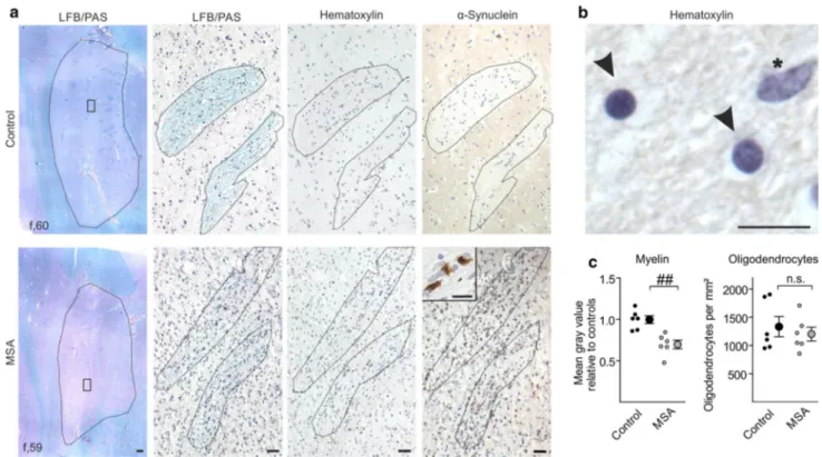
While myelin loss has previously been reported as path-ological feature present already in early stages of MSA [35, 38, 47], underlying causes for this loss have not yet been elucidated. To assess whether demyelination is a result of oligodendrocyte degeneration or rather oligo-dendrocytic dysfunction in terms of myelin formation, we quantified myelin and the density of oligodendrocytic cells in post-mortem tissue of MSA patients. Selection criteria for MSA patients were the parkinsonian pheno-type (predominance of striatonigral degeneration, MSA-P) based on clinical history and the abundant presence of α-synuclein-positive inclusions within the putamen (Fig. 1; for details of the cohort see Suppl. Table T1). A combined Luxol Fast Blue/Periodic Acid Schiff staining was used to visualize myelin (Fig. 1a). Oligodendrocytes were identified by morphology in hematoxylin-stained sections according to Salvesen and colleagues [41] (Fig. 1a, b). While myelin was reduced by 30 ± 5 % in MSA patients relative to age- and gender-matched con-trols, no significant change in the density of oligodendro-cytes was detected within fiber tracts crossing the puta-men (Fig. 1c). To confirm this dichotomy, we compared myelin with oligodendrocyte density in mice overex-pressing α-synuclein in oligodendrocytes under the con-trol of a murine Mbp promoter (high-expressing line 29; hereafter referred to as MBP29). In this well-established pre-clinical MSA mouse model, we analyzed the corpus callosum, which represents a prototypical and—in con-trast to the striatum—pure white matter region, showing most abundant α-synuclein expression in the forebrain and allowing a comprehensive quantification of myelin and oligodendrocytes (Suppl. Fig. S1a, b). Furthermore, mature oligodendrocytes strongly expressed α-synuclein in MBP29 mice, whereas OPCs did not show α-synuclein immunoreactivity (Suppl. Fig. S1c). In similarity to MSA patients, we observed a reduction of myelin by 44 ± 5 % accompanied by an 1.7-fold increased oligodendrocyte density in MBP29 mice compared to non-transgenic lit-termates (Fig. 2).
Impaired myelin formation upon oligodendrocytic α‑synuclein overexpression in an
oligodendrocyte‑neuron co‑culture
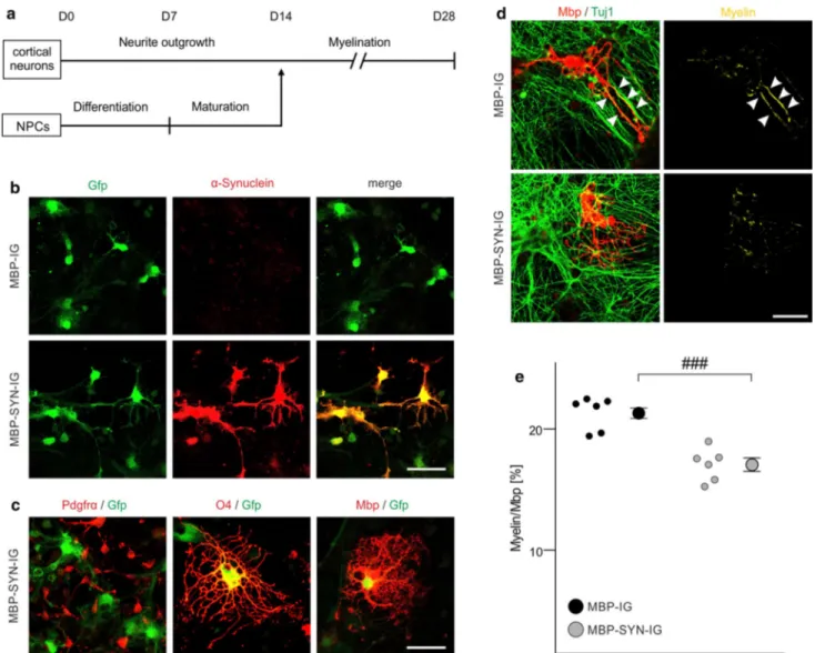
The intriguing dichotomy of a profound myelin deficit despite preserved or even increased oligodendrocyte num-bers in MSA patients (Fig. 1) and MBP29 mice (Fig. 2), respectively, suggests that an oligodendrocytic dysfunc-tion may underlie myelin loss in MSA. Thus, we next asked whether oligodendrocytic α-synuclein accumulation impacts the process of myelin formation. To analyze func-tional consequences of α-synuclein on myelination, we took advantage of a recently described approach to assess myelin sheath formation in vitro by co-culturing mouse embryonic stem cell-derived oligodendrocytes and cortical neurons [25] (Fig. 3a). We adapted the protocol to quantify myelination of individual oligodendrocytes. Withdrawal of the pro-myelinating thyroid hormone T3 from the culture medium significantly decreased myelin formation, demon-strating that this method is suitable to quantify myelination of individual oligodendrocytes (Suppl. Fig. S2). To study
the effect of intraoligodendrocytic α-synuclein on myelin formation, lentiviral vectors with the Mbp promoter as a regulatory element were generated (α-synuclein expression vector: MBP-SYN-IG; control vector: MBP-IG), yielding a high proportion of α-synuclein-overexpressing oligodendro-cytes (Fig. 3b). The usage of the Mbp promoter resulted in α-synuclein and/or Gfp expression in mature oligodendro-cytes, but not in OPCs (Fig. 3c). Upon transduction, there was no difference in the numbers of apoptotic, activated Caspase 3-positive oligodendrocytes between α-synuclein-overexpressing and control oligodendrocytes (Suppl. Fig. S3). A comprehensive and computer-assisted quantifica-tion on a single cell level revealed that α-synuclein overex-pression significantly reduced myelin sheath formation by 21 ± 3 % compared to oligodendrocytes transduced with the control vector lacking the α-synuclein-coding sequence (Fig. 3d, e). Importantly, Tuj1- and Mbp-positive pixels were not altered upon α-synuclein overexpression (Suppl. Fig. S4), indicating that there was no effect of Mbp pro-moter-driven α-synuclein overexpression on neuronal and oligodendrocytic survival in the co-culture.
Fig. 1 Putaminal myelin loss in multiple system atrophy (MSA)
without alterations in oligodendrocyte density. a Representative Luxol Fast Blue/Periodic Acid Schiff (LFB/PAS) stained images (left) depict the putamen (encircled) of a control (upper panel) and a MSA patient (lower panel). The putamen of the MSA patient showed a reduced Luxol Fast Blue staining intensity reflecting severe loss of myelin. Scale bar 500 µm. Boxed areas within the left images are magnified showing LFB/PAS-, hematoxylin-, and α-synuclein-stained striae. Note abundant α-synuclein-positive inclusions in the putamen
of the MSA patient (magnified as insert). Scale bars 50 µm, insert 10 µm. b Oligodendrocytes (arrowheads; round-shaped, dense chro-matin) were distinguished by morphology from astrocytes (asterisk; larger and more elongated in size, loose chromatin) in the hema-toxylin-stained sections. Scale bar 10 µm. c While myelin was sig-nificantly reduced in MSA patients (gray dots) compared to controls (black dots), oligodendrocyte density was unaltered (n = 6). Data are shown as mean ± standard error of mean. T test: ##p < 0.01, n.s. (not significant) p > 0.05. f female
Restored myelination by benztropine in vitro
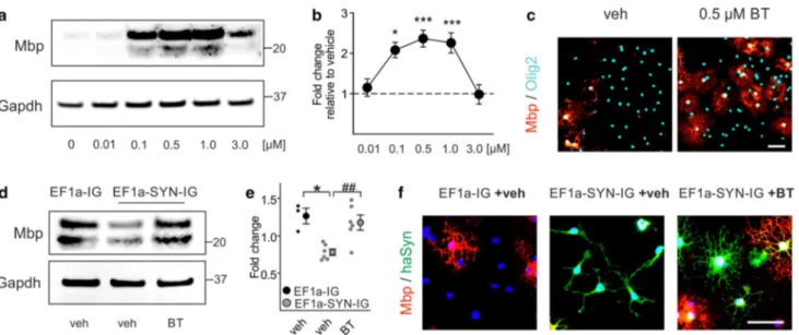
Next, we asked whether the interference of α-synuclein with myelin formation is reversible and thus, might rep-resent a novel interventional target for MSA. For this purpose, we chose benztropine, which has recently been described as a potent small molecule to enhance myelina-tion [7]. First, we validated the effect of benztropine on
myelin gene expression in non-modified primary OPC monoculture. Compared to vehicle, benztropine dose dependently increased Mbp protein in primary OPCs (Fig. 4a). Gene expression analysis revealed an effective dose in the range of 0.1–1.0 µM, peaking at 0.5 µM with an increase in Mbp expression by 2.4 ± 0.2 fold (Fig. 4b). Immunofluorescence against Mbp confirmed that benztro-pine at its most effective dose (0.5 µM) strongly increased the number of Mbp-positive cells (Fig. 4c). We next evalu-ated the effect of benztropine on Mbp expression in pri-mary oligodendrocytes overexpressing human α-synuclein. For this purpose, primary oligodendrocytes were lentivi-rally transduced with a constitutively active α-synuclein expression vector (EF1a-SYN-IG) or the control vector lacking the α-synuclein-coding sequence (EF1a-IG). As previously described [14], α-synuclein overexpression significantly reduced Mbp protein (Fig. 4d) and transcrip-tion (Fig. 4e) compared to control condition. Addition of benztropine increased Mbp expression in α-synuclein-overexpressing oligodendrocytes, restoring it to the level of control cells. Based on morphology, vehicle-treated α-synuclein-overexpressing oligodendrocytes were bi- or tripolar indicative for an immature phenotype, whereas benztropine induced the formation of multiple and highly arborized branches in α-synuclein-overexpressing oligo-dendrocytes reflecting an advanced maturation stage com-parable to control cells (Fig. 4f).
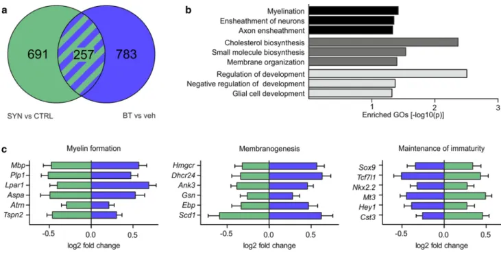
To globally analyze the effects of α-synuclein overex-pression and benztropine treatment on the myelinogenic capacity of oligodendrocytes, transcriptome profiles of vehicle- or benztropine-treated primary OPCs transduced with the α-synuclein expression vector (EF1a-SYN-IG) or the respective control vector (EF1a-IG) were determined by RNA sequencing (Fig. 5). The genes, which were dif-ferentially regulated by both α-synuclein and benztropine compared to respective controls, were subjected to a GO analysis (Fig. 5a, b). Enriched GO terms were implicated in myelin formation (GO:0042552 myelination, GO:0007272 ensheathment of neurons, GO:0008366 axon ensheathment), membranogenesis (GO:0006695 cholesterol biosynthetic process, GO:0044283 small molecule biosynthetic process, GO:0061024 membrane organization), and the maintenance of oligodendrocyte immaturity (GO:0050793 regulation of developmental process, GO:0051093 negative regulation of developmental process, GO:0021782 glial cell develop-ment). Intriguingly, transcripts related to myelin formation and membranogenesis were significantly underrepresented in α-synuclein-overexpressing cells and likewise upregulated by benztropine (e.g., Mbp, Plp1, Hmgcr, and Ank3) (Fig. 5c). In contrast, transcripts involved in maintaining the immatu-rity of OPCs were significantly enriched upon α-synuclein overexpression and accordingly downregulated by benztro-pine treatment (e.g., Nkx2.2, Sox9, and Tcf7l1).
Fig. 2 Severe myelin deficit despite increased oligodendrocyte
density in mice overexpressing human α-synuclein under a myelin basic protein-promoter (MBP29). MBP29 mice (3 months of age) showed a profound myelin deficit (illustrated by images of a Luxol Fast Blue staining and quantified within the corpus callosum; scale bars 500 µm). Density of callosal oligodendrocytes (identified by Olig2; scale bar: 50 µm) was increased by 1.7 fold. Data are shown as mean ± standard error of mean. T test (n = 5): ###p < 0.001. NTG non-transgenic mice
To extend the findings from primary oligodendrocyte monocultures, we next analyzed whether benztropine is also able to restore the myelination deficit of α-synuclein-overexpressing stem cell-derived oligodendrocytes in co-culture with cortical neurons. First, the myelinogenic effect
of benztropine was validated in non-modified stem cell-derived oligodendrocytes (Suppl. Fig. S5). Strikingly, benz-tropine treatment also increased myelin sheath formation of α-synuclein-overexpressing oligodendrocytes by 29 ± 4 % to the level of control cells (Fig. 6).
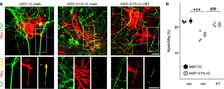
Fig. 3 Impaired myelination of α-synuclein-overexpressing mouse
embryonic stem cell-derived oligodendrocytes. a The experimen-tal paradigm to analyze myelination of individual oligodendrocytes in the mouse embryonic stem cell-derived oligodendrocyte-neuron co-culture is schematically illustrated according to [25]. b Stem cell-derived oligodendrocytes expressed human α-synuclein (when transduced with MBP-SYN-IG expression vector only; lower panel) and green fluorescent protein (Gfp) (also when transduced with the MBP-IG control vector; upper panel). Expression of α-synuclein overlapped with GFP expression in MBP-SYN-IG transduced oli-godendrocytes. Scale bar 20 µm. c Co-labeling with stage-specific oligodendrocytic markers revealed the specificity of myelin basic protein (Mbp) promoter-driven transgene expression for mature oli-godendrocytes (positive for the early maturation marker O4 and Mbp as a marker for terminal maturation), whereas transgene expres-sion was not detectable in platelet-derived growth factor receptor-α
(Pdgfrα)-positive oligodendrocyte progenitor cells. Scale bar 20 µm.
d Representative images of control (transduced with MBP-IG prior to
starting the co-culture) and α-synuclein-overexpressing (transduced with MBP-SYN-IG) Mbp-positive oligodendrocytes co-cultured with beta-III-tubulin (Tuj1)-positive cortical neurons are depicted. Myeli-nation (yellow; arrowheads) was regularly detected under control condition, whereas α-synuclein overexpression impaired myelin for-mation. Scale bar 20 µm. e Myelination of individual oligodendro-cytes (altogether: MBP-IG: 235 cells, MBP-SYN-IG: 215 cells) in the stem cell-derived co-culture was quantified (n = 6) by calculating the ratio of Mbp/Tuj1-positive pixels over total Mbp-positive pixels and is shown as percentage. α-Synuclein-overexpressing oligoden-drocytes (gray dots) formed significantly less myelin than controls (black dots). Data are shown as mean ± standard error of mean. T test: ###p < 0.001
Attenuated myelin deficit in MBP29 mice upon benztropine administration
After observing the restoration of the α-synuclein-induced myelination deficit by benztropine in vitro (Figs. 4,
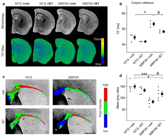
5, 6), we next administered benztropine at 2 mg/kg to MBP29 mice to assess whether benztropine is also able to enhance myelination in the presence of oligodendrocytic α-synuclein accumulation in vivo. T2-weighted magnetic resonance imaging revealed a reduced gray-white mat-ter contrast in vehicle-treated MBP29 mice compared to non-transgenic littermates, which corresponds to a mye-lin reduction (Fig. 7a). A significant gain of contrast was observed in benztropine-treated MBP29 mice. For quantifi-cation, T2*-weighted magnetic resonance imaging was per-formed because of its previously demonstrated sensitivity in detecting myelin loss in murine brains [27]. Compared to controls, vehicle-treated MBP29 mice showed a significant increase by 19 ± 3 % in the T2* relaxation time within the corpus callosum, confirming the myelin deficit (Fig. 7b). Treatment with benztropine significantly reduced the cal-losal T2* relaxation time in MBP29 mice by 9 ± 1 %, thus restoring the myelin deficit by almost 50 %. Matching this imaging approach, benztropine treatment significantly increased the mean Luxol Fast Blue signal intensity in the
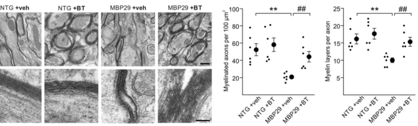
corpus callosum of MBP29 mice, ameliorating the myelin deficit by more than 50 % (Figs. 2, 7c, d). Results were further confirmed by ultrastructural analysis of myelin in the corpus callosum using electron microscopy (Fig. 8). In vehicle- and benztropine-treated non-transgenic mice, mye-lin showed a highly organized multilaminar structure. In contrast, myelin in vehicle-treated MBP29 mice appeared disorganized and substantially reduced. Both density of myelinated axons and myelin layers per axon were signifi-cantly reduced. In benztropine-treated MBP29, numbers of myelinated axons and myelin layers per axon were compa-rable to non-transgenic controls.
Taken together, these data demonstrate that the pro-mye-linating activity of benztropine attenuates the severe myelin deficit upon α-synuclein overexpression in MBP29 mice.
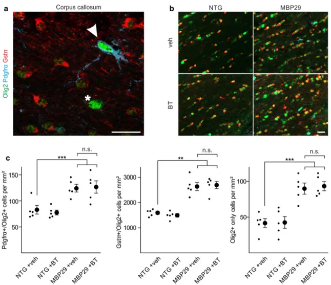
Unchanged oligodendrocyte dynamics in MBP29 mice upon benztropine administration
We next examined whether benztropine impacts the dynam-ics of oligodendrocytic populations in MBP29 mice. To this end, we histologically identified distinct oligodendrocytic sub-populations within the corpus callosum of vehicle- and ben-ztropine-treated MBP29 mice and non-transgenic littermates using maturation stage-specific markers (Fig. 9a, b): OPCs Fig. 4 Benztropine restores myelin basic protein (Mbp) expression in
α-synuclein-overexpressing primary oligodendrocyte progenitor cells (OPCs). a The dose-dependent effect of benztropine on Mbp expres-sion in primary OPCs is illustrated using Western blot. Gapdh signal served as control. b Benztropine significantly increased Mbp gene expression in a range of 0.1–1.0 µM as quantified by real-time PCR (n = 3–6). c Immunocytochemistry for Mbp and Olig2 illustrates the effect of 0.5 µM benztropine (BT) on OPC maturation in comparison to vehicle- (veh) treated cells. Scale bar: 20 µm. d The restoration of Mbp expression in α-synuclein-overexpressing cells (EF1a-SYN-IG
+veh) upon treatment with 0.5 µM benztropine (EF1a-SYN-IG +BT) to control levels (EF1a-IG +veh) is demonstrated by Western blot. Gapdh signal served as control. e Benztropine restored Mbp gene expression in α-synuclein-overexpressing OPCs as quantified by real-time PCR (n = 3–6). f α-Synuclein-overexpressing cells exhibited the typical morphology of OPCs (bi-/tripolarity), whereas treatment with benztropine induced Mbp expression and formation of multiple pro-cesses as signs of advanced maturity comparable to control condition. Scale bar 20 µm. Data are shown as mean ± standard error of mean. ANOVA: *p < 0.05, ***p < 0.001. T test: ##p < 0.01
(Pdgfrα/Olig2-positive) were distinguished from mature oli-godendrocytes (Gstπ/Olig2-positive), whereas a minority of Olig2-positive cells co-labeled for neither Pdgfrα nor Gstπ (Olig2 only). Mbp promoter-driven α-synuclein overexpres-sion in MBP29 mice resulted in a 1.5- to 2.2-fold increased density of all distinct subpopulations compared to non-trans-genic control animals (Fig. 9c). In contrast to α-synuclein overexpression, it is remarkable that benztropine did not affect oligodendrocyte density. Given that the proportional distribu-tion of oligodendrocytic subpopuladistribu-tions was unchanged in all groups (Suppl. Fig. S6), these findings suggest that treatment with benztropine directly acts on myelin formation, but does not increase OPC maturation in MBP29 mice.
Prevention of motor cortical neuronal loss without affecting microgliosis in MBP29 mice upon benztropine treatment
In addition to the severe loss of myelin, MBP29 mice recapitulate crucial neurodegenerative and inflammatory features of MSA [45]. Thus, we next examined whether
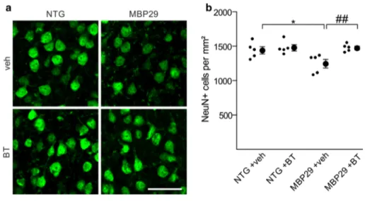
benztropine treatment had an impact on neuronal and microglial cell densities in MBP29 mice. To this end, we first quantified the density of NeuN-positive neurons within the motor cortex (Fig. 10). While vehicle-treated MBP29 mice showed a moderate (14 %), but significant reduction in neuronal cell density, benztropine treatment prevented neuronal loss in MBP29 animals.
We next assessed the effect of benztropine on motor cortical and callosal microglial cells in MBP29 mice by determining the density of Iba1-positive microglia (Suppl. Fig. S7). Microgliosis was apparent in the corpus callosum of vehicle-treated MBP29 mice (2.5-fold increase in Iba1-positive cells), whereas the motor cortex showed no appar-ent signs of microgliosis when compared to control mice (1.1-fold increase). Benztropine treatment neither affected callosal nor motor cortical Iba1 cell density in MBP29 mice.
Taken together, these findings suggest that benztropine treatment has a beneficial effect on survival of motor corti-cal neurons, but does not alter microglial cell densities in MBP29 mice.
Fig. 5 Restored myelinogenic capacity of
α-synuclein-overexpressing primary rat oligodendrocyte progenitor cells (OPCs) by benztropine. a A Venn diagram depicts differentially regulated genes revealed by RNA sequencing of primary rat OPCs (n = 3). Green circle: α-synuclein-overexpressing (SYN) versus control cells (CTRL). Blue circle: benztropine (BT) versus vehicle (veh)-treated α-synuclein-overexpressing cells. b Genes differentially regulated by both α-synuclein overexpression and benztropine treatment clas-sified into gene ontologies (GOs) implicated among others in myelin formation (black), membranogenesis (dark gray), and maintenance of oligodendrocyte immaturity (light gray). c Expression dynam-ics of representative genes identified by GO term enrichment are
listed. Benztropine (blue) oppositely regulated genes differentially expressed upon α-synuclein overexpression (green). Myelin forma-tion: Mbp myelin basic protein, Plp1 proteolipid protein 1, Lpar1 lysophosphatidic acid receptor 1, Aspa aspartoacylase, Atrn attractin, Tspn2 tetraspanin 2; Membranogenesis: Hmgcr 3-hydroxy-3-meth-ylglutaryl-CoA reductase, Dhcr24 24-dehydrocholesterol reductase, Ank3 ankyrin 3, Gsn gelsolin, Ebp emopamil binding protein (sterol isomerase), Scd1 stearoyl-CoA desaturase-1; Maintenance of imma-turity: Sox9 sex determining region Y-box 9, Tcf7l1 transcription fac-tor 7-like 1, Nkx2.2 Nk2 homeobox 2, Mt3 metallothionein 3, Hey1 hes-related family bHLH transcription factor with YRPW motif 1, Cst3 cystatin C
Discussion
Cellular pathomechanisms that underlie profound myelin loss and neurodegeneration in MSA are poorly understood. In this regard, our study provides compelling evidence for impaired myelin formation in MSA, which is directly associated with the oligodendrocytic accumulation of α-synuclein and not accompanied by loss of oligodendro-cytes. Considering that oligodendrocytic α-synuclein accu-mulation is the primary event during MSA pathogenesis, the deficit in myelin formation may be an early and crucial pathomechanism during disease progression. Given that efficient interventional strategies for MSA do not exist and by demonstrating that the interference of α-synuclein with myelin formation is reversible, our in vitro and in vivo data imply pro-myelinating strategies as a novel approach for an urgently needed therapy in MSA.
Using histological analyses of MSA post-mortem tis-sue, we showed that myelin loss is a widespread patho-logical feature in MSA. Loss of myelin, downregulation of myelin proteins [1, 35, 38, 47], and widespread lipid alterations [5, 11] have previously been reported in post-mortem brains of MSA patients, supporting our finding of a severe myelin deficit in MSA. Despite strong evidence for myelin degeneration in MSA, the underlying pathomecha-nisms of this myelin loss are not well understood. In the MSA cohort analyzed in the present study, the putamen
exhibited extensive oligodendrocytic α-synuclein pathol-ogy within white matter tracts, suggesting that oligoden-drocytic α-synuclein accumulation underlies loss of mye-lin. This is supported by the fact that a substantial myelin deficit was also present upon transgenic overexpression of α-synuclein in oligodendrocytes of MBP29 mice. In line, a similar myelin reduction has also been observed in trans-genic mice with α-synuclein overexpression under alterna-tive myelin gene promoters, namely the proteolipid protein and the 2′,3′-cyclic-nucleotide 3′-phosphodiesterase pro-moter [48, 52]. Notably, structural alterations, including nuclear shrinkage, cytosolic re-localization of myelin pro-teins, and oligodendrocytic swelling, were also observed in oligodendrocytes without α-synuclein accumulation and possibly contribute to myelin loss in MSA [47, 49]. More-over, we selectively used post-mortem material of MSA patients with striatonigral degeneration, limiting the inter-pretation of the present study to the parkinsonian variant of MSA (MSA-P).
Myelin loss in MSA patients and in transgenic MSA mice may be linked to either oligodendrocyte loss or impaired ability of oligodendrocytes to form new mye-lin sheaths. The observation of an unchanged or even increased oligodendrocyte density in human MSA post-mortem tissue and MBP29 mice suggests that rather a dysfunction of oligodendrocytes in terms of myelin sheath formation than pronounced loss of oligodendrocytes Fig. 6 Restored myelination deficit of α-synuclein-overexpressing
mouse embryonic stem cell-derived oligodendrocytes by benztropine.
a Representative images show that overexpression of α-synuclein
(transduced with MBP-SYN-IG prior to starting the co-culture) impaired myelin formation of stem cell-derived oligodendrocytes (stained for myelin basic protein, Mbp) co-cultured with cortical neurons (beta-III-tubulin, Tuj1). Addition of benztropine enhanced myelin formation of α-synuclein-overexpressing cells [comparable to vehicle (veh)-treated control cells, which were transduced with
MBP-IG]. Mbp-positive pixels co-localizing with Tuj1-positive pix-els were considered as myelin (shown in yellow). Scale bar 20 µm.
b Myelination of individual oligodendrocytes (in total 90–110 cells
per condition) was quantified (n = 3). α-Synuclein-overexpressing oligodendrocytes (gray dots) in the presence of vehicle (veh) formed significantly less myelin than controls (black dots, veh). Benztropine (BT) restored the myelination deficit of α-synuclein-overexpressing oligodendrocytes. Data are shown as mean ± standard error of mean. ANOVA: ***p < 0.001. T test: ##p < 0.01
underlies myelin loss in MSA. A recent stereological analysis, focusing on basal ganglia, has indicated that oli-godendrocytes are by far more preserved than neurons in MSA [41]. In line, we and others have observed increased numbers of OPCs in the striatum and the cerebellum of MSA patients, supporting the notion of preserved oli-godendrocytic cells in MSA [2, 36]. Similarly, we have shown that mice moderately overexpressing α-synuclein driven by the Mbp promoter (line 1) exhibit an increased number of OPCs accompanied by myelin loss [36, 45]. Given that the Mbp promoter did not drive α-synuclein overexpression in OPCs, the observed increase in OPC density of MBP29 mice indicates enhanced OPC prolifera-tion to compensate the myelin deficit.
Supporting these observations in MSA post-mortem and transgenic mouse forebrains, our in vitro data provide fur-ther evidence, that oligodendrocytic α-synuclein impairs myelin formation: α-synuclein-overexpressing oligoden-drocytes derived from mouse embryonic stem cells exhib-ited a reduced capacity to form myelin sheaths around axons of cortical neurons. Moreover, transcriptome profiles of primary rat OPC monocultures showed that α-synuclein overexpression in maturating oligodendrocytes impaired expression of gene families essentially involved in myelin sheath formation. The temporal dynamic of α-synuclein expression in cultured murine and human OPCs dur-ing maturation toward pre-myelinatdur-ing oligodendrocytes suggests that α-synuclein is physiologically restricted to Fig. 7 Amelioration of the myelin deficit in mice with
oligoden-drocytic overexpression of α-synuclein (MBP29) upon benztropine treatment. a Callosal myelin levels of non-transgenic (NTG) and MBP29 mice treated with vehicle (veh) or benztropine (BT, 2 mg/ kg) were analyzed using magnetic resonance imaging. Upper panel illustrates morphology as determined by T2-weighted magnetic reso-nance imaging, whereas lower panel depicts T2*-weighted magnetic resonance imaging (shown as color-coded maps), which was used for myelin quantification. b MBP29 mice (n = 3) showed a reduction in myelin levels detected by increased T2*-relaxation times within
the corpus callosum. Benztropine attenuated the myelin deficit in MBP29 confirmed by a reduction of T2*. c Representative pictures of a Luxol Fast Blue staining (color-coded for signal intensity: blue low, green intermediate, red high intensity), confirming the effects of α-synuclein overexpression and benztropine treatment on myelina-tion. Scale bar 500 µm. d MBP29 mice (n = 5) exhibited a reduc-tion in Luxol Fast Blue intensity, which was attenuated, but not com-pletely restored by benztropine. Data are shown as mean ± standard error of mean. ANOVA: *p < 0.05, ***p < 0.001. T test: #p < 0.05
immature oligodendrocytes [8, 40]. In addition, we have previously demonstrated that the sustained presence of intracellular α-synuclein delays the upregulation of myelin gene expression in monocultures of primary rat oligoden-drocytes [14], further supporting that α-synuclein expres-sion needs to be downregulated for timed and physiologi-cal oligodendrocyte maturation and myelination. Thus, our data further strengthen the view on α-synuclein as an inhibitor of oligodendrocyte maturation and myelination and suggest that pathologic oligodendrocytic α-synuclein accumulation in MSA impairs myelin formation.
In recent years, the view on myelin dynamics in the adult brain has fundamentally changed. The fact, that myelin is physiologically formed in the adult murine and human cen-tral nervous system throughout life, has only lately been rec-ognized [18, 37, 42, 53, 54]. In mice, adaptive myelination in the adult forebrain is mediated mostly by newly formed oligodendrocytes derived from OPCs, occurs upon neuronal activity, and is essentially required for motor skill learning [18, 37]. In line, motor training has also induced a remode-ling of white matter structures in humans [42]. Importantly, a continuous physiological myelin turnover during adult-hood, which is mostly mediated by preexisting oligoden-drocytes, has been detected in the human corpus callosum by determining the integration of nuclear bomb test-derived (14)C [53]. Therefore, impaired myelination in the presence of α-synuclein may result not only in compromised remyeli-nation, but also in reduced myelin turnover. In this context, it is important to note that disturbances in myelin homeo-stasis result in axonal and neuronal degeneration [51]. The impaired capacity to form myelin sheaths may thus repre-sent a fundamental and early pathological mechanism in MSA, favoring myelin loss and ultimately triggering wide-spread axonal dysfunction and neuronal loss.
To restore oligodendrocyte functionality, we used the small molecule benztropine, which has recently been iden-tified by a high-throughput screen to accelerate OPC mat-uration and directly promote myelin formation, thereby ameliorating functional deficits in pre-clinical mouse mod-els of multiple sclerosis [7]. We chose this small molecule to target α-synuclein-induced effects on oligodendrocyte function because (i) it is clinically used for the treatment of tremor in patients diagnosed with sporadic Parkinson’s disease and thus promises a fast translation into clinics; and (ii) benztropine has a dual mode of action as it promotes myelin gene expression in pre-myelinating oligodendro-cytes and directly enhances myelin formation in mature oli-godendrocytes [7]. Both processes are essentially required for myelination and are compromised by α-synuclein over-expression. While the effect of benztropine on myelin gene expression has been attributed to its antagonistic activity at the muscarinic acetylcholine receptors M1 and M3 [7], the mechanism by which benztropine induces myelin sheath formation is not yet known. Our observation, that benztro-pine increased myelination without changing the density of OPCs and mature oligodendrocytes in MBP29 mice, favors rather a direct action of benztropine on myelin sheath for-mation in this model. Importantly, benztropine even ame-liorated the myelination deficit in MBP29 mice by approxi-mately 50 %, although used at a low dose (2 mg/kg in comparison to the most efficient dose of 10 mg/kg accord-ing to Deshmukh and colleagues [7]). Higher dosages (5 and 10 mg/kg) were not tolerated due to the severe pheno-type of MBP29 mice with a premature death at 4–6 months of age [45]. Moreover, our experiments using the stem cell-derived oligodendrocyte-neuron co-culture also point toward a direct pro-myelinating effect of benztropine. Our transcriptome analysis suggests that benztropine readjusts Fig. 8 Structural restoration of myelin by benztropine in mice
over-expressing α-synuclein in oligodendrocytes (MBP29). Representa-tive electron micrographs of the corpus callosum at low (upper panel, scale bar 1 µm) and high (lower panel, scale bar 0.2 µm) magnifi-cation depict myelin sheaths of non-transgenic (NTG) and MBP29 mice treated with either vehicle (veh) or benztropine (BT). In
vehicle-treated MBP29 mice only, myelin appeared strongly reduced and dis-organized. Quantification of myelinated axons and myelin layers per axon revealed a significant myelin deficit in vehicle-treated MBP29 mice, which was restored by benztropine treatment to control level. Data are shown as mean ± standard error of mean. ANOVA (n = 6): **p < 0.01. T test: ##p < 0.01
disturbed lipid metabolism in oligodendrocytes with α-synuclein accumulation, thereby enhancing myelin for-mation. While not in the focus of the present work, future studies need to dissect the complex pharmacology and define molecular targets underlying the pro-myelinating activity of benztropine.
Benztropine treatment also prevented motor cortical neuronal cell loss in MBP29 mice, suggesting that mye-lin regeneration supports neuronal survival. Although we cannot exclude a direct effect of benztropine on neuronal cells, the preservation of neurons might be attributed to oligodendrocyte-derived factors and their importance for axonal maintenance and neuronal survival [29]. In line, protection of cortical neurons was also achieved upon
immunotherapeutic prevention of myelin loss in transgenic mice overexpressing α-synuclein in oligodendrocytes [33]. In this study, oligodendrocytic α-synuclein pathology was reduced via specific antibodies against α-synuclein. Impor-tantly, benztropine injections did not alter microglial cell numbers in MBP29 mice, supporting that neuronal pres-ervation is not linked to decreased neuroinflammation. In line, Deshmukh and colleagues have demonstrated that benztropine has no immunomodulatory effect in experi-mental mouse models for multiple sclerosis [7].
The surprising dichotomy of a substantial myelin defi-cit despite a preserved oligodendrocyte density opens novel avenues for restorative interventional strategies in MSA. Up to now, experimental and clinical trials for disease-modifying Fig. 9 No alteration of oligodendrocyte density by benztropine. a
Oligodendrocytic cells within the corpus callosum were identified using stage-specific markers [entire population: Olig2; oligoden-drocyte progenitor cells: platelet-derived growth factor receptor-α (Pdgfrα)/Olig2strong (arrowhead), mature oligodendrocytes:
glutathione-s-transferase-π (Gstπ)/Olig2weak, as well as cells
stain-ing for Olig2strong only (asterisk)]. Scale bar 20 µm. b A profound
increase of oligodendrocytic cells within the corpus callosum of mice
overexpressing α-synuclein in oligodendrocytes (MBP29) compared to non-transgenic controls (NTG) was observed. Scale bar 20 µm. c While MBP29 mice exhibited an almost twofold increased density of the entire spectrum of oligodendrocytic subpopulations, benztropine (BT) had no effect on oligodendrocytic cell density as compared to vehicle (veh)-treatment. Data are shown as mean ± standard error of mean. ANOVA (n = 5): **p < 0.01, ***p < 0.001. T test: not signifi-cant (n.s.) p > 0.05
therapies in MSA have focused on direct protection of neurons (e.g., riluzole [44]), prevention or clearance of α-synuclein aggregates (e.g., rifampicin [31]), reduction of neuroinflamma-tion (e.g., minocycline [10]), and neurorestorative approaches (e.g., systemic mesenchymal stem cell delivery [28]). However, all of these strategies showed only very moderate—if any— clinical efficacy (for review see [26]). We propose the myeli-nation deficit of dysfunctional oligodendrocytes as a novel and promising interventional target for MSA because of two rea-sons: firstly, α-synuclein accumulation within oligodendrocytes is considered as the primary and pathognomonic event during MSA pathogenesis [50]; and secondly, we showed that oligo-dendrocytes are preserved in MSA and—albeit being dysfunc-tional—their ability to form myelin was not irreversibly lost in the presence of α-synuclein. Consequently, restoring the capac-ity to form new myelin may provide a new basis for a therapeu-tic strategy even in early stages of MSA. It should be empha-sized that—although we observed prevention of neuronal cell loss in our model—we cannot state whether a pro-myelinating monotherapy will lead to a functional recovery in the context of MSA, given that the usage of the most severe pre-clinical mouse model for MSA, the MBP29 mouse line, did not allow a comprehensive behavioral assessment. Thus, additional pre-clinical studies have to explore whether a myelinogenic mon-otherapy or a synergistic combination of a pro-myelinating intervention with strategies targeted against other pathological hallmarks of MSA, including neuronal degeneration and neu-roinflammation, may be the basis for treating MSA patients.
To conclude, our findings shed new light into α-synuclein-linked pathomechanisms in MSA. Intrigu-ingly, the observed reversibility of the α-synuclein-induced
myelination deficit in primary rat OPCs, in mouse embry-onic stem cell-derived oligodendrocytes, and in MBP29 mice identified a new direction to treat MSA.
Acknowledgments This work was supported by the
Interdisci-plinary Center for Clinical Research (IZKF Erlangen, TP E18), the Bavarian State Ministry of Education and Culture, Science and Arts in the framework of the Bavarian Research Network Induced Pluripotent Stem Cells (ForIPS), the Deutsche Forschungsgemeinschaft (DFG grant INST 410/45-1 FUGG), and the NIH (AG5131, AG18440, NS092803). The study was supported in part by the G. Harold and Leila Y. Mathers Charitable Foundation, the JPB Foundation, the Leona M. and Harry B. Helmsley Charitable Trust. BE is an IZKF PhD student and was supported by the IZKF Erlangen to conduct experiments involving stem cells in the Laboratory of Genetics at the Salk Institute for Biological Studies, La Jolla, CA, USA. JCMS is supported by a research grant of the Deutsche Forschungsgemein-schaft (DFG grant no. SCHL 21021-1). The authors greatly acknowl-edge the NBB for providing human post-mortem tissue. Excellent technical assistance was provided by Holger Meixner, Someya Salem, Arianna Mei, Jazmin Florio, Maria Hirblinger, Petra Rothe, Angelika Diem, and Heike Friebel-Stange. We thank Beate Winner and Chi-chung Lie for scientific discussion and comments on the manuscript. Mary Lynn Gage is greatly acknowledged for editorial comments.
Compliance with ethical standards
Human brain samples used in this study were obtained from the NBB and have been collected from donors for or from whom a written in-formed consent for a brain autopsy and the use of the material and clinical information for research purposes had been obtained by the NBB. All animal procedures were conducted with approval of the animal care and use committees of the University of California San Diego, the Friedrich-Alexander-Universität Erlangen-Nürnberg, and the state of Bavaria.
Conflict of interest The authors declare that they have no conflict
of interest.
Fig. 10 Prevention of motor cortical neuronal cell loss in mice
overexpressing α-synuclein in oligodendrocytes by benztropine. a Representative images depict NeuN-positive neuronal cells within the motor cortex of non-transgenic controls (NTG) and α-synuclein transgenic mice (MBP29) treated with vehicle (veh) or benztropine (BT). Scale bar 20 µm. b Densities of motor cortical neurons are
shown (n = 5). In vehicle-treated MBP29 mice, a significant reduc-tion in neuronal cell density was observed. Benztropine treatment prevented the loss of cortical neurons in MBP29 mice. Data are shown as mean ± standard error of mean. ANOVA: *p < 0.05. T test: ##p < 0.01
References
1. Ahmed Z, Asi YT, Sailer A, Lees AJ, Houlden H, Revesz T, Hol-ton JL (2012) The neuropathology, pathophysiology and genetics of multiple system atrophy. Neuropathol Appl Neurobiol 38:4– 24. doi:10.1111/j.1365-2990.2011.01234.x
2. Ahmed Z, Asi YT, Lees AJ, Revesz T, Holton JL (2013) Iden-tification and quanIden-tification of oligodendrocyte precursor cells in multiple system atrophy, progressive supranuclear palsy and Parkinson’s disease. Brain Pathol 23:263–273. doi:10.1111/j.1750-3639.2012.00637.x
3. Anders S, Pyl PT, Huber W (2015) HTSeq–a Python framework to work with high-throughput sequencing data. Bioinformatics 31:166–169. doi:10.1093/bioinformatics/btu638
4. Asi YT, Simpson JE, Heath PR, Wharton SB, Lees AJ, Revesz T, Houlden H, Holton JL (2014) Alpha-synuclein mRNA expres-sion in oligodendrocytes in MSA. Glia 62:964–970. doi:10.1002/ glia.22653
5. Bleasel JM, Hsiao JH, Halliday GM, Kim WS (2013) Increased expression of ABCA8 in multiple system atrophy brain is asso-ciated with changes in pathogenic proteins. J Parkinsons Dis 3:331–339. doi:10.3233/JPD-130203
6. Bottenstein JE, Sato GH (1979) Growth of a rat neuroblastoma cell line in serum-free supplemented medium. Proc Natl Acad Sci 76:514–517. doi:10.1073/pnas.76.1.514
7. Deshmukh VA, Tardif V, Lyssiotis CA, Green CC, Kerman B, Kim HJ, Padmanabhan K, Swoboda JG, Ahmad I, Kondo T et al (2013) A regenerative approach to the treatment of multiple scle-rosis. Nature 502:327–332. doi:10.1038/nature12647
8. Djelloul M, Holmqvist S, Boza-Serrano A, Azevedo C, Yeung MS, Goldwurm S, Frisen J, Deierborg T, Roybon L (2015) Alpha-synuclein expression in the oligodendrocyte lineage: an in vitro and in vivo study using rodent and human models. Stem Cell Rep 5:174–184. doi:10.1016/j.stemcr.2015.07.002
9. Dobin A, Davis CA, Schlesinger F, Drenkow J, Zaleski C, Jha S, Batut P, Chaisson M, Gingeras TR (2013) STAR: ultrafast uni-versal RNA-seq aligner. Bioinformatics 29:15–21. doi:10.1093/ bioinformatics/bts635
10. Dodel R, Spottke A, Gerhard A, Reuss A, Reinecker S, Schimke N, Trenkwalder C, Sixel-Doring F, Herting B, Kamm C et al (2010) Minocycline 1-year therapy in multiple-system-atro-phy: effect on clinical symptoms and [(11)C] (R)-PK11195 PET (MEMSA-trial). Mov Disord 25:97–107. doi:10.1002/ mds.22732
11. Don AS, Hsiao JH, Bleasel JM, Couttas TA, Halliday GM, Kim WS (2014) Altered lipid levels provide evidence for myelin dys-function in multiple system atrophy. Acta Neuropathol Commun 2:150. doi:10.1186/s40478-014-0150-6
12. Eden E, Navon R, Steinfeld I, Lipson D, Yakhini Z (2009) GOrilla: a tool for discovery and visualization of enriched GO terms in ranked gene lists. BMC Bioinform 10:48. doi:10.1186/1471-2105-10-48
13. Eschlbock S, Krismer F, Wenning GK (2016) Interventional trials in atypical parkinsonism. Parkinsonism Relat Disord 22(Suppl 1):S82–S92. doi:10.1016/j.parkreldis.2015.09.038 14. Ettle B, Reiprich S, Deusser J, Schlachetzki JC, Xiang W, Prots
I, Masliah E, Winner B, Wegner M, Winkler J (2014) Intracel-lular alpha-synuclein affects early maturation of primary oli-godendrocyte progenitor cells. Mol Cell Neurosci 62:68–78. doi:10.1016/j.mcn.2014.06.012
15. Ettle B, Schlachetzki JC, Winkler J (2015) Oligodendroglia and myelin in neurodegenerative diseases: more than just bystand-ers? Mol Neurobiol. doi:10.1007/s12035-015-9205-3
16. Fanciulli A, Wenning GK (2015) Multiple-system atrophy. N Engl J Med 372:1375–1376. doi:10.1056/NEJMc1501657
17. Gaspard N, Bouschet T, Herpoel A, Naeije G, van den Ameele J, Vanderhaeghen P (2009) Generation of cortical neurons from mouse embryonic stem cells. Nat Protoc 4:1454–1463. doi:10.1038/nprot.2009.157
18. Gibson EM, Purger D, Mount CW, Goldstein AK, Lin GL, Wood LS, Inema I, Miller SE, Bieri G, Zuchero JB et al (2014) Neuronal activity promotes oligodendrogenesis and adaptive myelination in the mammalian brain. Science 344:1252304. doi:10.1126/science.1252304
19. Gilman S, Wenning GK, Low PA, Brooks DJ, Mathias CJ, Tro-janowski JQ, Wood NW, Colosimo C, Durr A, Fowler CJ et al (2008) Second consensus statement on the diagnosis of mul-tiple system atrophy. Neurology 71:670–676. doi:10.1212/01. wnl.0000324625.00404.15
20. Gow A, Friedrich VL Jr, Lazzarini RA (1992) Myelin basic pro-tein gene contains separate enhancers for oligodendrocyte and Schwann cell expression. J Cell Biol 119:605–616. doi:10.1083/ jcb.119.3.605
21. Griffiths I, Klugmann M, Anderson T, Yool D, Thomson C, Schwab MH, Schneider A, Zimmermann F, McCulloch M, Nadon N et al (1998) Axonal swellings and degeneration in mice lacking the major proteolipid of myelin. Science 280:1610– 1613. doi:10.1126/science.280.5369.1610
22. Groves AK, Barnett SC, Franklin RJ, Crang AJ, Mayer M, Blakemore WF, Noble M (1993) Repair of demyelinated lesions by transplantation of purified O-2A progenitor cells. Nature 362:453–455. doi:10.1038/362453a0
23. Irvine KA, Blakemore WF (2008) Remyelination protects axons from demyelination-associated axon degeneration. Brain 131:1464–1477. doi:10.1093/brain/awn080
24. Kahle PJ, Neumann M, Ozmen L, Haass C (2000) Physi-ology and pathophysiPhysi-ology of alpha-synuclein. Cell cul-ture and transgenic animal models based on a Parkinson’s disease-associated protein. Ann N Y Acad Sci 920:33–41. doi:10.1111/j.1749-6632-2000.tb06902.x
25. Kerman BE, Kim HJ, Padmanabhan K, Mei A, Georges S, Joens MS, Fitzpatrick JA, Jappelli R, Chandross KJ, August P et al (2015) In vitro myelin formation using embryonic stem cells. Development 142:2213–2225. doi:10.1242/dev.116517
26. Kuzdas-Wood D, Stefanova N, Jellinger KA, Seppi K, Schloss-macher MG, Poewe W, Wenning GK (2014) Towards transla-tional therapies for multiple system atrophy. Prog Neurobiol 118:19–35. doi:10.1016/j.pneurobio.2014.02.007
27. Lee J, Shmueli K, Kang BT, Yao B, Fukunaga M, van Gelderen P, Palumbo S, Bosetti F, Silva AC, Duyn JH (2012) The contri-bution of myelin to magnetic susceptibility-weighted contrasts in high-field MRI of the brain. Neuroimage 59:3967–3975. doi:10.1016/j.neuroimage.2011.10.076
28. Lee PH, Lee JE, Kim HS, Song SK, Lee HS, Nam HS, Cheong JW, Jeong Y, Park HJ, Kim DJ et al (2012) A randomized trial of mesenchymal stem cells in multiple system atrophy. Ann Neurol 72:32–40. doi:10.1002/ana.23612
29. Lee Y, Morrison BM, Li Y, Lengacher S, Farah MH, Hoffman PN, Liu Y, Tsingalia A, Jin L, Zhang PW et al (2012) Oligoden-droglia metabolically support axons and contribute to neurode-generation. Nature 487:443–448. doi:10.1038/nature11314 30. Love MI, Huber W, Anders S (2014) Moderated estimation of
fold change and dispersion for RNA-seq data with DESeq2. Genome Biol 15:550. doi:10.1186/s13059-014-0550-8
31. Low PA, Robertson D, Gilman S, Kaufmann H, Singer W, Biag-gioni I, Freeman R, Perlman S, Hauser RA, Cheshire W et al (2014) Efficacy and safety of rifampicin for multiple system atro-phy: a randomised, double-blind, placebo-controlled trial. Lancet Neurol 13:268–275. doi:10.1016/S1474-4422(13)70301-6 32. Low PA, Reich SG, Jankovic J, Shults CW, Stern MB, Novak
Natural history of multiple system atrophy in the USA: a pro-spective cohort study. Lancet Neurol 14:710–719. doi:10.1016/ S1474-4422(15)00058-7
33. Mandler M, Valera E, Rockenstein E, Mante M, Weninger H, Patrick C, Adame A, Schmidhuber S, Santic R, Schneeberger A et al (2015) Active immunization against alpha-synuclein ame-liorates the degenerative pathology and prevents demyelination in a model of multiple system atrophy. Mol Neurodegener 10:10. doi:10.1186/s13024-015-0008-9
34. Marchetto MC, Muotri AR, Mu Y, Smith AM, Cezar GG, Gage FH (2008) Non-cell-autonomous effect of human SOD1 G37R astrocytes on motor neurons derived from human embry-onic stem cells. Cell Stem Cell 3:649–657. doi:10.1016/j. stem.2008.10.001
35. Matsuo A, Akiguchi I, Lee GC, McGeer EG, McGeer PL, Kimura J (1998) Myelin degeneration in multiple system atro-phy detected by unique antibodies. Am J Pathol 153:735–744. doi:10.1016/S0002-9440(10)65617-9
36. May VE, Ettle B, Poehler AM, Nuber S, Ubhi K, Rockenstein E, Winner B, Wegner M, Masliah E, Winkler J (2014) alpha-Synu-clein impairs oligodendrocyte progenitor maturation in multiple system atrophy. Neurobiol Aging 35:2357–2368. doi:10.1016/j. neurobiolaging.2014.02.028
37. McKenzie IA, Ohayon D, Li H, de Faria JP, Emery B, Tohy-ama K, Richardson WD (2014) Motor skill learning requires active central myelination. Science 346:318–322. doi:10.1126/ science.1254960
38. Papp MI, Kahn JE, Lantos PL (1989) Glial cytoplasmic inclusions in the CNS of patients with multiple system phy (striatonigral degeneration, olivopontocerebellar atro-phy and Shy-Drager syndrome). J Neurol Sci 94:79–100. doi:10.1016/0022-510X(89)90219-0
39. Patrikios P, Stadelmann C, Kutzelnigg A, Rauschka H, Schmid-bauer M, Laursen H, Sorensen PS, Bruck W, Lucchinetti C, Lassmann H (2006) Remyelination is extensive in a subset of multiple sclerosis patients. Brain 129:3165–3172. doi:10.1093/ brain/awl217
40. Richter-Landsberg C, Gorath M, Trojanowski JQ, Lee VM (2000) alpha-synuclein is developmentally expressed in cultured rat brain oligodendrocytes. J Neurosci Res 62:9–14. doi:10.1002/1097-4547(20001001)62:1<9:AID-JNR2>3.0.CO;2-U
41. Salvesen L, Ullerup BH, Sunay FB, Brudek T, Lokkegaard A, Agander TK, Winge K, Pakkenberg B (2015) Changes in total cell numbers of the basal ganglia in patients with multiple sys-tem atrophy—A stereological study. Neurobiol Dis 74:104–113. doi:10.1016/j.nbd.2014.11.008
42. Scholz J, Klein MC, Behrens TE, Johansen-Berg H (2009) Train-ing induces changes in white-matter architecture. Nat Neurosci 12:1370–1371. doi:10.1038/nn.2412
43. Scholz SW, Houlden H, Schulte C, Sharma M, Li A, Berg D, Melchers A, Paudel R, Gibbs JR, Simon-Sanchez J et al (2009) SNCA variants are associated with increased risk for multiple system atrophy. Ann Neurol 65:610–614. doi:10.1002/ana.21685 44. Seppi K, Peralta C, Diem-Zangerl A, Puschban Z, Mueller J,
Poewe W, Wenning GK (2006) Placebo-controlled trial of rilu-zole in multiple system atrophy. Eur J Neurol 13:1146–1148. doi:10.1111/j.1468-1331.2006.01452.x
45. Shults CW, Rockenstein E, Crews L, Adame A, Mante M, Lar-rea G, Hashimoto M, Song D, Iwatsubo T, Tsuboi K et al (2005) Neurological and neurodegenerative alterations in a transgenic mouse model expressing human alpha-synuclein under oligodendrocyte promoter: implications for multiple system atrophy. J Neurosci 25:10689–10699. doi:10.1523/ JNEUROSCI.3527-05.2005
46. Simons M, Nave KA (2015) Oligodendrocytes: myelination and axonal support. Cold Spring Harb Perspect Biol. doi:10.1101/ cshperspect.a020479
47. Song YJ, Lundvig DM, Huang Y, Gai WP, Blumbergs PC, Hojrup P, Otzen D, Halliday GM, Jensen PH (2007) p25alpha relocalizes in oligodendroglia from myelin to cytoplasmic inclu-sions in multiple system atrophy. Am J Pathol 171:1291–1303. doi:10.2353/ajpath.2007.070201
48. Stefanova N, Kaufmann WA, Humpel C, Poewe W, Wenning GK (2012) Systemic proteasome inhibition triggers neurodegenera-tion in a transgenic mouse model expressing human alpha-synu-clein under oligodendrocyte promoter: implications for multi-ple system atrophy. Acta Neuropathol 124:51–65. doi:10.1007/ s00401-012-0977-5
49. Uyama N, Uchihara T, Mochizuki Y, Nakamura A, Taka-hashi R, Mizutani T (2009) Selective nuclear shrinkage of oligodendrocytes lacking glial cytoplasmic inclusions in multiple system atrophy: a 3-dimensional volumetric study. J Neuropathol Exp Neurol 68:1084–1091. doi:10.1097/ NEN.0b013e3181b67678
50. Wenning GK, Stefanova N, Jellinger KA, Poewe W, Schloss-macher MG (2008) Multiple system atrophy: a primary oli-godendrogliopathy. Ann Neurol 64:239–246. doi:10.1002/ ana.21465
51. Wilkins A, Kondo Y, Song J, Liu S, Compston A, Black JA, Wax-man SG, Duncan ID (2010) Slowly progressive axonal degen-eration in a rat model of chronic, nonimmune-mediated demyeli-nation. J Neuropathol Exp Neurol 69:1256–1269. doi:10.1097/ NEN.0b013e3181ffc317
52. Yazawa I, Giasson BI, Sasaki R, Zhang B, Joyce S, Uryu K, Trojanowski JQ, Lee VM (2005) Mouse model of multiple sys-tem atrophy alpha-synuclein expression in oligodendrocytes causes glial and neuronal degeneration. Neuron 45:847–859. doi:10.1016/j.neuron.2005.01.032
53. Yeung MS, Zdunek S, Bergmann O, Bernard S, Salehpour M, Alkass K, Perl S, Tisdale J, Possnert G, Brundin L et al (2014) Dynamics of oligodendrocyte generation and myeli-nation in the human brain. Cell 159:766–774. doi:10.1016/j. cell.2014.10.011
54. Young KM, Psachoulia K, Tripathi RB, Dunn SJ, Cossell L, Attwell D, Tohyama K, Richardson WD (2013) Oligo-dendrocyte dynamics in the healthy adult CNS: evidence for myelin remodeling. Neuron 77:873–885. doi:10.1016/j. neuron.2013.01.006