doi:10.3944/AOTT.2012.2682
Correspondence: Tu¤ba Kuru, PT. ‹stanbul Üniversitesi Sa¤l›k Bilimleri Fakültesi, Fizyoterapi ve Rehabilitasyon Bölümü, Millet Cad. 34390, Çapa, ‹stanbul, Turkey.
Tel: +90 212 - 291 50 31 e-mail: [email protected] Submitted: June 5, 2011 Accepted: February 17, 2012 ©2012 Turkish Association of Orthopaedics and Traumatology
Available online at www.aott.org.tr doi:10.3944/AOTT.2012.2682 QR (Quick Response) Code: Objective: The aim of this study was to compare the efficiency of Kinesio®taping and electrical stim-ulation in the treatment of patellofemoral pain syndrome.
Methods: Thirty patients (26 females, 4 females; mean age: 32.9±12.2 years) with patellofemoral pain
syndrome were equally divided into two groups; the KT group receiving Kinesio®taping and an exer-cise program, and the ES group receiving electrical stimulation and the same exerexer-cise program. All patients received stretching and strengthening exercises for the lower extremity under the supervision of a physiotherapist in the outpatient unit 3 times a week for 6 weeks (18 sessions). Patients were eval-uated for pain (visual analog scale), range of motion (using a goniometer), muscle strength (manual muscle test), functional condition (step test, triple-jump test, knee flexion test and Kujala patellofemoral score), and quality of life (SF-36) before and after treatment.
Results: Visual analog scale scores were reduced by 3.33 and 3.93 and Kujala patellofemoral scores
increased by 8.93 and 9.66 for the KT and ES groups, respectively. Both these improvements were statistically significant (p<0.05). While improvements were observed in functional tests, range of motion, and muscle strength values in both groups; there were no significant differences between the two groups (p>0.05). There were statistically significant improvements in the SF-36 scores in both groups (p<0.05) and these improvements were of a similar rate (p>0.05).
Conclusion: Kinesio® taping and electrical stimulation have similar effects on decreasing pain, improving functional condition, increasing muscle strength and improving quality of life and neither are superior in the treatment of patellofemoral pain syndrome.
Key words: Electrical stimulation; Kinesio®taping; patellofemoral pain syndrome.
Patellofemoral pain syndrome (PFPS) is a term com-monly used to describe anterior knee pain and is usual-ly aggravated by walking up/down stairs, deep-squat-ting, kneeling, prolonged sitting and standing up.[1-3]
PFPS especially affects young adults’ daily living activ-ities and leads to functional deficiency.[4]
Rehabilitation includes specific exercises thought to encourage vastus medialis obliquus (VMO) activity,
Comparison of efficiency of Kinesio
®taping and
electrical stimulation in patients with
patellofemoral pain syndrome
Tu¤ba KURU1, Ayfle YALIMAN2, E. Elçin DEREL‹3 1
Department of Physiotherapy and Rehabilitation, Faculty of Health Sciences, ‹stanbul University, ‹stanbul, Turkey;
2
Department of Physical Medicine and Rehabilitation, ‹stanbul Faculty of Medicine, ‹stanbul University, ‹stanbul, Turkey;
3
general quadriceps exercises and stretching tight later-al structures.[5]
Additionally, patient education, rest, activity modification, electromyographic biofeedback, neuromuscular electric stimulation (electrostimula-tion), therapeutic ultrasound, thermotherapy, patellar taping, bracing, shoe orthotics, knee sleeves, and non-steroidal anti-inflammatory drugs are generally includ-ed in the nonoperative treatment of PFPS.[6-9]
There is not much mention of Kinesio® taping (KT) for PFPS rehabilitation in the literature, although it is known to improve muscle function, help circulation, decrease pain, and is regarded by physio-therapists as a method that supports rehabilitation and modifies some physiological processes.[10,11] KT sup-ports joint function by exerting an effect on muscle function, enhancing lymphatic system activity and endogenous analgesic mechanisms as well as improv-ing microcirculation.[10]
Electrostimulation (ES) facilitates muscle action and strengthens muscles by activating motor units that induce action potentials in the motor nerve.[12] Electrostimulation has been used for quadriceps reha-bilitation in knee conditions.[13]
In PFPS management, electrostimulation is used for the muscles around the knee, especially the quadriceps.[6,14] Some researchers have suggested electrostimulation or biofeedback to facilitate strengthening VMO.[15-17]
The literature sug-gests that electrostimulation facilitates muscle action, that KT has an effect on muscle function, and that the VMO muscle should be considered during rehabilita-tion of PFPS patients.[10,13,18]
There is inconclusive evidence supporting the superiority of one physiotherapy intervention over the others in the treatment of PFPS. Many studies have demonstrated the role of electrostimulation in PFPS rehabilitation and various knee problems with the aim of strengthening quadriceps muscle or its different parts.[13,14,16,17]
The beneficial effects of KT on muscle strengthening have also been mentioned, although these lack strong evidence.
The aim of our study was to compare the effect of KT and electrostimulation on pain and function in patients with PFPS.
Patients and methods
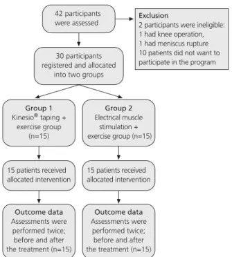
The present study was designed as a master’s thesis. Forty-two participants were referred and 30 partici-pants (71%) fulfilled the eligibility criteria and then were divided into two groups (Fig. 1). All participants completed the study.
Patients were diagnosed with PFPS by the Istanbul Faculty of Medicine’s Department of Physical Medicine and Rehabilitation. Patients with anterior or retropatellar knee pain during at least two activities (ascending-descending stairs, hopping-running, squat-ting, kneeling, prolonged sitting) for a period lasting longer than 3 months were included in the study. Patients with contraindications for exercise or electros-timulation practice, prior knee operations, knee insta-bility, meniscus rupture, abnormal foot, ankle prona-tion, Grade 3 or 4 osteoarthritis, patellofemoral pain longer than 10 years or initial onset of symptoms relat-ed to trauma were excludrelat-ed. Patients with knee pain caused by the hip or spine were also excluded.
Patients were divided into two equal groups, in order of their entry to the clinic with the first 15 (KT group; 20 affected extremities) receiving KT and an exercise program and the next 15 (ES group; 19 effect-ed extremities) receiving electrostimulation and the same exercise program. Nine patients had bilateral PFPS and a lower-extremity therapy program was per-formed for the 39 limbs.
Treatment was applied in both groups by a certified physiotherapist. Sessions were individual and lasted for 45 to 50 minutes, 3-times a week for 6 weeks (Table 1). Patients were asked to do exercises at home on non-treatment days and compliance was monitored via a daily log.
Fig. 1. Flow chart describing the progression of participants through the trial.
A 2-inch I strip of Kinesio®Tex Tape split to a Y was used proximal to the superior patellar border. With the patient in a supine with knee fully-extended position, the strip was applied starting approximately 10 cm below the anterior superior iliac spine (origin of rectus femoris) with an I base at zero tension. The tape was lined towards the kneecap at 50% tension, then the knee was flexed gradually and tails of Y were affixed at flexion (Fig. 2). The lateral tail of the Y portion was applied at 75% of available tension used over the later-al border, finishing with zero tension. The medilater-al tail was applied starting at 10% and finishing at zero ten-sion. Another 2-inch Y-shaped tape was used for the VMO. An I strip was applied to the VMO (at an approximately 50-55º angle to the femur’s long axis), its base starting at zero tension, then lined to the patel-la at 50% tension. The patel-lateral tail of the Y portion was applied at 75% tension, its medial tail was applied at 10% tension and was finished at zero tension.
Electrostimulation was delivered using a dual-chan-nel, portable electrostimulation unit (EMS 2000; BioMedical Life Systems, Inc., Vista, CA, USA) with the patient sitting and the knee flexed at approximately 30º. Two self-adhesive electrodes were used for each patient. One was placed approximately 4 cm superior and 3 cm medial to the superior-medial border of the patella (50 to 55º to vertical) for the VMO muscle and the other approximately 10 cm superior and 6 cm lateral to the superior border of the patella (10 to 15º to vertical) for the vastus lateralis (VL) muscle. Stimulation periods lasted 20 minutes (frequency: 40 Hz; pulse duration: 300 μs; duty cycle: 20:40). Stimulation intensity was the highest level comfortably tolerable for the patient.
Participants read and signed an informed consent form before the study and the Istanbul University, Istanbul Faculty of Medicine Ethics Committee approved the study.
Assessments were performed twice, at the start of the treatments and at the six-week end. A patient assessment form was completed at the first assessment session. Data including age, gender, height, weight, affected extremity, and education were collected. Body mass index (BMI) was calculated as mass (kg) divided by height squared (m2
).
Patellofemoral pain was assessed using the visual analog scale (VAS), comprising a 100 mm line with 0 representing ‘no pain’ and 10 representing ‘the worst pain’. The VAS is a reliable, valid and sensible scale for the assessment of pain in patients with PFPS.[19]
Knee extension strength was assessed using the manual muscle test standardized by the British Medical Research Council.[20]
The ‘Kujala Patellofemoral Score’, a self-reported questionnaire scoring system, was used to assess knee function. The scoring system values range from 100 (a normal, painless, fully functioning knee) to 0 (severe knee pain, dysfunction).[21,22]
Strengthening exercises
1. Quadriceps muscle isometric contraction in sitting 8-10 sets of 5 seconds hold
2. Terminal knee extension (0-30º) in sitting 3 sets of 10 repetitions 3. Isometric hip abduction in sitting 8-10 sets of 5 seconds hold 4. Straight leg extension in sitting (3 sets of 10 reps)
5. Straight leg extension with bridge exercise (3 sets of 10 reps) 6. Walking with dorsiflexed ankle (3 sets of 10 steps) 7. Squats to 30º knee flexion combined with gluteal muscle
contractions (3 sets of 10 reps)
Stretching exercises (8-10 sets of 10 seconds hold) 8. Quadriceps muscle stretches in prone position 9. Hamstring muscle stretching in sitting
10. Gastrocnemius muscle stretching against to wall while standing 11. Iliotibial tract stretching while standing
Table 1. Elements of exercise program for both groups.
Fig. 2. (a-b) Taping method.
Step, triple-jump and knee flexion tests were used to measure functional outcomes. The step test was per-formed by counting the number of steps up and down that patient could perform until patellofemoral pain onset using a 25-cm step.[13]
In the triple-jump test, we asked patients to stand on a painful limb, perform three consecutive hops, landing on the same foot. Then the total distance was measured.[23]
In the knee flexion test, we assessed knee flexion until initial pain onset while squatting. This was measured using a universal goniometer, aligned with the femur’s lateral aspect through the fibulae’s lateral aspect.[13]
Generic health-related quality of life (QoL) was assessed using the Turkish version of the Medical Outcomes Study Short Form 36 (SF-36) questionnaire, developed for use in clinical practice, research, health policy evaluations, and general population surveys by Ware.[24]
The validity and reliability of the Turkish ver-sion of the SF-36 has been well documented.[25]
Statistical analyses were performed using the SPSS v.12.0 for Windows. A p value of less than 0.05 was con-sidered statistically significant for all analyses for a two-tailed test. Comparison of treatment groups was per-formed using an intention-to-treat analysis of difference between baseline-final scores with 95% confidence intervals, independent sample t-tests or Mann-Whitney U tests. The paired sample t-test and Wilcoxon Signed-Rank test were used to compare outcomes before and after treatment. The Mann-Whitney U test was used to compare improvements and differences between groups.
Results
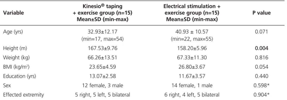
There were no significant differences between partici-pants in the two groups for any demographic character-istics, with the exception of height (Table 2). Although mean height values were significantly different in the
two groups, mean BMI showed no significant difference; so height differences were not considered.
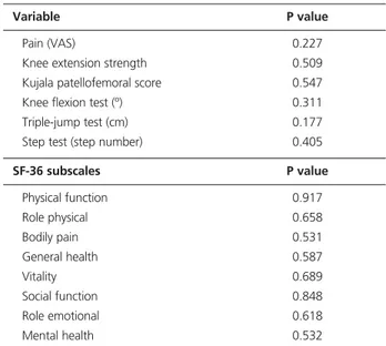
Baseline pain values, muscle strength, clinical and functional test results and SF-36 scores did not differ between the KT and ES groups (Table 3).
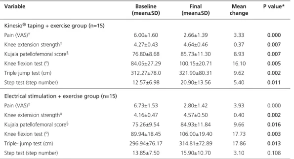
There were no significant differences in post-treat-ment pain levels between the groups (p=0.227). There were significant improvements in both groups’ pre-and post-VAS scores (Tables 3 pre-and 4).
There were no significant differences in knee exten-sion strength between groups (p=0.509); and the KT and ES groups showed significant improvements in the within-group analysis (p=0.007 and p=0.002, respective-ly) (Table 3).
No significant differences were found in post-treat-ment Kujala scores between groups (p=0.547). Within-group analysis revealed significant improvements between pre- and post-treatment values for both groups. There were no significant differences between groups for final knee flexion test scores (p=0.311). Within-group analysis showed significant improve-ments of 16º in the KT group (p=0.005) and 17º in the ES group (p=0.003).
No significant differences were found between the groups at the final triple-jump test scores (p=0.177). Within-group analysis showed significant post-treat-ment improvepost-treat-ments of 9 cm in the KT group (p=0.002) and 17 cm in the ES group (p=0.013).
There was no significant difference in the final step test scores between the groups (p=0.405). There were significant within-group improvements for the step test in the KT group (mean difference: 5 steps; p=0.011), but not in the ES group (mean difference: 3-steps; p=0.108) (Tables 3 and 4).
Kinesio®taping Electrical stimulation +
Variable + exercise group (n=15) exercise group (n=15) P value
Mean±SD (min-max) Mean±SD (min-max)
Age (yrs) 32.93±12.17 40.93 ± 10.57 0.071
(min=17, max=54) (min=22, max=55)
Height (m) 167.53±9.76 158.20±5.96 0.004
Weight (kg) 66.26±13.51 67.33±11.30 0.816
BMI (kg/m2) 23.65±4.59 26.80±3.67 0.054
Education (yrs) 13.07±2.58 11.67±3.57 0.440
Sex 12 female, 3 male 14 female, 1 male 0.598*
Effected extremity 5 right, 5 left, 5 bilateral 6 right, 4 left, 5 bilateral 0.904* *Chi-square test
SF-36 subscale scores showed no significant differ-ences between groups for post-treatment values (Table 5). Within-group analysis differences were significant for both groups in all subscale scores. General health (p=0.166) and vitality (p=0.066) subscale scores’ differ-ences at baseline and final evaluations were not signif-icant but others were statistically signifsignif-icant (Table 4) in both groups.
Discussion
Patients in both the KT and ES groups were treated individually with standardized treatment programs and improved in almost every outcome after 6 weeks. Our study showed no difference between the groups. Additionally, beneficial results following both treatment options were observed for pain, muscle strength, func-tion, and QoL. These results support the use of KT and electrostimulation as part of a rehabilitation program.
Electrostimulation for PFPS and various knee prob-lems aiming to strengthen the quadriceps muscle or its parts has been demonstrated in many studies.[13,14,26,27] Werner et al.[14]
showed that electrostimulation of the vastus medialis (VM) and stretching of the lateral thigh muscles may be useful for patients with patellofemoral complaints. They also demonstrated that the mass of the VM developed significantly and that two-thirds of the patients receiving treatment maintained these develop-ments in the affected extremity after 3.5 years. Callaghan
and Oldham[13]
assessed the effectiveness of the use of two different electrostimulation techniques in the reha-bilitation of 74 patients with PFPS. Improvements in muscle strength and function were statistically signifi-cant and there were signifisignifi-cant reductions in pain and muscle fatigue. Leroux et al. suggested using electros-timulation or biofeedback to facilitate VMO strengthen-ing.[16] Similarly, Steadman proposed electrostimulation for the VM with the goal of providing proper patella alignment in the patellofemoral joint.[17]
Parallel to the literature, the current study supports the use of electros-timulation to reduce pain and improve function for patients with PFPS.
Although there are many studies including the use of electrostimulation, sufficient data does not exist on the effects of KT use in patients with PFPS. Chen et al.[28]researched the efficacy of KT application and its effect on activation time and rate of the VL and VMO muscles. As a result of this research, they determined that KT accelerates VMO activation and does not affect VMO/VL activation ratio. They also suggested that the ‘tactile input’ provided by KT may alter the muscle strength.
Słupik et al.[10]
investigated the bioelectric effect of KT on the VM muscle. They showed that muscle bio-electrical activity increased 24 hours after taping and continued for 48 hours after the tape was removed.
Variable Baseline Final Mean P value*
(mean±SD) (mean±SD) change
Kinesio®taping + exercise group (n=15)
Pain (VAS)† 6.00±1.60 2.66±1.39 3.33 0.000
Knee extension strength‡ 4.27±0.43 4.64±0.46 0.37 0.007
Kujala patellofemoral score§ 76.80±8.68 85.73±11.30 8.93 0.007
Knee flexion test (º) 84.05±27.29 100.15±20.71 16.10 0.005
Triple jump test (cm) 312.27±78.0 321.90±80.31 9.62 0.002
Step test (step number) 12.57±6.98 20.90±13.56 5.40 0.011
Electrical stimulation + exercise group (n=15)
Pain (VAS)† 6.73±1.53 2.80±1.42 3.93 0.000
Knee extension strength‡ 4.16±0.47 4.57±0.50 0.40 0.002
Kujala patellofemoral score§ 75.26±9.54 84.93±11.84 9.66 0.016
Knee flexion test (º) 89.94±18.45 106.00±19.40 17.73 0.003
Triple- jump test (cm) 296.94±76.17 314.81±72.89 17.86 0.013
Step test (step number) 13.85±7.50 15.90±10.70 3.10 0.108
*Differences for changes in primary outcomes. Values below 0.05 were considered statistically significant.†Measured on a 10 cm visual analog scale (0= no pain; 10= maximum pain).‡Standardized manual muscle test grade on a 0 to 5 scale (0= no active con-traction can be detected; 5= normal muscle strength).§Scoring system values ranging from 0-100 (0 = severe knee pain and dys-function; 100= a normal, painless, fully functioning knee).
Significant improvements in pain reduction were obtained in both groups (p=0.000). A common expla-nation for pain relief in patients with PFPS is that improvement in quadriceps muscle provides patellar stability.[29]
Many physical therapy protocols emphasize VMO strengthening for its medial stabilizing effect on the patella. Zappala et al.[30]
and McConnell[31]
showed that the VMO keeps the patella normally aligned in the anatomical groove. The correction to normal function and movement puts less stress on the pain-sensitive patellar retinaculum and other tissues, and thus reduces pain.[32]
In our study, pain reduction can be explained by the strengthening of the VMO through short-arc and weight-bearing exercises and KT or elec-trical stimulation, as well as the lengthening of short-ened structures with stretching exercise.
Improvements in knee extension strength in the KT and ES groups were significant with one group not superior to the other. We can say that both treatment programs are effective for knee extension strength and quadriceps muscle strength, which are considered important for patients with PFPS.[8,30]
We assessed knee functions with clinical tests (knee flexion, triple-jump, step test). All outcome measures for these clinical tests showed significant improvement in both groups, with the exception of the step test in the ES group. However, there was no statistically sig-nificant difference between the two groups.
The Kujala patellofemoral score, described by Kujala et al. in 1993, is a reliable and sensitive scale for the func-tional assessment of knee complaints associated with patellofemoral structures.[19,21]
There were no significant differences between groups, although both groups showed improvements from pre- to post-treatment results.
Variable P value
Pain (VAS) 0.227
Knee extension strength 0.509
Kujala patellofemoral score 0.547
Knee flexion test (º) 0.311
Triple-jump test (cm) 0.177
Step test (step number) 0.405
SF-36 subscales P value Physical function 0.917 Role physical 0.658 Bodily pain 0.531 General health 0.587 Vitality 0.689 Social function 0.848 Role emotional 0.618 Mental health 0.532
Table 5. Intergroup analysis for post-treatment (final) values.
Variables Baseline Final Mean P value*
(mean±SD) (mean±SD) change
Kinesio®taping + exercise group (n=15)
Physical function 41.10±7.80 48.49±7.10 7.39 0.003 Role physical 33.79±8.96 45.39±8.79 11.60 0.009 Bodily pain 39.98±6.42 50.01±5.76 10.03 0.001 General health 40.29±7.52 43.79±6.91 3.50 0.021 Vitality 45.59±6.91 50.49±6.55 4.90 0.027 Social function 42.31±8.06 47.37±5.49 5.06 0.010 Role emotional 39.36±13.40 50.40±7.80 11.03 0.004 Mental health 39.52±8.92 44.07±5.97 4.54 0.012
Electrical stimulation + exercise group (n=15)
Physical function 39.26±11.02 48.05±8.34 8.79 0.001 Role physical 42.56±12.91 52.92±7.46 10.35 0.012 Bodily pain 43.22±10.37 51.59±5.11 8.37 0.010 General health 43.34±10.95 46.02±8.39 2.68 0.166 Vitality 43.70±10.29 47.80±9.75 4.10 0.066 Social function 43.75±9.13 49.16±9.12 5.41 0.015 Role emotional 42.66±15.00 52.49±8.41 9.82 0.026 Mental health 39.54±11.93 45.74±8.21 6.20 0.013
One of the most commonly used scales to objective-ly assess QoL is the Medical Outcomes Study Short Form, Health Survey (SF-36). We assessed our patients’ general health status with this scale, in addition to pain, function, and knee extension strength assessments. There were no significant differences between the groups and both groups had significantly improved between pre- and post-treatment QoL results.
The lack of an exercise-only group may be consid-ered to be a deficiency of our study, but the literature has already established that exercise is beneficial for PFPS. Therefore, testing the superiority of therapies in addi-tion to exercise seemed appropriate. However, future research to substantiate the efficacy of KT and electros-timulation compared to an exercise-only program is needed.
A limitation to our study was the absence of a power analysis. We did not perform this type of analy-sis as this was a theanaly-sis study performed with a limited timeframe.
There are advantages and disadvantages to both methods in their clinical application and the patients’ viewpoint. KT application in the clinic takes a short period of time. Tape can be used for 3 to 7 days and therapeutic effects continue throughout the day, giving a feeling of knee support. However, disadvantages of KT include the necessity of training on proper and effi-cient application, and possible patient complaints with the appearance of the tape under clothes. Disadvantage of ES application is that it requires a specific device, a physiotherapist, and more time spent with patients. Patient comments led us to believe that electrostimula-tion provided higher patient satisfacelectrostimula-tion, although this was not analyzed. This may be explained by cultural characteristics. When the costs of two methods are con-sidered, it may be inferred that electrostimulation is cheaper than taping.
In conclusion, combined with an exercise program, KT and electrostimulation have similar effects on pain reduction, functional condition improvement, increased muscle strength and improved QoL and have no superiority to the other in the treatment of PFPS. Conflicts of Interest: No conflicts declared.
References
1. Ng GY, Cheng JM. The effects of patellar taping on pain and neuromuscular performance in subjects with patellofemoral pain syndrome. Clin Rehabil 2002;16:821-7.
2. Qi Z, Ng GY. EMG analysis of vastus medialis obliquus/vas-tus lateralis activities in subjects with patellofemoral pain syn-drome before and after a home exercise program. J Phys Ther Sci 2007;19:131-7.
3. Warden SJ, Hinman RS, Watson MA Jr, Avin KG, Bialocerkowski AE, Crossley KM. Patellar taping and bracing for the treatment of chronic knee pain: a systematic review and meta-analysis. Arthritis Rheum 2008;59:73-83.
4. Çubukçu D, Sarsan A. Rehabilitation of patellofemoral pain syndrome. [Article in Turkish] Turkish Journal of Rheuma-tology 2008;23:18-23.
5. Worrell T, Ingersoll CD, Bockrath-Pugliese K, Minis P. Effect of patellar taping and bracing on patellar position as determined by MRI in patients with patellofemoral pain. J Athl Train 1998;33:16-20.
6. LaBella C. Patellofemoral pain syndrome: evaluation and treatment. Prim Care 2004;31:977-1003.
7. Brunker P, Khan K. Clinical Sports Medicine. 3rd ed. Australia: McGraw-Hill Company; 2007. p. 506-37. 8. Sanchis-Alfonso V, ed. Anterior knee pain and patellar
insta-bility. Singapore, KYO: Springer; 2006.
9. Aminaka N, Gribble PA. A systematic review of the effects of therapeutic taping on patellofemoral pain syndrome. J Athl Train 2005;40:341-51.
10. Słupik A, Dwornik M, Białoszewski D, Zych E. Effect of Kinesio taping on bioelectrical activity of vastus medialis mus-cle. Preliminary report. Ortop Traumatol Rehabil 2007;9: 644-51.
11. Yasukawa A, Patel P, Sisung C. Pilot study: investigating the effects of Kinesio Taping in an acute pediatric rehabilitation setting. Am J Occup Ther 2006;60:104-10.
12. Prentice WE. Therapeutic modalities in rehabilitation. 3rd ed. USA: McGraw-Hill; 2005.
13. Callaghan MJ, Oldham JA. Electric muscle stimulation of the quadriceps in the treatment of patellofemoral pain. Arch Phys Med Rehabil 2004;85:956-62.
14. Werner S, Arvidsson H, Arvidson I, Eriksson E. Electrical stimulation of vastus medialis and stretching of lateral thigh muscles in patients with patello-femoral symptoms. Knee Surg Sports Traumatol Arthrosc 1993;1:85-92.
15. Davlin CD, Holcomb WR, Guadagnoli MA. The effect of hip position and electromyographic biofeedback training on the vastus medialis oblique: vastus lateralis ratio. J Athl Train 1999;34:342-6.
16. Leroux A, Boucher JP, Poumarat G. Investigation of quadri-ceps femoris function through electrical stimulation. J Athl Train 1997;32:115-8.
17. Steadman JR. Nonoperative measures for patellofemoral problems. Am J Sports Med 1979;7:374-5.
18. Lin YF, Lin JJ, Jan MH, Wei TC, Shih HY, Cheng CK. Role of the vastus medialis obliquus in repositioning the patella: a dynamic computed tomography study. Am J Sports Med 2008;36:741-6.
19. Crossley KM, Bennell KM, Cowan SM, Green S. Analysis of outcome measures for persons with patellofemoral pain: which are reliable and valid? Arch Phys Med Rehabil 2004;85:815-22.
20. Medical Research Council of the United Kingdom. Aids to examination of the peripheral nervous system: Memorandum no. 45. Palo Alto, CA: Pendragon House; 1978.
21. Kujala UM, Jaakkola LH, Koskinen SK, Taimela S, Hurme M, Nelimarkka O. Scorring of patellofemoral disorders. Arthroscopy 1993;9:159-63.
22. Kuru T, Dereli EE, Yaliman A. Validity of the Turkish ver-sion of the Kujala patellofemoral score in patellofemoral pain syndrome. Acta Orthop Traumatol Turc 2010;44:152-6. 23. Keskula DR, Duncan JB, Davis VL, Finley PW. Functional
outcomes measures for knee dysfunction assessment. J Athl Train 1996;31:105-10.
24. Ware JE Jr, Sherbourne CD. The MOS 36-item short-form health survey. Med Care 1992;30:473-83.
25. Dündar P, Fidaner C, Fidaner H, Oral A, Eser S, Atman UC, et al. Comparing the Turkish versions of WHOQOL-BREF and SF-36. Convergent validity of WHOQOL-BREF and SF-36. Hippokratia 2002;6:37-43.
26. Draper V, Ballard L. Electrical stimulation versus electromyo-graphic biofeedback in the recovery of quadriceps femoris muscle function following anterior cruciate ligament surgery. Phys Ther 1991;71:455-64.
27. Mizusaki A, Almedia GJM, Atallah AN, Peccin MS, Cohen MC, Soares BGO. Electrical stimulation for rehabilitation
after soft tissue injury of the knee in adults. Cochrane Database of Systematic Reviews 2005;4. CD001826. 28. Chen WC, Hong WH, Huang TF, Hsu HC. Effects of
kine-sio taping on the timing and ratio of vastus medialis obliques and vastus lateralis muscle for person with patellofemoral pain. J Biomech 2007;40:318.
29. Steinkamp LA, Dillingham MF, Markel MD, Hill JA, Kaufman KR. Biomechanical considerations in patellofemoral joint rehabilitation. Am J Sports Med 1993;21:438-44. 30. Zappala FG, Taffel CB, Scuderi GR. Rehabilitation of
patellofemoral joint disorders. Orthop Clin North Am 1992; 23:555-66.
31. McConnell J. The physical therapist’s approach to patellofemoral disorders. Clin Sports Med 2002;21:363-87. 32. Grelsamer RP, McConnell J. The patella: a team approach.