Eurasian J Vet Sci, 2017, 33, 2, 130-132
130
Eurasian Journal
of Veterinary Sciences
CASE REPORT
Sustained polymorphic ventricular tachycardia in a calf
Amir Naseri¹*, Merve Ider¹, Mahmut Ok¹
Department of Internal Medicine, Faculty of Veterinary Medicine, Selçuk University, Konya, Turkey Received: 26.12.2016, Accepted: 06.02.2017
Abstract
Naseri A, Ider M, Ok M. Sustained polymorphic ventricular
tachycardia in a calf.
Sustained polymorphic ventricular tachycardia is a type of ventricular dysrhythmias which originated from more than one focus in ventricle and may lead to fatal arrhythmias like as ventricular fibrillation and sudden death. A seven days old female Holstein calve was referred to the Large Animals Hos-pital of the Faculty of Veterinary Medicine of Selcuk Univer-sity with a history of sudden collapse. Electrocardiography examination showed sustained polymorphic ventricular tachycardia. Cardiac damage suspicion confirmed by eleva-tion in serum cardiac troponin I (cTnI), creatine kinase-MB (CK-MB) and creatine phosphokinase (CPK) concentrations as cardiac damage enzymes.
Keywords: Calf, ventricular tachycardia, polymorphic, cardi-ac biomarkers.
Öz
Naseri A, Ider M, Ok M. Bir buzağıda sürekli polimorfik
ventriküler taşikardi.
Sürekli polimorfik ventriküler taşikardi, ventrikülerin bir-den fazla odağından kaynaklanan bir ventriküler aritmidir ve ventriküler fibrilasyon gibi ölümcül aritmilere ve ani ölü-me yol açabilölü-mektedir. Yedi günlük Holştayn dişi bir buzağı Selçuk üniversitesi, Veteriner Fakültesi, büyük hayvan has-tanesine ani kollaps şikayeti ile getirildi. Elektrokardiyografi bulguları sürekli polimorfik ventriküler taşikardi’yi gösterdi. Kardiyak hasar şüphesi kardiyak troponin I (cTnI), kreatin kinaz- MB (CK-MB) ve kreatin fosfokinaz (CPK) düzeylerin-deki artışla doğrulandı.
Anahtar kelimeler: Buzağı, ventriküler taşikardi, polimor-fik, kardiyak biyobelirteç.
Eurasian J Vet Sci, 2017, 33, 2, 130-132
DOI:10.15312/EurasianJVetSci.2017.148
Eurasian J Vet Sci, 2017, 33, 2, 130-132
131
Cardiac arrhythmias are abnormalities in the heart rate, rhythm, or conduction pattern (Smith 2009). From a clinical perspective, it is useful to categorized cardiac dysrhythmias by their site of origin (supraventricular or ventricular) and by their rate (bradydysrhythmias or tachydysrhythmias) (Bonagura and Miller 1985, Bonagura and Miller 1986). Most abnormal arrhythmias of horses and cattle are tachyarrhy-thmias (Smith 2009). Ventricular arrhytachyarrhy-thmias commonly occur in the severe cardiac disease and final stages of heart failure (Radostits et al 2007). Principal concern of ventricu-lar arrhythmias is hemodynamic consequences (Reed et al 2004) and usually if uncorrected may lead to ventricular fib-rillation and death (Radostits et al 2007).
A seven days old female Holstein calve was referred to the Large Animals Hospital of the Faculty of Veterinary Medi-cine of Selcuk University with a history of sudden collapse. In the primary examination, calf was comatos and in lateral recumbency. After admission to the intensive care unit, clini-cal, laboratory and electrocardiographic examinations were performed. In the first evolution, the cardiac rhythm was ir-regular and the heart rate interestingly was 2 fold (HR: 220/ min) upper the maximum limits of normal values. In the ECG study, base apex lead system was performed for explore the genus of arrhythmia.
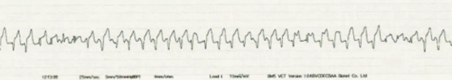
Recorded ECG traces showed sustained polymorphic vent-ricular tachycardia which characterized by rapid heartbeats accompanied by continuously bizarre QRS complexes and abnormal T waves (Figure 1, 2). Before preparing any
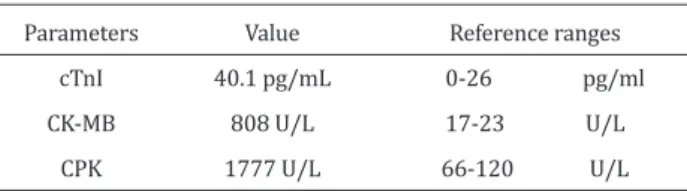
anti-arrhythmic treatment, ventricular tachycardia degenerated to ventricular fibrillation (Figure 3) and calf died. Laboratory findings showed significant increase in serum concentrati-ons of cTnI, CK-MB and CPK (Table 1).
Ventricular tachycardia (VT) is a cardiac arrhythmia charac-terized by a rapid rhythm originating in the ventricle (Smith 2009). A series of four or more premature ventricular cont-ractions is diagnostic of ventricular tachycardia (McGuirk and Muir 1985). Polymorphic ventricular tachycardia occurs when the ventricular premature complexes (VPCs) originate from more than one focus in the ventricle, creating abnormal QRS and T complexes of different orientations (Marr and Bo-wen 2010). Ventricular re-entry is an important mechanism for the development of sustained VT (Smith 2009).
Ventricular tachycardia can occur when there is myocardi-tis, myocardial necrosis, bacterial endocardimyocardi-tis, autonomic nervous system imbalance, hypoxia, ischemia, electrolyte or metabolic disturbances, sepsis or unknown causes (Smith 2009).
Cardiac injury in calves has been identified by increased cTnI in endotoxemia (Peek et al 2008) and myocarditis (Tunca et al 2008, Karapinar et al 2010). Additionally, elevation of CK-MB as cardiac damage biomarker in critically ill calves (Aydogdu et al 2016) and less specific cardiac damage iso-enzyme, CPK, may support the prospective injury of cardiac tissue (Kemp et al 2004, Peek et al 2008, Smith 2009). In the present case, association of sustained polymorphic
ventricu-Figure 1. ECG tracing shows abnormal QRS complexes and T waves (Lead I, 25 mm/sec and 10 mm/mV).
Figure 2. ECG tracing shows continuously polymorphic complexes at 200 beat/min heart rate (Lead II, 25 mm/sec and 10 mm/mV).
Figure 3. ECG showing ventricular fibrillation. There is no any coordinated contraction (Lead II, 25 mm/sec and 10 mm/mV)
Naseri et al Polymorphic ventricular tachycardia calf
Eurasian J Vet Sci, 2017, 33, 2, 130-132
132
lar tachycardia and elevation of cTnI, CK-MB and CPK enz-ymes levels may be confirmed development cardiac damage and poor prognosis.
References
Smith BP, 2009. Large Animal Internal Medicine, 4th edition, Mosby, Maryland Heights, Missouri, USA.
Bonagura J, Miller MS, 1985. Junctional and Ventricular Arrhythmias, J Equine Vet Sci, 5, 347-350.
Bonagura J, Miller MS, 1986. Common Conduction Distur-bances, J Equine Vet Sci, 6, 23-25.
Radostits OM, Gay CC, Hinchcliff KW, Constable PD, 2007,
Ve-Parameters cTnI CK-MB
CPK
Table 1. The Levels of cardiac biomarker of calf Value 40.1 pg/mL 808 U/L 1777 U/L Reference ranges 0-26 pg/ml 17-23 U/L 66-120 U/L
terinary Medicine, 10 edition, Saunders, Philadelphia, USA. Reed SM, Bayly WM, Sellon DC, 2004. Equine Internal
Medici-ne, 2nd edition, Saunders, Philadelphia, USA.
McGuirk SM, Muir WW, 1985. Diagnosis and treatment of cardiac arrhythmias. Vet Clin North Am Equine Pract, 1, 353-370.
Marr CM, Bowen IM, 2010. Cardiology of the Horse, 2nd edi-tion, Elsevier, Philadelphia, USA.
Peek SF, Apple FS, Murakami MA, Crump PM, Semrad SD, 2008. Cardiac isoenzymes in healthy Holstein calves and calves with experimentally induced endotoxemia, Can J Vet Res, 72, 356-361.
Tunca R, Sozmen M, Erdogan H, Citil M, Uzlu E, Ozen H, Gokce E, 2008. Determination of cardiac troponin I in the blood and heart of calves with foot-and-mouth disease, J Vet Di-agn Invest, 20, 598-605.
Karapinar T, Dabak DO, Kuloglu T, Bulut H, 2010. High cardiac troponin I plasma concentration in a calf with myocarditis, Can Vet J, 51, 397-399.
Aydogdu U, Yildiz R, Guzelbektes H, Coskun A, Sen I, 2016. Cardiac biomarkers in premature calves with respiratory distress syndrome, Acta Vet Hung, 64, 38-46.
Kemp M, Donovan J, Higham H, Hooper J, 2004. Biochemical markers of myocardial injury, Br J Anaesth, 93, 63-73.
Naseri et al Polymorphic ventricular tachycardia calf