SEMINARS IN LIVER DISEASE-VOL. 19, NO. 3, 1999
Genetic Aspects of Hepatocellular Carcinogenesis
MEHMET OZTURK, Ph.D
ABSTRACT:
Hepatocellular carcinoma (HCC) is linked etiologically to viruses (hepatitis B virus [HBV] and hep-
atitis C virus [HCV]), chemical carcinogens (i.e., aflatoxins), and other environmental and host ,factors causing
chronic liver injury. Some hepatoblastomas may be linked to inherited gene mutations, but adult hereditary HCC up-
pears to be rare. HCCs display gross genomic ultel-ations, including DNA rearrangements associated with HBV
DNA integration, loss cf heterozygosity, and, less importantly, chromosomal amplifications and loss of imprinting.
Many genes with somatic mutations have now been identified in these tumors. Most frequently involved genes are tu-
mor suppressor genes such as p53, M6P/IGF2R, p-catenin, p16INK4A, and retinoblastoma genes. Most identified
mutations are somatic, but germline mutations of pl61NK4A, APC, and BRCA2 have also been reported. Oncogenic
activation of several cellular genes such as cyclin D and cyclin A have been described in HCC, but the possible im-
plication of candidate viral oncogenes (i.e., X protein of HBV) is still debuted.
A
comprehensive analysis of all the
genetic changes described for HCC demonstrates that at least four different growth regulatory pathways are altered
in these tumors. However, each pathway appears to be implicated in a limited fraction of these tumors, suggesting
that HCCs are genetically heterogenous neoplasms. This genetic heterogeneity correlates with the heterogeneity qf
etiologic factors implicated in HCC.
KEY WORDS:
hepatocellular carcinoma, primary liver cancer, p53, p16INK4A, cyclin D, p-catenin, M6P/IGF2R
Hepatocellular carcinoma (HCC) cells often display
chromosomal changes such as polyploidy, loss of het-
erozygosity (LOH), allelic imbalance (AI), amplifica-
tions, and trans1ocations.l It has also been known for a
long time that hepatitis B virus (HBV) DNA causes chro-
mosomal rearrangements by integration into the host
genome.? It is expected that the chromosomal regions
that undergo tumor-specific changes harbor critical genes
involved directly (oncogenes and tumors suppressor
genes) or indirectly (DNA repair genes) in carcinogene-
sis. To date, a dozen genes, including p53, mannose-6-
phosphate/insulin-like factor 2 receptor (M6P/ IGF2R),
p-catenin, retinoblastoma ( R B I ) , p161NK4A, adeno-
matosis polyposis coli (APC), breast cancer gene 2
(BRCAZ), cyclin A, cyclin
D,
and insulin-like growth
factor 2 (IGF2) have been shown to be altered in HCC
and/or hepatoblastoma (Table 1). This list will probably
grow over the next years to include many more genes.
There are at least two reasons to explain the high num-
ber of altered genes in HCC. First, solid tumors of the
adult may need the accumulation of many genetic alter-
ations before they become clinically detectable. Indeed,
Objectives
Upon completion of this article, the reader should be able to: I) list the factors that are etiologically linked to hepatocellular carcinoma; 2) state the most frequently involved genes; and 3) recognize the four different growth regulatory pathways that are altered in these tumors. Accreditation
The Indiana University School of Medicine is accredited by the Accreditation Council for Continuing Medical Education to sponsor continuing medical education for physicians.
Credit
The Indiana University School of Medicine designates this educational activity for a maximum of 1.0 hours credit toward the AMA Physicians Recognition Award in category one.
Disclosure
Statements have been obtained regarding the author's relationships with financial supporters of this activity. There is no apparent conflict of interest related to the context of participation of the author of this article.
From the Department of Molecular Biology and Genetics, Bilkenr University, 06653 Ankara, Turkey
Reprint requests: Dr. Mehmet Ozturk, Dept. of Molecular Biology and Genetics, Bilkent University, 06533 Ankara, Turkey.
Copyright 0 1999 by Thieme Medical Publishers, Inc., 333 Seventh Avenue, New York, NY 10001, USA. Tel.: +1(2 12) 760-0888 x 132.0272-8087/1999/1098-8971 (1999) 19:03:0235-0242:SLDOOOlOX
SEMINARS IN LIVER DISEASE-VOL. 19, NO. 3,1999
TABLE 1. Genetic Alterations in Hepatocellular Carcinoma and Hepatoblastoma
Gene Mutation (%) Other Alterations References
~ 5 3 28 M6P/IGF2R 18-33 TGFRB2 0 RBI 15 p15INK4B 0 p16INK4A* 0-55 ~ 2 1 5 Cyclin Dt 11-13 Cyclin At 19 p-catenin 19-26 APC* 62s E-cadherin NR BRCA2* 5 Smad2 0-2 Smad4 0-6 hMLHl NR hMSH2 NR IGF2 NR K-ras 0-17 N-ras 0-16 H-ras 0-10 c-myct 0-50 N-myct 0
*Somatic and germline mutations. +Amplification.
*In hepatoblastoma only. HD, homozygous deletion. LOH, loss of heterozygosity. LOI, loss of imprinting.
the well-known "latent period" between the first expo-
sure to an etiologic agent (i.e., infection with HBV) and
the development of HCC is in favor of such a hypothe-
sis.3 Second, the multiplicity of genetic alterations in
HCC may indicate that different etiologic factors affect
different sets of target genes in hepatocytes. This etio-
logically defined genetic heterogeneity of HCC results
in a phenotypic heterogeneity of these tumors. In other
words, distinct but related growth regulatory pathways
are altered during hepatocarcinogenesis. As discussed
later, at least four different pathways are altered in hu-
man HCCs.
p53 GENE
Many reports now indicate that the p53 gene, which
is located on chromosome 17p, is mutated in about 30%
of HCCs worldwide (for a recent review, see ref. 4). All
reported p53 mutations in HCC are somatic. Therefore,
germline mutations of p53 appear not to predispose to
HCC. Both the frequency and the type of p53 mutations
are different depending on geographic location and sus-
pected etiology of these tumors (Table 2). An HCC-
specific codon 249 mutation (AGG
-+AGT) leading to
an arginine to serine substitution (R249S), suspected to
be induced by aflatoxins, was found in most HCCs from
geographic areas with high incidence of HCC and a
HBx interaction HD - LOH, HD HD HD, Methylation - - HBV integration - LOH LOH, Methylation LOH - - LOH LOH LO1 - Amplification Methylation - see Table 2 2 1 , 3 6 , 4 4 23 l I,* 45-50 50,51 50-54 55 56,57 58,59 30,31 11.32-35,37 60,61 62 24,25 24,25 63 63 3 8 4 3 90-95 9 0 , 9 1 , 9 3 , 9 5 , 9 6 7 3 , 9 1 , 9 3 , 9 5 , 9 7 , 9 8 99,100 99,101
high risk of exposure to aflatoxins.5-7 This mutation was
found in 50% of HCCs from Mozambique,8,9 50 to 75%
of HCCs from Qidong province of China,6.10J1 and 67%
of HCCs from Senegal.7 A worldwide study by Ozturk
et a1.8 suggested a close correlation between the pres-
ence of codon 249 mutations in HCC and high risk of
aflatoxin intake. This early study has now been largely
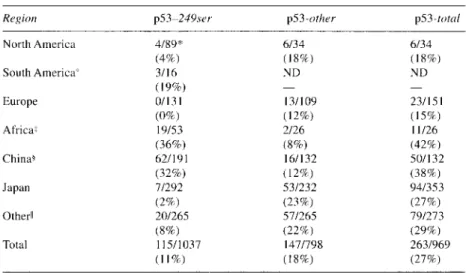
confirmed by others. As shown in Table 1, the codon
249 mutation is present in 36% of tumors from Africa
and 32% of tumors from China, respectively. These two
regions of the world are known for high incidence of
HCC, where both HBV and aflatoxins are recognized as
major etiologic factors. In contrast, the codon 249 muta-
tion is seen in less than 4% of HCCs from Japan, Eu-
rope, and North America, where HBV and hepatitis C
virus (HCV), but not aflatoxins, are the main etiologic
factors. The overall frequency of codon 249 mutations
in the world is 11
%.Mutations affecting other codons of
the p53 gene are detected in HCC, and their worldwide
frequency is 18%. The frequency of all p53 mutations in
HCC varies between 15% in Europe and 42% in China,
with a worldwide frequency of 27% (see Table 2 for a
detailed analysis of p53 mutations). Thus, p53 gene is
mutated in about a third of HCCs, but only a third of
these mutations can be etiologically linked to a high risk
of aflatoxin exposure. Therefore, p53 mutations can oc-
cur in HCC independent of aflatoxin risk, and in HBV
or HCV infection. However, Unsal et al.9 reported an
GENETIC ASPECTS OF HEPATOCARCINOGENESIS-OZTURK
TABLE 2. Frequency of p53 Mutations in Hepatocellular Carcinoma
Region p53-249srr p53-other p53-total
North America South America' Europe Africaz China+ Japan Other11 Total
Data were compiled from references 6, 7, 9-1 1, 45, 64-89, 9 8 and 102-104. The numbers d o not add up for two reasons: in some studies only the p53-249 mutation was reported; in some others only the information of the total number of mutations was reported.
"U.S.A. including Alaska. +Mexico only.
*South Africa, Mozambique, and Senegal only. +Mainland China and Hong Kong.
IAustralia, Singapore, South Korea, Taiwan, Thailand.
apparent association between the presence of
X
gene
coding sequences of HBV (HBx) and wild-type p 5 3 in
HCC. Based on this observation, a possible interference
of HBV with wild-typep53 function was suggested.Vn-
deed, recent studies showed that HBx protein encoded
by the
X
region of HBV interacts with wild-type p53
protein both physically and functionally.l2-15 These ob-
servations suggest that the suspected oncogenic activity
of HBx protein is linked to functional inactivation of
wild-type p53 protein, as observed with other viral pro-
teins with transforming activity. However, the interac-
tion of HBx with p53 was shown only experimentally. It
is presently unclear whether HBx-p53 interactions
really occur in HBV-infected hepatocytes and/or in
HCC cells with integrated HBV DNA sequences.
Frequent involvement of p53 mutations in HCC is
not surprising for several reasons. First, the p53 gene is
the only known gene to be mutated at a very high fre-
quency in tumors of different origin.16 Second, this pro-
tein is involved in different cellular processes (cell cy-
cle arrest, apoptosis, differentiation, angiogenesis, etc.),
all critically involved in the development of malig-
nancy.[' Under physiologic conditions, p53 protein is
complexed with MDM2 protein that promotes a rapid
degradation of p53. MDM2-p53 complexes are inhib-
ited either by pl9ARF (induced by both cellular and vi-
ral oncogenes) or by N-terminal phosphorylation of p.53
by DNA-dependent protein kinase. This leads to an ac-
cumulation and functional activation of p53 in cells,
leading to either cell cycle arrest by p21 or apoptosis by
bax induction. Thus, p53 protein appears to be involved
in a growth control response to abnormal oncogene ex-
pression and DNA damage.17 In patients with chronic
liver disease, the risks of oncogene activation and DNA
damage are elevated. As stated earlier, HBx may have
an oncogenic activity and aflatoxins are potent DNA
damaging agents.
p16/NK4A,
CYCLIN D, AND
RETINOBLASTOMA GENES
These three genes encode for proteins involved in
the regulation of the GI phase of the cell cycle. Cyclin D
forms active complexes with CDK4 protein, whereas
p16 protein is an inhibitor of CDK4 activity.18 The
retinoblastoma protein (pRb) is the main known sub-
strate of CDK4. In nonproliferating cells, pRb protein
forms complexes with E2F transcription factors. When
complexed to pRb, E2Fs are transcriptionally inactive.
Upon phosphorylation by CDK4, pRb is released from
its complexes and "free E2Fs" promote the initiation of
DNA synthesis.lYhese observations predict that the
loss of pRb protein or its aberrant phosphorylation will
lead to a loss of growth control at the GI phase of the
cell cycle. Increased phosphorylation of pRb may result
from an aberrant activation of CDK4 by either an excess
of cyclin D and/or a deficit in p16 protein. Recent stud-
ies demonstrated that all three genes, namely RBI,
SEMINARS IN LIVER DISEASE-VOL. 19, NO. 3, 1999
p161NK4A, and cyclin D, undergo structural alterations
in HCC. The retinoblastoma gene (RBI) is one of the tu-
mor suppressor genes studied in HCC just after the im-
plication of p53 in these tumors. LOH at the RBI gene
locus is quite frequent in HCC. In addition, RBI muta-
tions were observed in 15% of these tumors (Table 1
).The p161NK4A gene, which is located at chromo-
some 9p, codes for two alternatively spliced tran-
scripts.l7.18 One of the transcripts is for p16 protein,
an inhibitor of cyclin-dependent kinases
4
and 6.Ix
p161NK4A status in HCC has been studied indepen-
dently by several laboratories. Both germline and so-
matic mutations of pl6INKA were found in HCC pa-
tients. It was also reported that about 50% of HCC
display de novo methylation of p161NK4A, as observed
in other cancers (see Table
1
and references therein). It
is known that de
novo
methylation is a mechanism in-
volved in gene silencing.") Therefore, one can assume
that HCC cells with methylatedp16INK4A are unable to
express the gene, leading to the loss of a cyclin-depen-
dent kinase inhibitor protein.
As shown in Table 1, cyclin
D
and cyclin A genes
were shown to be amplified in 10-20% of HCCs. It is
noteworthy that RBI, pI6INKA, and cyclin genes are
mutated individually in 10 to 20% of HCCs. Although
this frequency is not high, their involvement in the same
growth regulatory pathway implies that when com-
bined, these mutations will lead a loss of growth control
in more than 30% of HCCs.
M6PlIGF2 RECEPTOR, SMAD2,
AND SMAD4 GENES
The
mannose-6-phosphate/insulin-like
growth fac-
tor 2 receptor (M6PIIGF2R) is involved in the activation
of transforming growth factor beta (TGF-P), whereas
SMAD2 and SMAD4 genes are intracellular mediators of
TGF-P, which induces both growth inhibition and apop-
totic cell death in hepatocytes.21-2Wfter the demonstra-
tion of LOH at the M6PIIGF2R gene locus by De Souza
et al.," several reports described that the MGP/IGF2R
gene is mutated in
18
to 33% of HCCs (Table
1).
SMAD2 and SMAD4 genes appear to be mutated in less
that 10% of these cancers.2452Vn contrast, no mutation of
TGF-P receptor type
I1
was found in HCC.z4 Taken to-
gether, these observations demonstrate that at least three
genes involved in TGF-P-mediated growth control are
altered in HCC and that overall the TGF-P pathway is
altered in about 25% of HCCs.
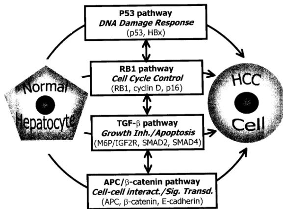
I
P53
pathway
I
FIG. 1. Main regulatory pathways altered in human hepatocellular carcinomas. The most frequently mutated genes of each pathway are also shown. The vertical arrowed lines connecting four pathways indicate that these pathways are related to each other and they should not be considered as independent and separate pathways of hepatocellular carcinogenesis.
GENETIC ASPECTS OF HEPATOCARCINOGENESIS-OZTURK
239
P-CATENIN,
APC,
AND E-CADHERIN GENES
The APC gene was initially identified in the famil-
ial adenomatous polyposis coli syndrome. Germline and
somatic mutations of APC have been detected in col-
orectal cancers.26 Some of these cancers display muta-
tions in the p-catenin gene instead of the APC gene.27
APC and p-catenin proteins have physical and func-
tional
p-Catenin also forms complexes
with E-cadherin.29 APC and E-cadherin may be in-
volved in intercellular interactions.28.29 In contrast,
p-catenin appears to play a role in transcriptional regu-
lation in addition to its participation in cell-to-cell inter-
a c t i o n ~ . ~ ~
Somatic mutations of p-catenin were ob-
served in 19-26% of HCCs.MO." These mutations that
occur at the N-terminal region of 6-catenin lead to an
accumulation of aberrant p-catenin proteins that stimu-
late the activity of a transcription factor.'8.30,31 Somatic
APC mutations may be rare in HCC, but they appear to
be quite frequent in hepatoblastomas.ll.32-" Finally, the
E-cadherin gene was shown to display frequent LOH
and de novo
methylation in HCC (Table I). Thus, it is
possible that E-cadherin function is lost in some HCCs.
Taken together, these observations indicate that the
"P-cateninlAPC pathway" is altered in more than 30%
of HCCs.
OTHER GENETIC ALTERATIONS
As shown in Table 1, ras and myc oncogenes are not
frequently mutated in human HCC. Loss of genomic im-
printing and bi-allelic expression of the IGF2 gene was
shown in hepatoblastomas and in some HCCs.3g43
Among other known genes, BRCA2 p21, andp15INK4B
appear to be involved only rarely in these tumors. MLHl
and MSH2, two genes involved in DNA mismatch repair,
have not been studied for possible mutations in HCC
(Table 1).
CONCLUDING REMARKS
Recent studies clearly indicate that many genes un-
dergo somatic aberrations (point mutations, amplifica-
tions, loss of imprinting, de novo methylation, etc.) in
HCC. The number of aberrant genes is high, but the fre-
quency of individual gene mutations is low. However,
these mutations are not random. They tend to cluster at
genes involved in important growth regulatory path-
ways. Even though the picture is still imperfect, our
present knowledge of the molecular genetics of HCC
leads us to four main pathways that are altered in HCC:
the p 5 3 pathway involved in DNA damage response, the
RBI pathway involved in cell cycle control, the TGF-fi
pathway involved in growth inhibition and apoptosis,
and the P-cateninlAPC pathway involved in morpho-
genesis and signal transduction. As illustrated in Figure
1, these pathways should not be considered as indepen-
dent pathways. They are most probably related to each
other and may even represent individually a distinct
step of hepatocellular carcinogenesis. Unfortunately,
our knowledge of the order of events for the initiation
and stepwise progression of HCC is still incomplete.
Acknowledgment: Supported
by
grants fromTUEITAK,
TUBA (Turkey),
and TWAS.ABBREVIATIONS
HCC
HBV
HCV
M6PIIGF2R
APC
p16INK4A
p151NK4B
BRCA2
HBx
LOH
RB
1
P R ~
IGF2
p l9ARF
MDM2
~2
1
CDK4
E2F
TGF-P
MLHl
MSH2
hepatocellular carcinoma
hepatitis B virus
hepatitis C virus
mannose-6-phosphate/insulin-like
growth factor I1 receptor
adenomatosis polyposis coli gene
gene coding for a 16-kDa inhibitor of
cyclin-dependent kinase 4 enzyme
gene coding for a 15-kDa inhibitor of
cyclin-dependent kinase 4 enzyme
breast cancer susceptibility gene 2
X
protein of hepatitis B virus
loss of heterozygosity
retinoblastoma gene
protein encoded by the retinblastoma
gene
insulin-like growth factor I1
protein encoded by an alternatively
spliced form of transcript from
p161NK4A gene
human homolog of mouse double mu-
tant gene 2
21 -kDa cyclin-dependent kinase in-
hibitor protein also called CIPl
cyclin-dependent kinase 4
a group of transcription factors regu-
lated by the retinoblastoma family of
pocket proteins
transforming growth factor
P
gene encoding a protein involved in
DNA mismatch repair
gene encoding for another protein in-
volved in DNA mismatch repair
REFERENCES
1 . Ozturk M. Biology of hepatocellular carcinoma. In: AK Rustgi, ed. Gastrointestinal Cancers, Biology, Diagnosis, and Therapy. Philadelphia, PA: Lippincott-Raven, 1995, pp 5 11-525
SEMINARS IN LIVER DISEASE-VOL. 19, NO. 3 , 1999 2. Buendia MA. Hepatitis B viruses and hepatocellular carcinoma.
Adv Cancer Res 1992;59: 167-226
3. Idilman R, De Maria N, Colantoni A, Van Thiel DH. Pathogene- sis of hepatitis B and C-induced hepatocellular carcinoma. J Vi- ral Hepat 1998;5:285-299
4. Puisieux A, Ozturk M. TP53 and hepatocellular carcinoma. Pathol Biol (Paris) 1997;45:864-870
5. Bressac B, Kew M, Wands J, Ozturk M. Selective G to T muta- tions of p53 gene in hepatocellular carcinoma from southern Africa. Nature 1991 ;350:42943 1
6. Hsu IC, Metcalf RA, Sun T, et al. Mutational hotspot in the p53 gene in human hepatocellular carcinomas. Nature 1991; 350:427428
7. Coursaget P, Depril N, Chabaud M, et al. High prevalence of mutations at codon 249 of the p53 gene in hepatocellular carci- nomas from Senegal. Br J Cancer 1993;67: 1395-1397
8. Ozturk M, Puisieux A, Kew M, et al. p53 mutation in hepato- cellular carcinoma after aflatoxin exposure. Lancet 1991;338: 1356-1 359
9. Unsal H, Yakicier C, Marcais C, et al. Genetic heterogeneity of hepatocellular carcinoma. Proc Natl Acad Sci USA 1994; 9 1 :822-826
10. Li D, Cao Y, He L, Wang NJ, Gu JR. Aberrations of p53 gene in human hepatocellular carcinoma from China. Carcinogenesis
1993;14:169-173
11. Fujimoto Y, Hampton LL, Wirth PJ, et al. Alterations of tumor suppressor genes and allelic losses in human hepatocellular car- cinomas in China. Cancer Res 1994;54:281-285
12. Greenblatt MS, Feitelson MA, Zhu M, et al. Integrity of 1153 in hepatitis B x antigen-positive and -negative hepatocellular carci- nomas. Cancer Res 1997;57:426432
13. Wang XW, Forrester K, Yeh H, et al. Hepatitis B virus X protein inhibits p53 sequence-specific DNA binding, transcriptional ac- tivity, and association with transcription factor ERCC3. Proc Natl Acad Sci USA 1994;9 1 : 2230-2234
14. Ueda H, Ullrich SJ, Gangemi JD, et al. Functional inactivation but not structural mutation of p53 causes liver cancer. Nat Genet 1995;9:4147
15. Elmore LW, Hancock AR, Chang SF, et al. Hepatitis B virus X protein and p53 tumor suppressor interactions in the modulation of apoptosis. Proc Natl Acad Sci USA 1997;94:14707-14712 16. Greenblatt MS. Bennett WP, Hollstein M, Harris CC. Mutations
in the p53 tumor suppressor gene: clues to cancer etiology and molecular pathogenesis. Cancer Res 1994;54:48554878 17. Prives C. Signalling to p53: breaking the MDM2-p53 circuit.
Cell 1998;95:5-8
18. Reed SI. Control of the GIIS transition. Cancer Surv 1997; 29:7-23
19. Johnson DG, Schneider-Broussard R. Role of E2F in cell cycle control and cancer. Front Biosci 1998;3:447-448
20. Razin A. CpG methylation, chromatin structure and gene silenc- ing-a three-way connection. EMBO J 1998;17:4905-4908 21. De Souza AT, Hankins GR, Washington MK, Orton TC, Jirtle
RL. M6P/IGF2R gene is mutated in human hepatocellular carcinomas with loss of heterozygosity. Nat Genet
1995; 11 : 4 4 7 4 4 9
22. Thorgeirsson SS, Teramoto T, Factor VM. Dysregulation of apoptosis in hepatocellular carcinoma. Semin Liver Dis 1998; 18:IIS-122
23. Derynck R, Zhang Y, Feng XH. Smads: Transcriptional activa- tors of TGF-beta responses. Cell 1998;95:737-740
24. Kawate S, Takenoshita S, Ohwada S, et al. Mutation analysis of transforming growth factor type I1 receptor, smad2, and smad4 in hepatocellular carcinoma. Int J Oncol 1999; 14: 127-13 1 25. Yakicier MC, Irmak MB, Romano A, Kew M, Ozturk M. Smad2
and Smad4 gene mutations in hepatocellular carcinoma. Onco- gene (in press).
26. Kinzler KW, Vogelstein B. Lessons from hereditary colorectal cancer. Cell 1996;87: 159-170
27. Morin PJ, Sparks AB, Korinek V, et al. Activation of beta- catenin-Tcf signaling in colon cancer by mutations in beta- catenin or APC. Science 1997;275: 1787-1790
28. Peifer M. Beta-catenin as oncogene: the smoking gun. Science 1997;275:1752-1753
29. Hirohashi S. Inactivation of the E-cadherin-mediated cell adhe- sion system in human cancers. Am J Pathol 1998;153:333-339 30. de La Coste A, Romagnolo B, Billuart P, et al. Somatic muta-
tions of the beta-catenin gene are frequent in mouse and human hepatocellular carcinomas. Proc Natl Acad Sci USA 1998;95: 8847-885 1
31. Miyoshi Y, Iwao K, Nagasawa Y, et al. Activation of the beta- catenin gene in primary hepatocellular carcinomas by somatic alterations involving exon 3. Cancer Res 1998;58:2524-2547 32. Horii A, Nakatsuru S, Miyoshi Y, et al. Frequent somatic muta-
tions of the APC gene in human pancreatic cancer. Cancer Res 1992;52:6696-6698
33. Kurahashi H, Takami K, Oue T, et al. Biallelic inactivation of the APC gene in hepatoblastoma. Cancer Res 1995;55:5007-5011 34. Giardiello FM, Petersen GM, Brensinger JD, et al. Hepatoblas-
toma and APC gene mutation in familial adenomatous polypo- sis. Gut 1996;39:867-869
35. Gruner BA, DeNapoli TS, Andrews W, Tomlinson G, Bowman L, Weitman SD. Hepatocellular carcinoma in children associated with Gardner syndrome or familial adenomatous polyposis. J Pediatr Hematol Oncol 1998;20:274-278
36. Piao Z, Choi Y, Park C, Lee WJ, Park JH, Kim H. Deletion of the M6P/IGF2r gene in primary hepatocellular carcinoma. Cancer Lett 1997; 1 2 0 : 3 9 4 3
37. Oda H, Imai Y, Nakatsuru Y, Hata J, Ishikawa T. Somatic muta- tions of the APC gene in sporadic hepatoblastomas. Cancer Res 1996;56:3320-3323
38. Rainier S, Dobry CJ, Feinberg AP. Loss of imprinting in hepato- blastoma. Cancer Res 1995;55: 1836-1838
39. Li X, Adam G, Cui H, Sandstedt B, Ohlsson R, Ekstrom TJ. Ex- pression, promoter usage and parental imprinting status of in- sulin-like growth factor I1 (IGF2) in human hepatoblastoma: un- coupling of IGF2 and H I 9 imprinting. Oncogene 1995;11:221- 229
40. Takeda S, Kondo M, Kumada T, et al. Allelic-expression imbal- ance of the insulin-like growth factor 2 gene in hepatocellular car- cinoma and underlying disease. Oncogene 1996;12: 1589- 1592 41. Kim KS, Lee YI. Biallelic expression of the H19 and IGF2 genes
in hepatocellular carcinoma. Cancer Lett 1997; 11 9: 143- 148 42. Li X, Kogner P, Sandstedt B, Haas OA, EkstromTJ. Promoter-spe-
cific methylation and expression alterations of igf2 and h19 are in- volved in human hepatoblastoma. Int J Cancer 1998;75: 176-1 80 43. Li X, Nong Z, Ekstrom C, et al. Disrupted IGF2 promoter con-
trol by silencing of promoter PI in human hepatocellular carci- noma. Cancer Res 1997;57:2048-2054
44. Yamada T, De Souza AT, Finkelstein S, Jirtle RL. Loss of the gene encoding mannose 6-phosphatelinsulin-like growth factor I1 receptor is an early event in liver carcinogenesis. Proc Natl Acad Sci USA 1997;94:10351-10355
45. Murakami Y, Hayashi K, Hirohashi S, Sekiya T. Aberrations of the tumor suppressor p53 and retinoblastoma genes in human hepatocellular carcinomas. Cancer Res 1991;51:5520-5525 46. Nakamura T, Iwamura Y, Kaneko M, et al. Deletions and re-
arrangements of the retinoblastoma gene in hepatocellular carci- noma, insulinoma and some neurogenic tumors as found in a study of 121 tumors. Jpn J Clin Oncol 1991;21:325-329 47. Nishida N, Fukuda Y, Kokuryu H, et al. Accumulation of allelic
loss on arms of chromosomes 13q, 16q and 17p in the advanced stages of human hepatocellular carcinoma. Int J Cancer 1992;s 1 :862-868
GENETIC ASPECTS OF HEPATOCARCINOGENESIS-OZTURK
24 1
48. Zhang X, Xu HJ, Murakami Y, et al. Deletions of chromosome 13q, mutations in retinoblastoma I, and retinoblastoma pro- tein state in human hepatocellular carcinoma. Cancer Res 1994;54:4177-4182
49. Ashida K, Kishimoto Y, Nakamoto K, et al. Loss of heterozygosity of the retinoblastoma gene in liver cirrhosis accompanying hepa- tocellular carcinoma. J Cancer Res Clin Oncol 1997; 123: 4 8 9 4 9 5 50. Iolascon A, Giordani L, Moretti A, Basso G, Borriello A, Della Ra- gione F. Analysis of CDKN2A, CDKN2B, CDKN2C, and cyclin Ds gene status in hepatoblastoma. Hepatology 1998; 27:989-995 51. Lin YW, Chen CH, Huang GT, et al. Infrequent mutations
and no methylation of CDKN2A (PI 6lMTS I) and CDKN2B (p151MTS2) in hepatocellular carcinoma in Taiwan. Eur J Can- cer 1998;34: 1789-1795
52. Kita R, Nishida N, Fukuda Y, et al. Infrequent alterations of the p16INK4A gene in liver cancer. Int J Cancer 1996;67: 176-180 53. Chaubert P, Gayer R, Zimmermann A, et al. Germ-line muta-
tions of the p l 6 I N K 4 ( M T S I ) gene occur in a subset of pa- tients with hepatocellular carcinoma. Hepatology 1997; 25: 1376-138 1
54. Kim JR, Kim SY, Kim MJ, Kim JH. Alterations of CDKN2 ( M T S l / p l 6 I N K 4 A ) gene in paraffin-embedded tumor tissues of human stomach, lung, cervix and liver cancers. Exp Mol Med 1998;30: 109-1 14
55. Furutani M, Arii S, Tanaka H, et al. Decreased expression and rare somatic mutation of the CIPI/WAFI gene in human hepato- cellular carcinoma. Cancer Lett 1 9 9 7 ; l l l : 191-197
56. Zhang YJ, Jiang W, Chen CJ, et al. Amplification and overex- pression of cyclin Dl in human hepatocellular carcinoma. Biochem Biophys Res Commun 1993; 196: 1010-1016
57. Nishida N, Fukuda Y, Komeda T, et al. Amplification and over- expression of the cyclin D l gene in aggressive human hepatocel- lular carcinoma. Cancer Res 1994;54:3 107-3 1 10
58. Wang J, Chenivesse X, Henglein 9, Brechot C. Hepatitis B virus integration in a cyclin A gene in a hepatocellular carcinoma. Na- ture 1990;343:555-557
59. Chao Y, Shih YL, Chiu JH, et al. Overexpression of cyclin A but not Skp 2 correlates with the tumor relapse of human hepatocel- lular carcinoma. Cancer Res 1998;58:985-890
60. Slagle BL, Zhou YZ, Birchmeier W, Scorsone KA. Deletion of the E-cadherin gene in hepatitis B virus-positive Chinese hepa- tocellular carcinomas. Hepatology 1993;18:757-762
61. Kanai Y, Ushijima S, Hui AM, et al. The E-cadherin gene is si- lenced by CpG methylation in human hepatocellular carcino- mas. Int J Cancer 1997;71:355-359
62. Katagiri T, Nakamura Y, Miki Y. Mutations in the BRCA2 gene in hepatocellular carcinomas. Cancer Res 1996;56:45754577 63. Macdonald GA, Greenson JK, Saito K, Cherian SP, Appelman
HD, Boland CR. Microsatellite instability and loss of heterozy- gosity at DNA mismatch repair gene loci occurs during hepatic carcinogenesis. Hepatology 1998;28:90-97
64. De Benedetti VM, Welsh JA, Trivers GE, et al. p53 is not mu- tated in hepatocellular carcinomas from Alaska Natives. Cancer Epidemiol Biomarkers Prev 1995;4:79-82
65. Shieh YS, Nguyen C, Vocal MV, Chu HW. Tumor-suppressor p53 gene in hepatitis C and B virus-associated human hepatocel- lular carcinoma. Int J Cancer 1993;54:558-562
66. Goldblum JR, Bartos RE, Carr KA, Frank TS. Hepatitis B and alterations of the p53 tumor suppressor gene in hepatocellular carcinoma. Am J Surg Pathol 1993; 17: 1244-1 25 1
67. Kazachkov Y, Khaoustov V, Yoffe B, Solomon H, Klintmalm GB, Tabor E. p53 abnormalities in hepatocellular carcinoma from United States patients: Analysis of all 1 1 exons. Carcino- genesis 1996; 17:2207-22 12
68. De Benedetti VM, Welsh JA, Yu MC, Bennett WP. p53 muta- tions in hepatocellular carcinoma related to oral contraceptive use. Carcinogenesis 1996;17: 145-159
69. Honda K, Sbisa E, Tullo A, et al. p53 mutation is a poor prognostic indicator for survival in patients with hepatocellular carcinoma un- dergoing surgical tumour ablation. Br J Cancer 1998; 77:776-782 70. Soini Y, Chia SC, Bennett WP, et al. An aflatoxin-associated mu-
tational hotspot at codon 249 in the p53 tumor suppressor gene occurs in hepatocellular carcinomas from Mexico. Carcinogene- sis 1996;17:1007-1012
71. Kennedy SM, Macgeogh C, Jaffe R, Spurr NK. Overexpression of the oncoprotein p53 in primary hepatic tumors of childhood does not correlate with gene mutations. Hum Pathol 1994;25: 43 8-442
72. Kubicka S, Trautwein C, Schrem H, Tillmann H, Manns M. Low incidence of p53 mutations in European hepatocellular carcino- mas with heterogeneous mutation as a rare event. J Hepatol
1995;23:412-419
73. Kress S, Jahn UR, Buchmann A, Bannasch P, Schwarz M. p53 mutations in human hepatocellular carcinomas from Germany. Cancer Res 1992;52:3220-3223
74. Pontisso P, Belluco C, Bertorelle R, et al. Hepatitis C virus in- fection associated with human hepatocellular carcinoma: Lack of correlation with p53 abnormalities in Caucasian patients. Cancer 1998;83: 1489-1 494
75. Challen C, Lunec J, Warren W, Collier J , Bassendine MF. Analy- sis of the p53 tumor suppressor gene in hepatocellular carcino- mas from Britain. Hepatology 1992; 16: 1362-1 366
76. Yumoto Y, Hanafusa T, Hada H, et al. Loss of heterozygosity and analysis of mutation of p53 in hepatocellular carcinoma. J Gas- troenterol Hepatol 1995; 10: 179-1 85
77. Debuire B, Paterlini P, Pontisso P, Basso G, May E. Analysis of the p53 gene in European hepatocellular carcinomas and hepato- blastomas. Oncogene 1993;8:2303-2306
78. Scorsone KA, Zhou YZ, Butel JS, Slagle BL. 1153 mutations cluster at codon 249 in hepatitis B virus-positive hepatocellular carcinomas from China. Cancer Res 1992;52: 1635-1638 79. Yang M, Zhou H, Kong RY, et al. Mutations at codon 249 of p53
gene in human hepatocellular carcinomas from Tongan, China. Mutat Res 1997;38 1 :25-29
80. Ng 1 0 , Srivastava G, Chung LP, Tsang SW, Ng MM. Overex- pression and point mutations of p53 tumor suppressor gene in hepatocellular carcinomas in Hong Kong Chinese people. Can- cer 1994;74:30-37
81. Lunn RM, Zhang YJ, Wang LY, et al. p53 mutations, chronic hepatitis B virus infection, and aflatoxin exposure in hepatocel- lular carcinoma in Taiwan. Cancer Res 1997;57:3471-3477 82. Diamantis ID, McGandy C, Chen TJ, Liaw YF, Gudat F, Bianchi
L. A new mutational hot-spot in the p5.3 gene in human hepato- cellular carcinoma. J Hepatol 1994;20:553-556
83. Sheu JC, Huang GT, Lee PH, et al. Mutation ofp5.3 gene in hepa- tocellular carcinoma in Taiwan. Cancer Res 1992;52:6098- 6100 84. Shi CY, Phang TW, Lin Y, et al. Codon 249 mutation of the p53 gene is a rare event in hepatocellular carcinomas from ethnic Chinese in Singapore. Br J Cancer 1995;72: 1 4 6 1 4 9
85. Hollstein MC, Wild CP, Bleicher F, et al. p53 mutations and afla- toxin B1 exposure in hepatocellular carcinoma patients from Thailand. Int J Cancer 1993;53:5 1-55
86. Hayashi H, Sugio K, Matsumata T, et al. Tanaka S , Sugimachi K The mutation of codon 249 in the p53 gene is not specific in Japanese hepatocellular carcinoma. Liver 1993; 13:279-28 1 87. Nose H, Imazeki F, Ohto M, Omata M. p53 gene mutations and
17p allelic deletions in hepatocellular carcinoma from Japan. Cancer 1993;72:355-360
88. Tanaka S, Toh Y, Adachi E, Matsumata T, Mori R, Sugimachi K. Tumor progression in hepatocellular carcinoma may be medi- ated by p53 mutation. Cancer Res 1993;53:2884-2887
89. Vesey DA, Hayward NK, Cooksley WG. p53 gene in hepa- tocellular carcinomas from Australia. Cancer Detect Prev 1994;18: 123-130
242
90. Tsuda H, Hirohashi S, Shimosato Y, Ino Y, Yoshida T, Terada M. Low incidence of point mutation of c-Ki-ras and N-rris onco- genes in human hepatocellular carcinoma. Jpn J Cancer Res 1989;80: 196- 199
91. Tada M, Omata M, Ohto M. Analysis of ras gene mutations in human hepatic malignant tumors by polymerase chain reaction and direct sequencing. Cancer Res 1990;SO: 1 121-1 124 92. Stork P, Loda M, Bosari S, Wiley B, Poppenhusen K, Wolfe H.
Detection of K-rus mutations in pancreatic and hepatic neo- plasms by non-isotopic mismatched polymerase chain reaction. Oncogene 1991 ;6:857-862
93. Challen C, Guo K, Collier JD, Cavanagh D, Bassendine MF. In- frequent point mutations in codons 12 and 61 of ras oncogenes in human hepatocellular carcinomas. J Hepatol 1992;14:342- 346
94. Lin SY, Chen PH, Wang CK, et al. Mutation analysis of K-rus oncogenes in gastroenterologic cancers by the amplified created restriction sites method. Am J Clin Pathol 1993;100:686-689 95. Leon M, Kew MC. Analysis of ras gene mutations in hepatocel-
lular carcinoma in southern African blacks. Anticancer Res 1995;15:859-861
96. Zhang XK, Huang DP, Qiu DK, Chiu JF. The expression of c-myc and c-N-rus in human cirrhotic livers, hepatocellular car- cinomas and liver tissue surrounding the tumors. Oncogene
1990;5:909-9 14
SEMINARS IN LIVER DISEASE-VOL. 19, NO. 3, 1999
97. Ogata N, Kamimura T, Asakura H. Point mutation, allelic loss and increased methylation of c-Ha-rrrs gene in human hepatocel- lular carcinoma. Hepatology 1991 ; 13:3 1-37
98. Bjersing L, Andersson C, Lithner F. Hepatocellular carcinoma in patients from northern Sweden with acute intermittent por- phyria: morphology and mutations. Cancer Epidemiol Biomark- ers Prev 1996;5:393-397
99. Tsuda H, Shimosato Y, Upton MP, et al. Retrospective study on amplification of N-myc and c-myc genes in pediatric solid tu- mors and its association with prognosis and tumor differentia- tion. Lab Invest 1988;59:321-327
100. Abou-Elella A, Gramlich T, Fritsch C, Gansler T. c-myc amplifi- cation in hepatocellular carcinoma predicts unfavorable progno- sis. Mod Pathol 1996;9:95-98
101. Mares J, Polanska V, Gorgens H, et al. Oncogene amplification and ex- pression in pediatric solid tumors. Neoplasms 1998; 45: 1 2>127 102. Nishida N, Fukuda Y, Kokuryu H, et al. Role and mutational het-
erogeneity of the p53 gene in hepatocellular carcinoma. Cancer Res 1993;53:368-372
103. Oda T, Tsuda H, Scarpa A, Sakamoto M, Hirohashi S. p53 gene mutation spectrum in hepatocellular carcinoma. Cancer Res 1992;52:6358-6364
104. Kang YK, Kim CJ, Kim WH, Kim HO, Kang GH, Kim YI. 1753 mutation and overexpression in hepatocellular carcinoma and dysplastic nodules in the liver. Virchows Arch 1998;432:27-32