THE ROLE OF IRE1 IN METAFLAMMATION
AND ATHEROSCLEROSIS
A DISSERTATION SUBMITTED TO
THE GRADUATE SCHOOL OF ENGINEERING AND SCIENCE
OF BILKENT UNIVERSITY
IN PARTIAL FULFILLMENT OF THE REQUIREMENTS FOR
THE DEGREE OF
DOCTOR OF PHILOSOPHY
IN
MOLECULAR BIOLOGY AND GENETICS
By
Özlem TUFANLI May, 2017
i
THE ROLE OF IRE1 IN METAFLAMMATION
AND ATHEROSCLEROSIS
By Özlem TUFANLI May, 2017
We certify that we have read this dissertation and that in our opinion it is fully adequate, in scope and in quality, as a thesis for the degree of Doctor of Philosophy.
________________________ Ebru Erbay (Advisor) ________________________ Özgür Şahin ________________________ Urartu Özgür Şafak Şeker
________________________ Çağdaş Devrim Son
________________________ Erkan Yılmaz
Approved for the Graduate School of Engineering and Science
__________________ Ezhan Karaşan
ii
ABSTRACT
THE ROLE OF IRE1 IN METAFLAMMATION
AND ATHEROSCLEROSIS
Özlem TUFANLI
Ph.D. in Molecular Biology and Genetics Advisor: Ebru ERBAY
May 2017
Chronic metabolic overloading of anabolic and catabolic organelles such as the endoplasmic reticulum (ER) and mitochondria is a major cause of inflammation in obesity. ER serves as a critical metabolic center for protein, lipid and calcium metabolism. ER’s vital functions are maintained by a conserved, adaptive stress response known as the Unfolded Protein Response (UPR), which strives to re-establish homeostasis. Irremediable ER stress, however, can push the UPR to initiate pro-inflammatory and pro-apoptotic signaling. UPR activation is seen in all stages of atherosclerotic plaque formation and ER stress is causally associated with atherosclerosis. A profound interest in therapeutically limiting ER stress in a variety of human diseases has driven the discovery of small molecules that can modulate specific UPR signaling arms. These UPR modulators can also become tools to understand the distinct contribution of UPR branches to atherogenesis. In my studies I utilized a specific inhibitor for Inositol-requiring enzyme-1 (IRE1), a dual kinase and endoribonuclease (RNase) in the UPR, to define IRE1’s RNA substrates in macrophages. Using RNA sequencing, I discovered that IRE1’s RNase activity regulates many pro-atherogenic and pro-inflammatory genes in macrophages. The outcome of my studies provides compelling evidence that IRE1, through its target XBP1, regulates the inflammatory response to lipid excess. The data shows that specific inhibitors of IRE1’s RNase activity can uncouple lipid-induced ER stress from immune response in both mouse and human macrophages by blocking mitochondrial reactive oxygen species production and NLRP3 inflammasome activation. Furthermore, administrating two small molecule inhibitors of IRE1’s RNase activity to hypercholestrolemic
ApoE-iii
deficient (ApoE-/-) mice led to profound suppression of pro-atherogenic cytokine levels in the circulation and blunted T helper-1 type immune response, thus alleviating atherosclerosis. These results demonstrate that therapeutic fine-tuning of IRE1’s RNase activity with small molecule inhibitors could be developed further for atherosclerosis.
Keywords: Metaflammation, unfolded protein response, mitochondrial oxidative stress,
iv
ÖZET
IRE1’IN METAFLAMASYON VE ATEROSKLEROZDAKİ
ROLÜ
Özlem TUFANLI
Moleküler Biyoloji ve Genetik, Doktora Tez Danışmanı: Ebru ERBAY
Mayıs 2017
Obezitede endoplazmik retikulum (ER) ve mitokondri gibi anabolik ve katabolik organellere kronik olarak ve metabolik olarak yüklenilmesi, inflamasyona neden olan ana etmenlerden biridir. ER, protein, lipit ve kalsiyum metabolizması için kritik bir metabolik merkez olarak hizmet vermektedir. ER, Katlanmamış Protein Yanıt (KPY) olarak bilinen, evrimsel olarak korunmuş, adaptif bir stres tepkisini başlatarak hayati fonksiyonların ve homeostazın yeniden kurulmasını sağlamaktadır. Bununla birlikte, uzun süreli, üstesinden gelinemeyen stres durumlarında, ER KPY'yi, pro-inflamatuar ve pro-apoptotik sinyal yolaklarını başlatması için uyarabilir. KPY aktivasyonu ve aterosklerosis arasında sıkı bir bağ vardır, çünkü aterosklerotik plak oluşumunun her aşamasında KPY aktivasyonu görülmektedir. Belirli KPY enzimlerini modüle edebilen küçük moleküllerin keşfedilmesiyle, metabolik hastalıklarda ER stresin terapötik olarak sınırlanması büyük ilgi uyandırmaktadır. Bu KPY modülatörleri, her bir, KPY yolağının aterosklerosis indüksiyonuna farklı katkısını anlamak için yeni araçlar sağlayabilir. Bu çalışmamda, IRE1'in RNaz substratlarını makrofajlarda tanımlamak için KPY'de bir kinaz ve endoribonükleaz (RNaz) özelliği olan inositol gerektiren enzim-1 (IRE1)’in çok özel bir RNaz inhibitörünü kullandım. RNA dizilimi yöntemini kullanarak, IRE1'in RNA etkinliğinin makrofajlardaki birçok pro-aterojenik ve pro-inflamatuar genini düzenlediğini keşfettim. Çalışmalarım, IRE1'in RNaz hedefi olan XBP1 aracılığıyla lipit fazlalığının neden olduğu inflamatuar cevabı düzenlediğine dair güçlü kanıtlar sağlanmaktadır. Veriler, IRE1'in RNaz aktivitesine özgü inhibitörlerinin, mtROS üretimini ve NLRP3 inflamazom aktivasyonunu bloke ederek hem fare hem de insan makrofajlarında inflamasyon tepkisini lipit kaynaklı ER stres tepkisinden ayırabildiğini göstermektedir.
v
Ayrıca, IRE1'in RNaz aktivitesinin iki farklı küçük molekül inhibitörünün, hiperkolestrolemik ApoE eksikliği olan (ApoE- / -) farelere uygulanması, hem dokularda
hem de T yardımcı-1 tipi bağışıklık tepkisinde pro-aterojenik sitokin seviyelerinin bastırılmasına yol açmıştır. Böylece aterosklerozun ilerlemesi engellenmiştir. Bu sonuçlar, küçük molekül inhibitörleri ile IRE1'in RNaz aktivitesinin terapötik ayarlamasının ateroskleroz tedavisi için daha da geliştirilebileceğini ortaya koymaktadır
Anahtar sözcükler: metaflamasyon, katlanmamış protein yanıtı, mitokondriyal
oksidatif stres, reaktif oksijen türleri, endoplazmik retikulum stres, inflamazom, ateroskleroz
vi
vii
Acknowledgement
First of all, I would like to express my gratitude to my supervisor Assist. Prof. Dr. Ebru ERBAY for her guidance and support. As a first PhD. student of her laboratory, I am very happy and honored to contribute the foundation of this lab. I am very grateful to my advisor Assist. Prof. Dr. Ebru ERBAY’s mentorship and help in publishing my discoveries and teaching me the scientific approach.
I also want to thank Bilkent University and METU’s valuable scientists. All the things that I learned during my education life enlightened my scientific way.
I would like to express my deepest appreciations to the thesis committee members, Assist. Prof. Dr. Özgür Şahin and Assist. Prof. Dr. Urartu Özgür Şafak Şeker for their availability and valuable contributions during meetings. Furthermore, I would like to thank Assoc. Prof. Dr. Erkan Yılmaz and Assoc. Prof. Dr. Çagdaş Devrim Son for guidance during my dissertation and for their contribution as members of my thesis jury.
A special thanks to our collaborators, Prof. Dr. Christian Weber and his team for guidance in atherosclerosis experiments and Prof. Dr. Peter Walter and his postdoctoral fellow, Diego Acosta-Alvear, for their assistance for RNA-Seq data analysis presented in this study. They also helped in the preparing of manuscript and provided many valuable inputs to the design of our experiments.
During my thesis, we were like a family in our laboratory. Firstly, I want to thank to our lab’s previous members, Şeyma Demirsory and Büşra Yağabasan. We studied hard during tiring days and nights together. Also, we shared fun memories both in the laboratory and outside of work. We learned lots of things together. Also with new additions to our laboratory, our family enlarged. Thus, I wanted to express my special thanks to İnci Şimşek Onat, Begüm Kocatürk, Pelin Telkoparan, Hamid Sayed Muhammed, İsmail Çimen, Aslı Dilber Yıldırım, Zehra Yıldırım, Aslı Ekin Doğan,
viii
Zehra Veli and Buket Gültekin. There are not enough words to express the extent of my gratitude to them. Whenever I was in trouble, I knew I could consent with them and solve problems together. During our tiring and dense working ours, Buket Gültekin, both our lab manager and a close friend, always supported me. Also, I want to thank to our lab’s past members, Seda Koyuncu and Erdem Murat Terzi, for their friendship.
I want to thank to veterinarians Z. Gamze Aykut and Mojtaba Beyramzadeh for their technical support with animal experiments.
Furthermore, I feel very lucky to have some special friends. Ayşegül Örs and Banu Bayyurt, who always were great friends and offered their endless support. I want to thank Verda Bitirim, Gözde Güçlüler, Defne Bayık. We spent many funny moments together during our PhD studies at Bilkent University.
I also want to express my gratitude for the funding agencies that supported my thesis work; Scientific and Technological Research Council of Turkey (TUBITAK)-German Federal Ministry of Education and Research (BMBF) Intensified Cooperation Grant 110S293, TUBITAK Grant 113Z023 and European Research Council (ERC) Starting Grant 336643.
This is the most difficult part for me to write. I know whatever I write here will not be complete. My family and my loved ones are my life. Without them, I would not have reached to this stage.
ix
Contents
ABSTRACT ... II ÖZET ... IV ACKNOWLEDGEMENT ... VII CONTENTS ... IX LIST OF FIGURES ... XV LIST OF TABLES ... XVIII ABBREVIATIONS ...XIX CHAPTER 1. INTRODUCTION ... 22 1.1. The Endoplasmic Reticulum ... 22Endoplasmic Reticulum Function ... 23 1.1.1.
Endoplasmic Reticulum Stress and UPR ... 25 1.1.2.
1.2. Unfolded Protein Response Signaling... 27
IRE1 ... 27 1.2.1. PERK ... 30 1.2.2. ATF6 ... 31 1.2.3.
1.3. Endoplasmic Reticulum Stress and Immune Response ... 32
UPR Connection with Inflammatory Pathways ... 35 1.3.1.
UPR Connection with Inflammasome Activation ... 38 1.3.2.
1.4. Atherosclerosis ... 43
Initiation and Progression of Atherosclerosis ... 44 1.4.1.
ER stress in Atherosclerosis ... 49 1.4.2.
x
Therapeutic Approaches in Atherosclerosis ... 51
1.4.3. 1.5. Hypothesis and Aim of the Study ... 55
CHAPTER 2. MATERIALS AND METHODS ... 57
2.1. Materials... 57
Reagents ... 57
2.1.1. Cell culture chemicals and reagents ... 59
2.1.2. Antibodies ... 59 2.1.3. Solutions ... 61 2.1.4. Bacterial Strains ... 62 2.1.5. Cell lines and Their Growth Conditions ... 62
2.1.6. IRE1 RNase Inhibitors ... 62
2.1.7. 2.2. Methods ... 63
Cell Culture and Treatments ... 63
2.2.1. Preparation of Palmitate-Bovine Serum Albumin Complex ... 64
2.2.2. Preparation of Cholesterol Crystals ... 64
2.2.3. Quantification of Mitochondrial ROS Production (mtROS) ... 64
2.2.4. Western Blot Analysis ... 64
2.2.5. Transfection ... 65
2.2.6. RNA Interference ... 65
2.2.7. RNA Isolation and Quantitative RT-PCR ... 66
2.2.8. Mitochondrial Calcium Measurement ... 67
2.2.9. RNA-Sequence: Library Preparation and Sequencing... 67
2.2.10. RNA Sequencing Data Processing ... 68
2.2.11. Flow Cytometric Analysis of Intracellular Cytokine Staining ... 69 2.2.12.
xi
Measurement of Secreted IL-1β and IL-18 and CCL2 Cytokines ... 69 2.2.13.
Animal Study Design ... 70 2.2.14.
Staining of Cryosections ... 70 2.2.15.
En Face Aorta Staining ... 71 2.2.16. Plasma Measurements ... 72 2.2.17. Statistical Analysis ... 72 2.2.18. CHAPTER 3. RESULTS ... 73 3.1. Regulation of pro-atherogenic gene expression by IRE1’s endoribonuclease activity ... 73
Transcriptome analysis in macrophages treated with an IRE1 3.1.1.
endoribonuclease inhibitor ... 73
Regulation of pro-atherogenic gene expression in IRE1 or sXBP1 3.1.2.
overexpressing IRE-/- mouse embryonic fibroblasts ... 77
3.2. Regulation of IL-1β and CCL2 levels under lipid induced ER stress conditions in both mouse and human macrophages. ... 79
Regulation of IL-1β levels under lipid-induced ER stress conditions in 3.2.1.
bone-marrow derived macrophages. ... 79
Regulation of IL-1β levels in human peripheral blood mononuclear cells 3.2.2.
(PBMC) under lipid induced ER stress. ... 82
The impact of IRE1 and XBP1 silencing by siRNA on lipid-induced IL-1β 3.2.3.
production in bone marrow derived macrophages ... 83
Regulation of CCL2 levels by IRE1 during lipid-induced ER stress in bone 3.2.4.
marrow derived macrophages. ... 84
Regulation of CCL2 levels by IRE1 during lipid-induced ER stress in 3.2.5.
xii
Confirming IRE1-dependent regulation of CCL2 by suppressing the 3.2.6.
expression of IRE1 and XBP1 ... 86
3.3. Regulation of NLRP3 inflammasome activation with IRE1 ... 87
The role of IRE1 in the regulation of caspase-1 activation by lipid stress 87 3.3.1.
IRE1 RNase activity specifically regulates NLRP3 inflammasome 3.3.2.
activation ... 89
Regulation of lipid-induced mitochondrial oxidative stress by IRE1-XBP1 3.3.3.
axis ... 91
The impact of IRE1 on TXNIP induction and calcium mobilization from 3.3.4.
the ER to the mitochondria ... 94
IRE1-dependent regulation of mitochondrial matrix protease LONP1 and 3.3.5.
mtROS production ... 95
The impact of IRE1-XBP1 axis on the induction of LONP1 mRNA 3.3.5.1.
expression by lipid-induced ER stress ... 96
The impact of LONP1 on lipid-induced mtROS production ... 97 3.3.5.2.
3.4. In vivo applications of IRE1 inhibitors in ApoE-/- mice ... 98
The impact of IRE1 RNase inhibitor, STF-083010, on atherosclerosis in 3.4.1.
ApoE-/- mouse ... 99
Analysis of body weight and blood glucose in animal groups ... 99 3.4.1.1.
Regulation of XBP1 splicing and the canonical RIDD targets by 3.4.1.2.
STF083010 treatment in vivo ... 100
Assessment of toxicity in livers from STF-083010- treated ApoE-/- 3.4.1.3.
mice ... 101
Analysis of en face plaque area in STF-083010-treated ApoE-/- mice 3.4.1.4.
on Western diet ... 102
Analysis of plaque area in aortic root sections from STF-0803010-3.4.1.5.
xiii
Analysis of plaque composition in aortic root sections ... 105 3.4.1.6.
The effect of STF-083010 treatment on macrophage population in 3.4.1.7.
aortic root sections of ApoE-/- mice on Western diet ... 105
Analysis of IL-1β levels in both aortic root sections and tissues in 3.4.1.8.
STF-08030-treated ApoE-/- mice on Western diet ... 106
Analysis of collagen content and fibrous cap thickness in aortic root 3.4.1.9.
lesions from STF-0803010-treated ApoE-/- mice on Western diet ... 108
Analysis of total lesion area and necrotic core area in aortic root 3.4.1.10.
sections of STF-0803010-treated ApoE-/- mice on Western diet ... 110
Analysis of Th-1 immune response in spleen tissues of mice ... 110 3.4.1.11.
Analysis of lipoprotein profiles of STF-083010-treated ApoE-/- mice 3.4.1.12.
on Western diet ... 113
In vivo application of 4µ8c in ApoE
mouse model of atherosclerosis . 115 3.4.2.
Analysis of body weight and blood glucose in 48c-treated ApoE -/-3.4.2.1.
mice on Western diet... 115
Evaluating the impact of in vivo 48c treatment on IRE1 RNase and 3.4.2.2.
kinase activities ... 116
Assessment of liver toxicity that could be related to in vivo 4µ8c 3.4.2.3.
treatment in ApoE-/- mice on Western diet ... 117
Analysis of en face plaque area ... 117 3.4.2.4.
Analysis of plaque area in aortic root sections of 48c-treated ApoE -/-3.4.2.5.
mice on Western diet... 118
Analysis of IL-1β expression levels in bone marrow of 4µ8c-treated 3.4.2.6.
ApoE-/- mice on Western diet ... 119
CHAPTER 4. DISCUSSION ... 121 CHAPTER 5. FUTURE PERSPECTIVES ... 131
xiv
BIBLIOGRAPHY ... 134
APPENDICES ... 159
APPENDIX A ... 159
xv
List of Figures
Figure 1.1. Three main branches of unfolded protein response ... 27
Figure 1.2. IRE1 assembly and activation mechanism with ER stress ... 28
Figure 1.3. Evolution of unfolded protein response proteins ... 32
Figure 1.4. Connection between ER stress, metabolic diseases and inflammation ... 35
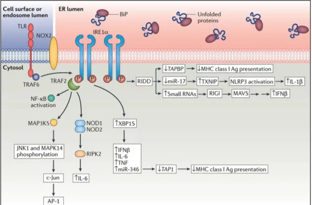
Figure 1.5. Close relation between IRE1 signaling pathways and immune response pathway ... 37
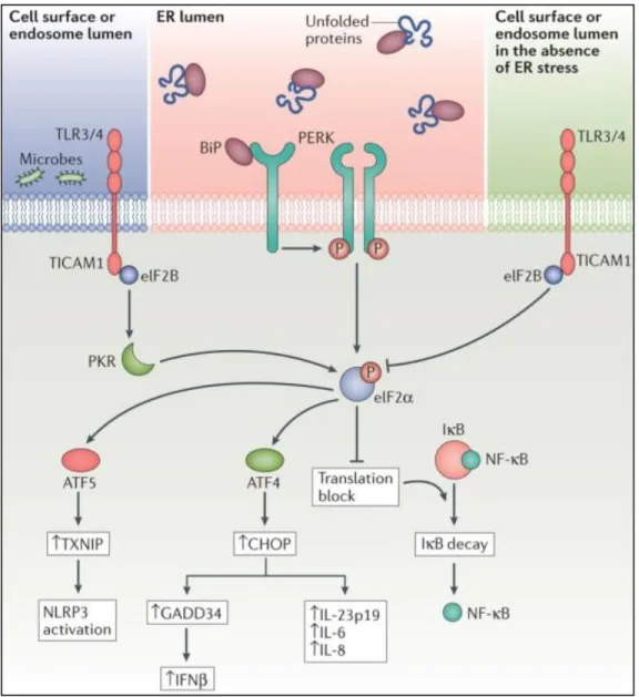
Figure 1.6. Close relation between PERK signalling pathways and immune response pathways ... 38
Figure 1.7. Progression of atherosclerosis in blood vessels ... 45
Figure 3.1. IRE1 RNase activity regulates the expression of multiple pro-atherogenic genes in BMDM cells. ... 74
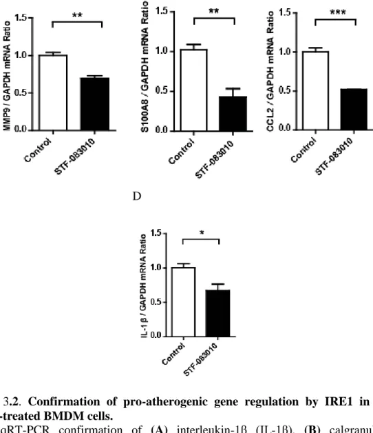
Figure 3.2. Confirmation of pro-atherogenic gene regulation by IRE1 in STF-083010-treated BMDM cells. ... 75
Figure 3.3. Confirmation of pro-atherogenic gene regulation by IRE1 in 4µ8C treated BMDM cells. ... 76
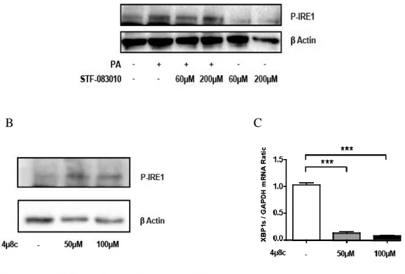
Figure 3.4. IRE1 RNase inhibitors, STF-083010 and 4µ8c, successfully inhibit IRE1 RNase function without affecting IRE1 kinase function. ... 77
Figure 3.5. Forced expressions of IRE1 and XBP1s in the IRE1-/- MEFs induce the expression of pro-atherogenic genes. ... 78
Figure 3.6. Reduction of lipid-induced IL-1β production in BMDM cells with IRE1 inhibitors. ... 80
xvi
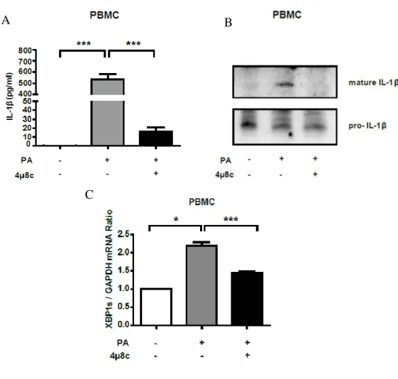
Figure 3.8. IRE1 RNase inhibition reduces IL-1β production in lipid-stressed PBMCs. ... 82
Figure 3.9. Silencing of IRE1 and XBP1s by siRNA reduces IL-1β levels in BMDM cells. ... 84
Figure 3.10. IRE1 RNase inhibitors block CCL2 production in BMDM cells. ... 85
Figure 3.11. 4µ8c treatment blocks lipid-induced CCL2 production in PBMC cells. .... 86
Figure 3.12. Silencing of IRE1 and XBP1 with siRNA leads to suppression of lipid-induced CCL2 levels in BMDMs. ... 87
Figure 3.13. IRE1 inhibition blocks lipid-induced activation and secretion of caspase- in BMDMs. ... 89
Figure 3.14. IRE1 RNase inhibition reduces NLRP3 inflammasome activation... 91
Figure 3.15. IRE1-XBP1 axis regulates mtROS production in response to lipid stress. 93
Figure 3.16. Lipid-induced upregulation of mtROS production is independent of changes in TXNIP levels or Ca2+ mobilization into the mitochondria. ... 95
Figure 3.17. IRE1 RNase activity regulates mitochondrial LONP1 protease level in BMDM cells. ... 97
Figure 3.18. Silencing of LONP1 reduces lipid-induced mtROS production in BMDM cells. ... 98
Figure 3.19. Experimental designs of STF-083010 treatment in mice ... 99
Figure 3.20. STF-083010 treatment inhibits IRE1 RNase activity in vivo. ... 101
Figure 3.21. STF-083010 treatment does not induce liver toxicity in ApoE-/- mice on Western diet. ... 102
Figure 3.22. STF-083010 treatment leads to a marked reduction in en face plaque area. ... 103
Figure 3.23. STF-083010 treatment reduces plaque area in aortic roots of ApoE-/- mice on Western diet. ... 104
xvii
Figure 3.24. STF-083010 treatment reduces macrophage population without altering apoptotic cell numbers in the proximal aortic root cryosections in ApoE-/- mice fed Western diet. ... 106
Figure 3.25. STF-083010 treatment reduces IL-1β levels in both aortic root sections and spleen tissues. ... 107
Figure 3.26. STF-083010 treatment increases collagen content in plaques without altering SMC content and increasing fibrous cap thickness in ApoE-/- mice on Western diet. ... 109
Figure 3.27. STF-083010 treatment does not affect the size of necrotic core area in ApoE-/- mice on Western diet. ... 110
Figure 3.28. STF-083010 treatment suppresses Th1 type immune response induced by hyperlipidemia in ApoE-/- mice on Western diet. ... 112
Figure 3.29. STF-083010 treatment leads to reduction in plasma IL-18 levels induced by hyperlipidemia in ApoE-/- mice. ... 113
Figure 3.30. Lipoprotein profiles do not change between STF-083010-treated and vehicle-treated (control) ApoE-/- mice on Western diet. ... 114
Figure 3.31. Experimental design of 4µ8c and DMSO treatments in ApoE
mice ... 115
Figure 3.32. 4µ8c treatment inhibits IRE1 RNase activity in vivo. ... 116
Figure 3.33. 4µ8c treatment is not toxic in liver tissues of ApoE-/-
mice on Western diet. ... 117
Figure 3.34. 4µ8c treatment leads to reduction in en face plaque area. ... 118
Figure 3.35. 4µ8c treatment reduces plaque area in aortic roots of ApoE
mice on Western diet. ... 119
Figure 3.36. 4µ8c treatment leads to a signficant reduction in IL-1β mRNA expression in bone marrow of ApoE-/- mice on Western diet. ... 120
xviii
List of Tables
Table 2.1. Chemicals, reagents, kits used for general laboratory purposes ... 57
Table 2.2. Chemicals, reagents, kits and media used in cell culture ... 59
Table 2.3. Antibodies used in study ... 60
Table 2.4. Solutions used in study ... 61
Table 2.5. Real Time PCR primers ... 66
Table 3.1. Body weight and blood glucose in treated animal groups ... 100
Table 3.2. Body weight and blood glucose in treated animal groups ... 116
Table A1. Top 25 up-regulated genes with STF-083010 treatment in mouse BMDM cells….………159
Table A2. Top 25 down-regulated genes with STF-083010 treatment in mouse BMDM cells……….161
Table A3. Down-regulated genes with stf-083010 treatment in mouse BMDM cells (P<0.05; Fold change≤ 1.5)………163
Table A4. Up-regulated genes with stf-083010 treatment in mouse BMDM cells (P<0.05; Fold change ≥ 1.5)………...170
xix
Abbreviations
Abbreviation Explanation 4µ8c 7-Hydroxy-4-methyl-2- oxo-2H-1-benzopyran-8-carboxaldehyde Ab Antibody Ag AntigenAIM absent in melanoma-2
receptors
APC Allophycocyanin
ASC apoptosis associated
speck-like protein containing a CARD ATF Activating transcription
factor
ATP Adenozin triphosphate
BM-DM Bone marrow-derived
macrophages
CARD Caspase recruitment
domains DAMPs danger-associated molecular patterns DAPI 4',6-diamidino-2-phenylindole DDX58 (RIGI)
DEAD box proteins
DMEM Dulbecco's Modified
Eagle's Medium
DNA Deoxyribonucleic acid
RNA Ribonucleic acid
ECL enhanced
chemiluminescence
EDTA Ethylenediaminetetraacet
ic acid
Abbreviation Explanation
eIF2α Eukaryotic Initiation
Factor 2
ELISA The enzyme-linked
immunosorbent assay ER Endoplasmic reticulum ERAD ER associated degradation FACS Fluorescence-activated cell sorting
FBS fetal bovine serum
FDA Food and Drug
Administration
GADD34 growth arrest and DNA
damage 34
GAPDH
glyceraldehyde-3-phosphate dehydrogenase
GSK Glycogen synthase kinase
HAC1 homolog of mammalian
XBP1 HEPES 4-(2-hydroxyethyl)-1-piperazineethanesulfonic acid i.p. Intraperitonel IFN Interferon
IPA Ingenuity Pathway
Analysis
IRE1 inositol-requiring
enzyme-1
ISR integrated signal
response
IκB inhibitor of NF-κB
xx Abbreviation Explanation
KLF Kruppel-like factor
LDL Low density lipoprotein
LP Lipoprotein
LPS Lipopolysaccharide
MAM mitochondria
associated membranes MAPK mitogen-activated protein
kinase
MAVS mitochondrial antiviral
signaling protein
MDP muramyl
dipeptidemitochondrial antiviral signaling protein
MEF Mouse fibroblast cells
MerTK MER Proto-Oncogene,
Tyrosine Kinase MHC major histocompatibility complex MMP Matrix metallaproteinase MOMA Monocyte/Macrophage Marker Antibody
MSU uric acid crystals
NBD
NF-κB Nuclear factor-kappa B
NK Naturel killer
NLR Nucleotide binding and
oligomerization domain (NOD) -like receptors
PAMP Pathogen-associated
molecular patterns PBS Phosphate buffer saline PDGF Platelet-derived growth
factor
Abbreviation Explanation
PERK protein kinase RNA-like ER kinase
PMSF phenylmethane sulfonyl
fluoride
PRRs pattern recognition
receptors
RHOD-2AM Rhod-2 Acetoxymethyl
ester
ROS Reactive oxygen
species
RPMI Roswell Park
Memorial Institute
RT-PCR Reverse transcriptase
PCR
siRNA Small interfering RNA
SMC Smooth muscle cell
STAT signal transducer and activator of transcription STF-083010 N-[(2-Hydroxy-1- naphthalenyl)methylene]-2-thiophenesulfonamide TBS Tris-buffered saline Th Helper T cell TLR Toll-like Receptor
TNF Tumor Necrosis Factor
TRAF TNF receptor associated
factor
Treg Regulatory T cells
TUDCA auroursodeoxycholic acid
TUNEL Terminal
deoxynucleotidyl transferase dUTP nick end labeling
TXNIP Thioredoxin Interacting
Protein
UPR Unfolded protein
xxi Abbreviation Explanation
UPRE unfolded protein response
element
22
Chapter 1.
Introduction
1.1. The Endoplasmic Reticulum
Endoplasmic reticulum (ER) is one of the largest organelles in the cell surrounded with a single, continuous membrane. ER is composed of interconnected branching tubules and flattened sacs. This membranous structure surrounds the nucleus and extends throughout the cytosol with dynamic tubules and sacs (1, 2). It contains multiple domains divided by their location and function. One of the major domains is juxtaposed to the nuclear envelope that separates the genetic material from the cytosol and regulates the transport of important molecules such as RNA and proteins. The other one is the peripheral ER, which extends from the nucleus to the cytosol and is comprised of cisternal sheets and dynamic tubules. Some regions of ER membrane are associated with ribosomes during protein synthesis and have been named the ‘rough’ ER. Hence, the ER regions free from ribosomes have become known as the smooth ER. There are also some ER regions very closely associated with the mitochondria and named the mitochondria associated membranes (MAMs) that play important roles in metabolic and inflammatory regulations (1, 3). Plasma membrane and secreted proteins are folded in the ER and transported to the Golgi apparatus. During this transportation, an intermediate compartment between Golgi and ER is created mainly as a continuum of the ER and contains vesicles and tubules that partake in intracellular trafficking (3). With the development of high resolution imaging techniques, the biological importance and the
23
complex architecture of ER is better understood. Also ongoing studies that aim to identify functions of new ER proteins and ER domains will generate a complete understanding of this highly important organelle (1).
Endoplasmic Reticulum Function 1.1.1.
ER has four major functions in the cell; proper protein synthesis and folding, degradation of improper or misfolded proteins, biogenesis of important lipids and regulation of Ca2+metabolism.
Protein Synthesis and Folding. ER is one of the major sites of protein synthesis in the cell. Ribosome-mRNA complexes are positioned on ER membranes after initiation of the synthesis of the polypeptide chain and the translation progresses on these ribosomes. Nascent polypeptides enter the ER through an ER membrane-spanning channel. Two types of proteins are synthesized by the ER; transmembrane proteins and soluble proteins. After translocation of proteins into the ER membrane through N-terminal signal sequences, hydrophobic residues called stop-transfer signals allow transmembrane protein folding and finally direct them to the ER membrane. Newly synthesized transmembrane proteins are then targeted into different destinations such as the plasma membrane; membrane bound organelles or ER membranes. On the other hand, proteins destined to be secreted or to remain in the lumen of membrane-bound organelles are directed into the ER lumen right after the cleavage of ER signal peptide sequence. Proper folding and many modifications occur in ER such as N-linked glycosylation, disulfide bond formation and oligomerization take place in the ER. These modifications have a crucial role in defining the intracellular destination of the proteins as well as the functions of the soluble proteins (2, 4). However, many proteins can be misfolded and cannot reach their correct 3 dimensional conformations. Hence, they remain in the ER as insoluble aggregates. These misfolded proteins can be cleared with the help of an ER associated degradation (ERAD) pathway (5).
24
ER-Associated Degradation ER has an elaborative quality-control system that can detect improperly processed or misfolded proteins within the ER lumen. If for any reason the proteins cannot achieve their functional forms, they are translocated to the cytosol for degradation through ER-associated degradation (ERAD) pathway. These proteins are de-glycosylated, ubiquitinylated and finally, degraded by the proteasome. The ERAD pathway thus provides an efficient, selective waste removal, so it can be viewed as a quality-control process (5).
Lipid Biogenesis In addition to its role in protein secretion, ER plays an important part in membrane lipid biogenesis. ER and Golgi can together form an endomembrane compartment that provides a special site for lipid synthesis. Lipids are transferred to the regions near Golgi on ER and exposed to modifications there (3). Especially the synthesis of phospholipids and cholesterol takes place on the ER and contributes to new cell membrane production. The first step of lipid bilayer enlargement begins with the addition of two fatty acids to glycerol phosphate to obtain phosphatidic acid and occurs on the cytosolic leaflet of the ER membrane. The following steps, that consist of determining the chemical nature and the addition of the head groups to lipids, are performed in endomembrane compartments that are formed together with ER and Golgi (3). Furthermore, ER-mitochondria contact sites are also enriched with some important enzymes that have a role in some lipid biosynthesis pathways. Especially, both ER and mitochondria are required for the synthesis of phosphatidylethanolamine (PE) and phosphatidylcholine (PC), the most abundant phospholipids of the cell. Hence contact sites of these two organelles allow lipid biosynthesis and lipid transfer between these organelles (6, 7).
In addition to its role in lipid bilayer biogenesis and assembly, ER can control transcriptional regulation of lipid metabolism-related genes. Under certain conditions, inactive transcription factors that are found on the ER are translocated first to the Golgi
25
for processing and maturation and then to the nucleus to initiate the expression of the genes required for lipid biosynthesis and uptake (8-10). For example SREBP-1c and SREBP-2 which are the key transcription factors in hepatic lipogenesis and cholesterol biosynthetic pathways are normally localized to the ER and sense lipid changes on the ER (9).
Ca2+ Metabolism ER also plays a central role as an intracellular Ca2+ store (11). Calcium concentrations in the cytosol and extracellular compartments are maintained tightly (~100 nM and is ~2 mM respectively). The ER lumen Ca2+ concentration changes
between ~100-800 M.(12) This high amount of ER calcium concentration, compared to
cytosol calcium concentrations, clearly shows the pivotal role of ER as a major storage for Ca2+. There are several calcium channels, ryanodine receptors and inositol 1, 4, 5-trisphosphate (IP3) receptors (IP3R) on ER and all of them regulate the transfer of Ca2+ from ER to cytosol when needed (2, 12). Calcium is a very important molecule that can regulate a wide variety of processes from protein localization, function and association (with other proteins, organelles or nucleic acids) and the reshaping of ER under certain conditions (such as physiological responses to neurotransmitter release and muscle contraction) (2, 13). Furthermore, changes in Ca2+ concentrations or Ca2+ transfer between cytosol and mitochondria have a crucial role in metabolic regulation. For example Ca2+ oscillations can regulate mitochondrial metabolism such as ATP production and mitochondrial permeability so regulate cell death. Also release of Ca2+ from ER can regulate redox state of mitochondrial chaperones (14-16). The Ca2+ transfer from ER to mitochondria deserves special emphasis and will be mentioned in a later section of this thesis.
Endoplasmic Reticulum Stress and UPR 1.1.2.
Most secretory and transmembrane proteins that enter the ER lumen as nascent unfolded chains fold and mature by ER resident chaperones, foldases, and cofactor proteins (4).
26
Generally, a small portion of proteins cannot be folded properly due to highly complex stochastic processes in the ER (17, 18). In those cases, the unfolded proteins are detected in the ER and initiate a highly efficient response known as ERAD that results in their removal (17). Many environmental and physiological factors such as increased rate of protein synthesis, genetic abnormalities, depletion of ATP, oxidative stress, viral infection, increased temperature, alterations in Ca2+ level and exposure to excess cholesterol or fatty acids (such as in obesity and dyslipidemia) and aging can lead to disturbances in ER functions, leading to increases in the accumulation of misfolded proteins that exceed ER folding capacity (18, 19). Hence, in such conditions, ER homeostasis is disturbed, a situation known ER stress. To cope with ER stress, cells provoke a strong adaptive response called as unfolded protein response (UPR) or the ER stress response (20, 21). UPR is a collection of signaling networks that include a wide variety of transcriptional and translational events mediated by three transmembrane ER stress sensors; inositol-requiring enzyme-1 (IRE1), the protein kinase RNA (PKR)-like ER kinase (PERK), and the activating transcription factor 6 (ATF6) (Figure 1.1). Under ER stress conditions, chaperones that normally associate with UPR sensors dissociate to engage with unfolded proteins, leading to the activation of UPR sensors and downstream signaling. Depending on the duration or the dose of the stressors, ER activates transcriptional or translational processes either to reestablish homeostasis in the cell or to initiate apoptotic pathways (20, 22, 23).
27
Figure 1.1. Three main branches of unfolded protein response
1.2. Unfolded Protein Response Signaling
Unfolded protein response is mediated by three major stress sensors located on the ER membranes; IRE1, PERK and ATF6 in response to certain environmental or cellular stress conditions.
IRE1 1.2.1.
IRE1 is the first identified and major ER stress sensor located on ER membranes. It is highly conserved from yeast to mammalian cells. The metazoan IRE1 has two homologues, IRE1α and IRE1β. IRE1α is expressed in all cells and tissues, whereas IRE1β is expressed only in the gastrointestinal and respiratory tracts. The function of
28
IRE1β is still unknown and UPR signaling is mainly mediated through IRE1α (24, 25) IRE1 molecules include 3 important domains; a luminal domain, a transmembrane domain and a cytosolic domain that contains two enzymatic activities, namely endoribonuclease (RNase) and kinase. When IRE1’s luminal domain senses the unfolded proteins, this leads to autophosphorylation by IRE1’s kinase domain and oligomerization. Activation of its kinase domain further activates its RNase domain. (Figure 1.2). Through its RNase and kinase domains, IRE1 contributes to re-establishing ER homeostasis. IRE1 and its targets, mainly XBP1 transcription factor, can perform a wide variety of functions in the cell (26). Next, I will present a review of the functions of IRE1 protein in two categorizes; RNase domain functions and kinase domain functions.
29
RNase Domain Function After activation, IRE1α can initiate a wide variety of signaling pathways ranging from pro-survival to apoptotic and depending on the nature of the stimuli (27). The best known RNase substrate of IRE1α is the mRNA encoding X-box binding protein 1 (XBP1). Pre-mature XBP1 (U) mRNA exists in a specific stem loop structure in the cytosol. Active IRE1 can recognize this stem loop structure and through an unconventional splicing event, remove 26 nucleotides in the intron of XBP1 (U). This spliced XBP1(S) produces a larger and active protein due to a frame-shift (28). XBP1 protein contains both a DNA binding domain and a transcriptional activation domain. This mature form of XBP1(S), by binding to the unfolded protein response element (UPRE) regions, regulates the transcription of UPR target genes such as chaperones, protein disulfide isomerases (PDIs) and the components of ERAD pathway (29, 30). In addition, XBP1 can regulate ER’s membrane expansion by increasing the transcription of genes involved in phospholipid biosynthesis (31). All of these XBP1 functions show that XBP1 is an important molecule in relieving stress in the ER stress.
Furthermore, there are factors and ways that affect XBP1 activity. For example, the stability of XBP1(S) is increased by binding of small ubiquitin-like modifier (SUMO) ubiquitin-conjugating enzyme 9 (UBC9) in the cytosol (32). On the other hand XBP (U) translation can be paused due to a peptide motif on its carboxyl terminus, and this makes it more susceptible to splicing by IRE1 (33). Under unstressed conditions, XBP1(S) forms a complex with XBP1 (U) protein, and then undergoes proteasomal degradation (34).
IRE1 also has another function to degrade specific mRNAs that are associated with ribosome on ER membrane for translation. By degrading these mRNAs, IRE1 tries to reduce protein load under stress conditions (35, 36). IRE1 can specifically target 5’-UGCU-3’ sequence in the stem loop structures of mRNAs. However, there are some mammalian regulated IRE1-dependent decay (RIDD) target genes that do not contain this consensus sequence. Basal RIDD activity is needed for cellular homeostasis and cell recovery; however, sustained activation of RIDD is associated with the induction of
30
apoptotic pathways (20, 37). RIDD activity has been shown to selectively degrade some microRNAs that can repress apoptotic pathways. Increasing RIDD activity leads to apoptosis through degradation of premature mir-17, specifically targeting caspase-2 mRNA. Caspase 2 cleaves the pro-apoptotic BH3 only protein BH3 interacting domain death agonist (BID) that activates BCL2 associated X, apoptosis regulator (BAX) and BCL2 antagonist/killer 1 (BAK) (38).
Kinase Domain Function To date, IRE1 is only known to phosphorylate itself (39). Through its kinase domain IRE1 can associate with some adaptor proteins to regulate different pathways. For example, TNF receptor associated factor 2 (TRAF2) binds to phosphorylated IRE1 and initiates the mitogen-activated protein kinase (MAPK) pathway to regulate inflammatory and apoptotic pathways (40). Also phosphorylated IRE1 can activate NF-κB via binding with inhibitor of NF-κB (IκB) kinase (41).
PERK 1.2.2.
PERK is another major ER stress sensor located on the ER membranes. It is composed of luminal (stress sensing), transmembrane and cytoplasmic (kinase) domains. PERK can autophosphorylate and dimerize under ER stress conditions (42). The best known substrate of PERK is eukaryotic translation initiation factor 2α (eIF2α). Phosphorylated eIF2α leads to general inhibition of translation of capped mRNAs in the cell and blocks further accumulation of unfolded proteins in the ER (42, 43). However, during ER stress there is a small subset of mRNAs that are preferentially translated. For example, activating transcription factor 4 (ATF4), preferentially translated during ER stress, is an important transcriptional factor, which can regulate the expression of genes required for amino acid import, metabolism and resistance to oxidative stress. PERK-ATF4 signaling is thought to play an important role in cytoprotection at the early stage of ER stress. However, if ER stress is irremediable or severe, ATF4 can also induce the transcription of C/EBP-homologous protein (CHOP), a transcription factor, which drives the stressed
31
cells to apoptosis (42, 44). CHOP leads to the activation of apoptotic pathways by inhibiting B cell leukemia 2 (BCL2) family members. On the other hand CHOP has a role in the dephosphorylation of eIF2α by upregulating the expression of protein phosphatase 1 regulatory subunit 15A (PP1R15A (also known as growth arrest and DNA damage 34 (GADD34). PP1R15A removes the blockage on protein translation and stimulates the translation of proteins necessary to reinstate homeostasis in the ER (45). Also, another PERK substrate is NF-κB repressing factor B (NRF2), which regulates transcription of antioxidant proteins and helps establish ER homeostasis (46). Finally, PERK can activate NF-κB and regulate inflammatory pathways (41). Thus, PERK performs opposing functions of cytoprotection, apoptosis and inflammation under specific cellular conditions.
ATF6 1.2.3.
ATF6, an ER transmembrane protein with a transcription factor function, controls the third arm of the UPR. ATF6α and ATF6β are two homologous proteins. They are transmembrane protein containing multiple domains with different functions; luminal BIP-associated domain, cytosolic domain with bZIP motif, and transmembrane domain with a Golgi target sequence (47, 48). Under stress conditions, ATF6 dissociates from the ER membrane and is transported to the Golgi via coat protein complex II (COPII)-containing vesicles. The cytoplasmic domain of ATF6 is phosphorylated and cleaved by membrane-bound transcription factor site 1 and site 2 proteases (S1P and S2P, respectively). This process is called regulated intramembrane proteolysis. After cleavage of its luminal domain by SP1 and transmembrane domain by SP2, the phosphorylated N-terminal domain of ATF6 (N) is released from Golgi and activates the expression of UPR target genes like ATF4 and XBP1(S) (28, 49, 50). These target genes contribute to ER quality control, protein folding, secretion, ER expansion, chaperone synthesis and ERAD (51-54). ATF6 together with ATF4 and XBP1(S) can transcriptionally regulate overlapping genes and modulate ER stress response or initiation of cell death pathways under different ER stress conditions (55). Some studies showed that ATF6 could play a
32
role in cell protection. For example, mice lacking Atf6 displayed increased susceptibility to ER stress induced cell death and also embryonic lethality was observed when both Atf6Atf6are deleted (51, 52, 56).
On the other hand, ATF6 also plays a role in the regulation of NF-κB under special toxin induced ER stress conditions that depend on mTOR related dephosphorylation of Protein Kinase B (AKT) protein (57). However, the relationship between ATF6 and inflammatory response needs further investigation.
1.3. Endoplasmic Reticulum Stress and Immune Response
Figure 1.3. Evolution of unfolded protein response proteins
The IRE1 branch of the UPR is the only arm that is conserved from yeast to mammals. (Figure1.3). Saccharomyces cerevisiae IRE1 also has the conserved endoribonuclease
33
activity that splices HAC1 (homolog of mammalian XBP1) mRNA. This signaling controls the transcriptional response to regulate the folding capacity of ER in yeast. On the other hand Schizosaccharomyces pombe IRE1’s endoribonuclease activity only performs the RIDD function, because there is no HAC1 coding gene in this organism (58-60). In pathogenic fungi, UPR has a role in adapting the fungi to its host’s microenvironment by regulating pathogen virulence through the secreted toxic compounds (61).
In plants, on the other hand, ATF6 and IRE1 dominate the UPR, because there is no PERK protein. Here, a more prominent RIDD activity of IRE1 tries to compensate for the missing PERK-regulated translational control. UPR activation controls protein load during seed development and under abiotic stress conditions such as salt or heat shock stress (58, 62, 63). Furthermore, IRE1 mounts an antibacterial defense by inducing the secreted foldases that function in protein folding as well as secreted hydrolases (64).
Caenorhabditis elegans has all three sensors of the UPR (65). The IRE1-XBP1 axis serves to sustain larval survival under pathogen threat. IRE1 arm does not directly control infection; instead it reinforces ER’s functions involved in adaptive immunity against the detrimental pathogen. Also XBP1 resolves ER toxicity that is caused by severe inflammatory conditions (66).
In metazoans, there is a more complex UPR pathway with three sensors. It has close-relationship with other stress-controlled translational regulatory pathways known as the integrated signal response (ISR). Bacterial or viral infections can activate UPR or ISR pathways as a host defense (59, 67).
Initially, UPR activation aims to resolve ER stress, but under prolonged ER stress conditions UPR can activate many pro-apoptotic and inflammatory pathways. Chronic exposure to metabolic stress factors such as excess of lipids, glucose or inflammatory
34
agents such as cytokines can trigger or sustain UPR activity in many cell types, but especially in cells specialized for the secretion of large amounts of proteins (such as pancreatic beta cells, adipocytes, oligodendrocytes) and immune factors (such as B cells and macrophages) (68-73). Many metabolic stress factors affect not only the ER but also a closely associated organelle, the mitochondria, inducing UPR, oxidative stress and inflammation (74, 75). How ER and mitochondria are coupled in stress is an area of intense research. For example, ER stress-induced calcium transfer from the ER to the mitochondria can induce reactive oxygen species (ROS) production from the latter organelle (76). These organelles also exchange ROS, lipids and other molecules that may synchronize their response to the stressor (76, 77). Activation of ER stress by metabolic stress as in the case of obesity and dyslipidemia, and consequent induction of inflammation can also contribute to impairment of cellular and systemic lipid and glucose metabolism, resulting in cell death or dysfunction, insulin resistance and diabetes (73, 78) (Figure 1.4). This vicious loop between ER stress and inflammation can accelerate the progression of many metabolic diseases, such as atherosclerosis, obesity, type 2 diabetes and neurodegenerative diseases (79-82).
35
Figure 1.4. Connection between ER stress, metabolic diseases and inflammation
UPR Connection with Inflammatory Pathways 1.3.1.
The connection between UPR and inflammation underlies many complex diseases. Environmentally-induced or genetic defects in the UPR branches have been associated with inflammatory phenotypes such as inflammatory bovine disease, lung respiratory disease, type 1 diabetes or other metabolic diseases (58, 83). Inflammatory pathways such as the downstream of the Toll-like receptor (TLR) and stress kinase signaling pathways share similar consequences with the UPR such as the induction of reactive oxygen species (ROS) and activation of the nuclear factor-κB (NF-κB) (84-86). NF-κB is an important transcription factor in inflammation (87). It is normally found in an inactive state and associated with its inhibitor (IκB) in the cytosol. With stimulation, the phosphorylation of IκB by a kinase IKK triggers its release from the inhibitory protein and translocation into the nuclei, where it initiates the transcription of pro-inflammatory
36
genes (88). ER stress can also lead to NF-κB activation (89). All branches of UPR have been implicated in the regulation of NF-κB. The PERK activation leads to translation inhibition of IκB, which is constitutively expressed in cells under normal conditions and is rapidly degraded under inflammatory stimuli (90). IRE1 activation indirectly leads to IκB phosphorylation. The TNF receptor-associated factor 2 (TRAF2) is recruited to the phosphorylated domain of IRE1, followed by the recruitment of IκB kinase (IKK). Here, IKK phosphorylates inhibitory IκB leading to its degradation. Thus NF-κB dissociates from its inhibitory partner and becomes active. On the other hand, ATF6 plays a role in NF-κB activation through its impact on the mTOR-AKT pathway. Silencing of ATF6 in liver immune cells reduced immune response that was caused by ER stress through the alleviation of NF-κB activation and restoration of AKT pathway. Nonetheless more detailed studies on this subject are needed (57, 91) .
In addition to their roles in NF-κB activation, IRE1 and PERK can coordinate inflammatory responses by many other ways. For example, IRE1-TRAF2 association can regulate production of interleukin-6 (IL-6). IL6 is an important cytokine dependent on the binding of TRAF2 protein to oligomerization domain (NOD)-containing proteins 1 and 2 (NOD1 and NOD2). That association can activate NF-ҡB dependent IL6 expression (92). Also IRE1-TRAF2 association activates mitogen-activated protein kinase (MAPK) kinase 5 (MAP3K5). MAP3K5 then phosphorylates and activates MAPK8 (or JUN N-terminal kinase (JNK1) and MAPK14 (93, 94). Subsequently, JNK1 phosphorylates and activates Activator protein 1 (AP-1) that is an important transcription factor. Additionally, IRE1 through XBP1, can regulate the expression of several pro-inflammatory cytokines such as interleukin 6 (IL-6), tumor-necrosis factor-α (TNFand interferon β (IFNβ) (95, 96). Furthermore, an XBP1-induced miRNA (miR)-346 can regulate major histocompatibility complex (MHC) class I Ag presentation. This microRNA targets mRNAs encoding the transporter of antigenic peptides-1 (TAP1) also called ATP-binding cassette (ABC) transporter, and human leukocyte antigen (HLA) class I proteins. These targets have important roles in antigen presentation in adaptive immune response and transport of peptides to ER respectively
37
(97). In addition IRE1’s RIDD function plays a role in some inflammatory pathways. RIDD activity can degrade TAP binding protein (TAPBP) mRNA and reduce antigen presentation (98). Small RNAs that are produced by RIDD activity can also induce IFNβ production through activation of ATP-dependent RNA helicase DDX58 (DEAD box proteins or RIGI) or mitochondrial antiviral signaling protein (MAVS) (99).
Figure 1.5. Close relation between IRE1 signaling pathways and immune response pathway
Furthermore, CHOP, induced through PERK-ATF4 signaling, can also contribute to IFNβ production. CHOP is also involved in ER stress-induced IL-23 expression in dendritic cells (84, 100).
38
Figure 1.6. Close relation between PERK signalling pathways and immune response pathways
UPR Connection with Inflammasome Activation 1.3.2.
Inflammasome Background. Pathogen-associated molecular patterns (PAMPs) and danger-associated molecular patterns (DAMPs) are recognized by germline encoded pattern recognition receptors (PPRs). The PPRs recognize a variety of stimuli and constitute an essential component of the innate immune response that controls the clearing of pathogens or damaged cells (101-103). The PRRs are expressed by cells that
39
serve in the immune system including macrophages, monocytes, dendritic cells, neutrophils, epithelial cells and also in airway smooth muscle (ASM) cells. Based on their genetic and functional features, PPRs can be classified into different classes. Nucleotide binding and oligomerization domain (NOD) -like receptors (NLRs), absent in melanoma-2 receptors (AIMs) and recently identified pyrin are the main PPRs that can lead to the assembly of the multi-protein inflammatory complexes, i.e. inflammasomes, in the cytosol (104, 105).
NLRs recognize both PAMPs and DAMPs. NLRs contain 3 main domains; an N terminal effector domain, a C-terminal leucine-rich sensor domain and a central nucleotide-binding and oligomerization domain (NBD). Based on the N-terminal domain, NLRs can be further subdivided. If the N-terminal domain includes a pyrin domain, it is called NLRP. Likewise, if it includes a caspase activation and recruitment domain (CARD), it is called NLRC. NLRs can activate the inflammasome multiprotein complex and induce an inflammatory response against cellular infection or tissue damage (104, 105). NLRP1, NLRP3 and NLRC4 are well-documented members of NLRs that assemble into an inflammasome complex in the cytosol. After the assembly of a multimeric inflammasome complex, zymogen pro-caspase-1 is recruited and cleaved. Following which, cleaved, mature caspase-1 can proteolytically process pro-IL-1β and IL-18 cytokines and induce inflammatory cell death. This pathway significantly differs from apoptosis in that it involves caspase-1-mediated cell swelling and lysis that results in the loss of membrane integrity (106-109). Bioactive IL-1β and IL-18 cytokines have important roles in both innate and adaptive inflammatory responses (108, 110, 111). They can stimulate further secretion of pro-inflammatory cytokines, leading to recruitment of more inflammatory cells to the inflamed region. These cytokines also regulate T helper (Th) cell polarization especially towards to Th1 and Th17 subtypes (110). Different NLRs are found at different locations in the cell and can be activated by different stimuli. This difference in localization can result from some tissue specific co-factors or different splicing variants of molecules that need to be studied further (112). For example, even though NLRP1 is a cytosolic receptor, it is also active in the nucleus
40
of neurons and lymphocytes (112). AIM2 was first defined as an IFNγ inducible protein that can oligomerize similarly to the other NLRs and form an inflammasome complex, however it was discovered that AIM2 can directly bind to nucleic acids in the cytoplasm via its hematopoietic IFN-inducible nuclear proteins with 200-amino acids (HIN200) domain and oligomerize via its nucleotide-binding and oligomerization (NACHT) domain. Also, the PTD domain of AIM2 interacts with apoptosis-associated speck-like protein containing a CARD (ASC) adaptor protein and then recruits caspase-1 zymogen for activation like the other inflammasomes (113). Bacterial ligands such as muramyl dipeptide (MDP) or anthrax lethal toxin produced by Bacillus anthrax activate the NLRP1 inflammasome (114). NLRC4 serves in immune defense but not by directly binding to its pathogen activators. It can be induced by a family of proteins called NLR family, apoptosis inhibitory proteins (NAIPs). NAIPs can sense their ligands and activate NLRC4. Traditional activators of NLRC4 inflammasome are bacterial flagellin or multiple proteins that belong to bacterial type 3 secretion systems (115, 116). NLRP3 is expressed by wide a variety of cell types such as granulocytes, monocytes, macrophages, dendritic cells, T and B cells, epithelial cells, osteoblasts, fibroblasts and melanoma cells. It is also activated by a broad array of ligands. NLRP3 triggers can be categorized as pathogen-associated ligands (such as cell wall components of bacteria (peptidoglycans), bacterial and viral nucleic acids, microbial toxins, nigericin (Streptomyces hygroscopicus), aerolysin (Aeromonas hydrophila), maitotoxin (Marine dinoflagellates), gramicidin (Bacillus brevis) and -toxin (Staphylococcus aureus), environmental crystalline pollutants (such as silica, asbestos, and alum crystals), and danger-associated molecules such as ATP, serum amyloid, uric acid crystals (MSU), and ROS (117) . Also, some metabolic products such as saturated free fatty acids also lead to NLRP3 inflammasome activation (118). In experimental conditions, the NLRP3 inflammasome activation requires a priming step that leads to an increase in NLRP3 and pro-IL-1β expression levels in the cell through activation of the NF-κB pathway. For example, Lipopolysaccharide (LPS) in the cell wall components of gram-positive bacteria can provide this priming cue to the cell via binding TLR4 receptors and activating the NF-κB pathway (105, 119). After priming, there are common signaling events that are shared by the various triggers that activate NLRP3 inflammasome such as
41
induction of potassium (K) efflux through the ATP-gated ion channels, increase in cellular calcium levels, and increased release of ROS, DNA and cardiolipin from the mitochondria, which can be sensed by the NLRP3 resulting in its translocation from the mitochondria associated ER membranes to the cytosol (120-124). There is still a debate about the importance of these mechanisms for NLRP3 activation, because not all of these signaling events can be activated by all NLRP3 agonists.
ER Stress and Inflammasome Relationship ER stress and UPR activation are observed in obesity-induced fatty liver diseases as well as alcohol-induced liver injury. Also, inflammasome activation has been shown to contribute to the pathogenesis of the same diseases (125, 126). Emerging evidence points to a strong relationship between ER stress and inflammasome activation in liver diseases. A chemical chaperone, Tauroursodeoxycholic acid (TUDCA)-treated, genetically obese mice (ob/ob), challenged with tunicamycin to promote ER stress-associated liver injury and severe steatosis, displayed reduced signs of ER stress and inflammasome activation (125, 126). ER stress-induced progression of liver steatosis was attenuated when caspase-1 was silenced in these mice (125). Although these studies mainly focused on liver disease, they clearly showed that ER stress induced liver steatosis through the activation of the inflammasome. However, the pathways that link ER stress and inflammasome activation remain to be elucidated. For example, Tschopp’s group suggested a new mechanism that links ER stress and inflammasome activation independent of classical UPR pathways but dependent on reactive oxygen species production and potassium efflux induction (127).
Another recent observation related to inflammasome activation, stated that UPR-induced thioredoxin-interacting protein (TXNIP) protein which could serve as a functional link between ER stress and inflammasome. TXNIP was discovered to be the top glucose-regulated gene in pancreatic cells and its expression increased in diabetes (128). Recently, TXNIP was shown to play a role in cellular apoptosis and insulin production. Mainly through interaction with thioredoxin in mitochondria, TXNIP regulates the redox state. All these functions make TXNIP a good candidate for therapy of diabetes (129,
42
130). Interestingly, in the study conducted by Papa et al., TXNIP levels were found to be upregulated under ER stress conditions and this effect depended on IRE1 RIDD activity. IRE1 RIDD activity can degrade the microRNA mir17, which specifically targets and suppresses TXNIP expression. TXNIP can also bind to and activate NLRP3 inflammasome components. Thus it was suggested that IRE1 could regulate inflammasome activation through TXNIP upregulation (131). Furthermore, under ER stress conditions caused by direct ER stress inducers, PERK activation also resulted in transcriptional upregulation of TXNIP. Increased TXNIP resulted in the production of mitochondrial ROS species and NLRP3 activation (132, 133). There is also another study binding IRE1 with inflammasome activation through TXNIP regulation. In this study, as a result of IRE1α-TXNIP signaling, TXNIP led to mitochondrial damage and TXNIP activated caspase-2 and Bid proteins found in mitochondria, further leading to the release of mitochondrial DNAs. Released DNA bound and activated NLRP3 inflammasome (134). On the contrary, Lebeaupin et al. showed that in obese mice that were challenged with LPS and tunicamycin, both separately and together, no changes in TXNIP levels occurred in liver even though ER stress led to NLRP3 inflammasome activation (126). Among the other actors that have been described with the aim of elucidating the link between ER stress and inflammasome activation is glycogen synthase kinase 3 beta (GSK-3β). A previous study showed that GSK-3β could be activated by tunicamycin and regulated IL-1β and TNF-α transcription in an IRE1-dependent manner (135). Based on these studies, ER stress relation with inflammasome activation mainly depends on NLRP3 inflammasome formation. There is also a study showing that IRE1 and PERK can regulate NLRP1 gene transcription, which is dependent on ATF4 (136). More studies are needed to clearly delineate the molecular relation between ER stress and inflammasome complexes under ER stress conditions induced through different stressors, but especially metabolic inducers.
43
1.4. Atherosclerosis
The global, epidemic rise in obesity and associated diseases such as insulin resistance, type 2 diabetes, fatty liver disease, and dyslipidemia are also major risk factors for atherosclerotic vascular disease (82, 137). These diseases present a major threat to public health and account for one third of deaths worldwide every year (138). The term arteriosclerosis was first introduced by Jean Lobstein, a surgeon and pathologist, in 1829 (139). Then, pathologists tied inflammatory changes to the developing atherosclerotic plaques. While Rokitansky claimed that mechanical injury and toxins are the main reasons of atherosclerosis and the following inflammation. Rudolf Virchow described for the first time the primary role that inflammation plays in atherogenesis (140, 141). In 1920, Windaus showed that cholesterol crystals and calcified connective tissue are prominent features of atherosclerotic plaques, and further studies by Anitschkow and Chaltow supported these observations by showing that atherosclerosis is indeed induced by feeding rabbits with a high cholesterol-rich diet (142, 143). Thus, these researchers identified cholesterol as one of the possible risk factors in atherosclerosis. And, in later years these atherosclerosis risk factors were expanded to include smoking, high blood pressure, high emotional stress, improper diet, diabetes and certain infections (144).
In 1993, Ross summarized in his “response to injury hypothesis” that mechanical injury, toxins and free radicals led to changes in endothelium and the initiation of atherosclerosis (145). Alternative theories by Steinberg and others postulated the “altered lipoprotein hypothesis” and “retention of modified LDL hypothesis”. In their explanations, these researchers emphasized that the retention and modification of lipoprotein in the intima of the arterial wall attracted and activated monocytes and smooth muscle cells that formed the lesions (146). However, the genetics of atherosclerosis show that it is a far more complex metabolic disease and its pathogenesis cannot be restricted simply to a lipid deposition problem (144).
44
Initiation and Progression of Atherosclerosis 1.4.1.
Atherosclerosis is initiated with the accumulation of apoliprotein B (ApoB)-containing lipoproteins (LPs) in the intima region of arterial walls and especially in arterial branches where there is a disturbed blood flow. The core regions of ApoB-LPs are composed of neutral lipids such as triglycerides and cholesteryl fatty acyl esters and this core is surrounded by a phospholipid monolayer and proteins (147). These lipoproteins are synthesized in the liver as very low density lipoprotein (VLDL). These are converted into more dense ApoB-LPs called low density lipoproteins (LDL) with the addition of triacylglycerol in the liver which are atherogenic particles retained in the arterial intima regions (148). In arterial branching points where there is a disturbed laminal flow, these LPs are deposited on the arterial wall and transformed into various oxidized forms. After these oxidative modifications on the LPs, they behave more like a DAMP in the sense that they can activate a persistent, low-grade inflammation. With the activation of endothelial cells lining the arteries begins the recruitment of monocytes into sub-endothelial intima regions. Also, the inflammatory response induced in the plaque activates the plaque-resident smooth muscle cells (SMCs) as well as to the recruitment of other inflammatory cells such as T cells B cells, dendritic cells and mast cells (149, 150).
During this establishment period of atherosclerosis, cellular, extracellular and lipid materials accumulate in the sub-endothelial regions. After the establishment of lesions, as a protective mechanism, a fibrous cap forms over the lesions, providing a barrier between blood platelets and pro-thrombotic material in the lesions. Also, this measure helps maintain the continuity of the luminal blood flow by preventing plaque rupture. On the other hand, some types of lesions over time trigger thrombus formations and lead to serious occlusions and complications. This type of plaques is called ‘vulnerable plaques’. They usually display a lipid-filled necrotic core due to increased accumulation of apoptotic cells resulting from defective phagocytic clearance. In those regions the
45
fibrous cap thickness is noted to be thinner and over time the lesion’s stability decrease making it prone to rupture (151-153) (Figure 1.7).
Figure 1.7. Progression of atherosclerosis in blood vessels
During atherosclerosis progression from initiation to rupture, three main cell types contribute; endothelial cells, intimal SMCs, and inflammatory cells (mainly macrophages). These cells contribute both to the progression of disease (vulnerable plaques) and eventually to rupture (154).
Endothelial cells (ECs) Atherosclerosis development occurs in a nonrandom pattern because lesions are typically located around the branched or curved areas of the blood vessels. The atherosclerosis-susceptible areas display low time-average shear stress, high oscillatory shear index and spatial gradient in shear stress. The unbranched arteries that are more protected against these forces, or exposed to uniform laminar flow, rarely develop atherosclerosis. There are observed structural, molecular and functional differences between the endothelial cells of atherosclerosis-susceptible and atherosclerosis-resistant regions of the arteries. While endothelial cells of atherosclerosis-susceptible regions have a cuboidal morphology, the endothelial cells in the atherosclerosis-resistant regions align coaxially along with the laminal flow direction and have an ellipsoidal shape. Also, the latter secrete more glycocalyx. In the branched