The findings of this study were presented as an abstract at the 17th Annual Meeting of the European Association of Echocardiography, EUROECHO 2013 in Istanbul.
Address for correspondence: Dr. Sadettin Selçuk Baysal, Şanlıurfa Mehmet Akif İnan Eğitim Araştırma Hastanesi, Esentepe 63300, Şanlıurfa-Türkiye
Phone: +90 414 318 60 00 Fax: +90 414 318 68 12 E-mail: [email protected] Accepted Date: 06.06.2016 Available Online Date: 05.09.2016
©Copyright 2017 by Turkish Society of Cardiology - Available online at www.anatoljcardiol.com DOI:10.14744/AnatolJCardiol.2016.7035
Sadettin Selçuk Baysal, Bahar Pirat, Kaan Okyay, Uğur Abbas Bal, Melek Zekiye Uluçam,
Derya Öztuna
1, Haldun Müderrisoğlu
Department of Cardiology, Faculty of Medicine, Başkent University; Ankara-Turkey
1Department of Biostatistics, Faculty of Medicine, Ankara University; Ankara-Turkey
Treatment-associated change in apelin concentration in patients with
hypertension and its relationship with left ventricular diastolic function
Introduction
Apelin is an endogenous peptide first isolated from bovine stom-ach extracts, and is reported to have a variety of functions in the cardiovascular system. Apelin has been identified as endogenous ligand for the G-protein coupled receptor apelin receptor (APJ), which has structural similarities with the angiotensin II type 1 re-ceptor (1, 2). It has been shown that the apelin/APJ system plays a role in the pathophysiology of atherosclerosis, coronary artery dis-ease, heart failure, hypertension, and pulmonary hypertension (3). It has been reported that plasma apelin concentration is decreased in patients with hypertension (4, 5). The apelin/APJ pathway influ-ences blood pressure by an endothelium-dependent, nitric oxide (NO)-mediated mechanism that causes arterial vasodilatation (6).
The relationship between apelin concentration and left ven-tricular (LV) diastolic function in patients with essential
hyper-tension is not completely understood, although recently it has been reported that circulating apelin concentration is associ-ated with LV systolic and diastolic impairment (5).
We hypothesized that plasma apelin concentration would be lower in patients with untreated arterial hypertension than pa-tients with normal blood pressure and that apelin concentration would rise in response to effective treatment of hypertension independent of the drug choice. We also sought to examine the relationship between apelin concentration and LV diastolic func-tion before and after treatment.
In order to achieve these goals, we 1) compared the plasma apelin concentration of patients with hypertension and healthy controls; 2) examined the change in plasma apelin concentration in patients with newly diagnosed hypertension after 1 month of drug therapy, and compared the effect of two anti-hypertensive drug regimens on apelin concentration (telmisartan or
amlodip-Objective: We examined the change in apelin concentration and its relationship with left ventricular diastolic function in patients treated for hypertension.
Methods: Ninety treatment-naive patients with newly diagnosed hypertension and 33 age- and sex-matched control subjects were prospectively enrolled. Patients with hypertension were randomized to treatment either with telmisartan 80 mg or amlodipine 10 mg. Apelin concentration was measured and echocardiography was performed at baseline and after 1 month of treatment.
Results: The data of 77 patients and 33 controls were analyzed. Mean age, gender, baseline blood pressure, apelin levels, and echocardiographic measurements were similar between the treatment groups (p>0.05 for all). Apelin concentration was significantly lower in patients with hyper-tension than in controls. There was a significant increase in apelin level after 1 month of treatment in both groups (0.32±0.17 vs. 0.38±0.17 ng/dL in telmisartan group, p=0.009, and 0.27±0.13 vs. 0.34±0.18 ng/dL in amlodipine group, p=0.013). Diastolic function improved significantly in both groups (p<0.05) but was not significantly associated with change in apelin concentration.
Conclusion: Apelin concentration increased significantly after 1 month of effective treatment with telmisartan or amlodipine to a similar extent. Change in apelin concentration was not associated with improvement in diastolic function. (Anatol J Cardiol 2017; 17: 125-31)
Keywords: apelin, hypertension, diastolic function
ine); and 3) examined the relationship between plasma apelin concentration and improvements in LV diastolic function after 1 month of treatment.
Methods
Patients
We prospectively enrolled 90 patients (50 men and 40 wom-en), aged over 18 years referred to our outpatient cardiology clin-ic and diagnosed with stage 1 or 2 essential hypertension based on the European Society of Cardiology guidelines (7). Thirty-three age- and sex-matched participants with normal blood pressure who were admitted to our outpatient cardiology clinic with atypi-cal chest pain or palpitation served as the control group. The local Ethics Committee approved the study protocol, and writ-ten informed consent was obtained from each participant. At the first visit, each patient underwent a medical evaluation, includ-ing clinical history, physical examination, routine blood analysis, lipid profile, electrocardiography, and echocardiography. Blood pressure was measured with an appropriately sized arm cuff and a manual mercury sphygmomanometer after 5 minutes of rest. The same physician performed all measurements, and the mean of two sitting measurements at 2-minute intervals was used. Hy-pertension was defined as a systolic blood pressure (SBP) ≥140 mm Hg and/or a diastolic blood pressure (DBP) ≥90 mm Hg on repeated measurements. Apelin concentration has been shown to be associated with several conditions, including coronary ar-tery stenosis, diabetes, obesity, aortic stenosis, chronic kidney disease, and heart failure (8–13). Therefore, patients who had a history of coronary artery disease, secondary hypertension, dia-betes mellitus, moderate or severe valvular heart disease, body mass index above 30 kg/m2, LV ejection fraction <45%, or serum
creatinine concentration >1.6 mg/dL were excluded. None of the patients were taking any antihypertensive medication at base-line. Patients diagnosed with essential hypertension were ran-domly assigned to treatment with either 80 mg telmisartan or 10 mg amlodipine orally once daily. Randomization was performed using a random number table. In addition to routine blood analy-sis, another blood sample for apelin measurement was obtained from each patient and stored in our laboratory for later analysis. Thirteen patients were excluded from the final analysis: five were lost to follow-up (three in the amlodipine group and two in the telmisartan group); five stopped treatment due to side effects (three in the amlodipine group and two in the telmisartan group); and three required additional drug therapy to bring blood pres-sure under control (one in the amlodipine group and two in the telmisartan group).
After 1 month of treatment, patients with hypertension were recalled for echocardiography and blood sampling for measure-ment of follow-up plasma apelin concentration. Ambulatory blood pressure monitoring was performed to evaluate the ef-fectiveness of antihypertensive therapy using an oscillometric device (Tracker NIBP2, DelMar Reynolds Medical, Morrisville,
USA) that recorded blood pressure every 30 minutes during the day and every 45 minutes at night.
Blood biochemistry and measurement of plasma apelin concentration
All blood samples were taken in the morning after 12 hours of fasting. Complete blood count, blood glucose levels, lipid pro-file, liver enzyme, and creatinine concentrations were analyzed on the same day. For apelin analysis, blood samples were im-mediately centrifuged, and plasma was separated and stored at –80°C for analysis as a batch after randomization. Plasma apelin concentration was quantified by enzyme-linked immunosorbent assay (Human apelin-12 EIA Kit, Phoenix Pharmaceuticals, USA), with a minimum detectable concentration of 0.07 ng/mL, intra-assay variation of ±10% and inter-intra-assay variation of ±15%.
Echocardiography
Echocardiographic imaging was performed using Philips So-nos 7500 Ultrasound System equipment with a 2–4-MHz multifre-quency transducer (Andover, MA, USA). The same experienced echocardiographer performed all the studies with the patients in left lateral decubitus position. Cardiac chamber dimensions and wall thicknesses were measured from parasternal long-axis and apical four-chamber views according to the American Society of Echocardiography guidelines (14). Left atrial (LA) volumes were measured from the apical four-chamber view using the biplane area-length method, and indexed for body surface area. Left ventricular end-systolic and end-diastolic volumes, and ejection fraction, were estimated using a modified Simpson’s biplane method. Left ventricular mass (LVM) was calculated according to the Devereux formula, and corrected for body surface area to determine the LVM index (LVMI).
Transmitral pulsed Doppler was recorded from the apical four-chamber view, with the sample volume positioned at the tips of the mitral valve leaflets. Peak early filling (E) and atrial filling (A) velocities, the deceleration time of the peak E wave (MDT), the duration of the mitral A wave (A time), and isovolumic relaxation time (IVRT) were measured from the transmitral flow pattern, and the E/A ratio was calculated.
Pulsed-wave tissue Doppler imaging was performed on the lateral and septal sites of the mitral annulus from the apical four-chamber view. Mean values of three consecutive beats were used for analysis. Peak systolic myocardial velocity (s), and peak early and late diastolic velocities (e’ and a’), were measured. Mean values of the tissue Doppler measurements from the lat-eral and septal sites were calculated. The ratio of mitral inflow early diastolic velocity to mitral annular peak early diastolic ve-locity (E/e’) was calculated.
The myocardial performance index (MPI, also known as the Tei-Doppler index), an index of combined systolic and diastolic function, was defined as the sum of the isovolumic contraction time and the isovolumic relaxation time divided by the ejection time (15). The MPI for the LV was calculated from the
pulsed-wave tissue Doppler recordings on the lateral (lateral MPI) and septal (septal MPI) sites of the mitral annulus from the apical four-chamber view.
Statistical analysis
The frequency (percent) of categorical variables, and mean±standard deviation for continuous variables are pre-sented as descriptive statistics. The nature of the distribution of continuous variables was evaluated using the Shapiro-Wilk test. The chi-squared test and Kruskal-Wallis variance analysis were used to compare more than two independent groups for cat-egorical variables and continuous variables, respectively. Bon-ferroni corrected Mann-Whitney U test was used after Kruskal-Wallis variance analysis in the case of significant differences between groups. The paired t-tests were performed to compare two dependent groups. In the presence of covariates, covari-ance analysis was undertaken to compare independent groups for continuous variables. The Spearman correlation coefficient was used to assess the degree of association between two vari-ables. All statistical analyses were performed using SPSS 17.0 software program (SPSS Inc., Chicago, IL, USA). A p value <0.05 was considered statistically significant.
Results
Data obtained from 77 patients (of median age 48 years, 44 of whom were men) with hypertension and 33 controls (21 men, median age: 49 years) were analyzed. Thirty-eight
pa-tients were randomized to treatment with amlodipine, and 39 to treatment with telmisartan. The demographic and clinical characteristics of the study population at baseline were similar between control and treatment groups except blood pressure measurements which were lower in the control group (Table 1). Baseline mean apelin concentration and hemoglobin level was significantly higher in the control group than the treatment groups (Table 1).
Baseline echocardiographic parameters are shown in Table 2. The LVMI, interventricular septum thickness, and posterior wall thickness were similar between the telmisartan and am-lodipine groups at baseline, and were significantly higher than the control group (Table 2). Left ventricular end-diastolic diam-eter was higher in the control group than the treatment groups, whereas EF was similar among 3 groups. The mean lateral and septal annular peak early diastolic velocities (lat-sep e’) at base-line were 8.9±1.8 cm/s and 8.3±2.3 cm/s in the telmisartan and amlodipine groups, respectively (p=0.239). Indices of LV diastolic function before treatment were similar between the telmisartan and amlodipine groups (Table 2).
The mean plasma apelin concentration before treatment in the telmisartan, amlodipine, and control groups was 0.27±0.13 ng/mL, 0.32±0.17 ng/mL, and 0.56±0.17 ng/mL, respectively
Table 1. Demographic, clinical and laboratory characteristics of telmisartan and amlodipine groups at baseline
Telmisartan Amlodipine Control P
group (n=39) group (n=38) group (n=33)
Age, years 48±11 48±9 49±5 0.735 Female, n,% 17 (43) 16 (42) 12 (36) 0.810 Height, cm 170±9 170±9 165±5 0.078 Weight, kg 75±12 78±11 72±5 0.089 BMI, kg/m2 25.9±2.4 26.8±2.1 26.4±1.5 0.175 Glucose, mg/dL 95±7 93±6 92±4 0.182 HDL, mg/dL 46±11 49±18 40±8 0.163 LDL, mg/dL 145±41 130±42 134±25 0.227 Triglyceride, mg/dL 126±32 131±34 118±27 0.342 Creatinine, mg/dL 0.7±0.1 0.7±0.1 0.7±0.1 0.333 Hemoglobin, mg/dL 14.4±1.3 15.0±1.4 15.8±1.3 0.0011 Apelin, ng/mL 0.27±0.13 0.32±0.17 0.56±0.17 <0.0011 SBP, mm Hg 154±7 154±10 118±7 <0.0011 DBP, mm Hg 92±6 95±9 78±4 <0.0011
Values are expressed as mean±standard deviation. BMI - body mass index; BSA - body surface area; DBP - diastolic blood pressure; HDL - high-density lipoprotein;
LDL - low-density lipoprotein; SBP - systolic blood pressure. 1- Significant difference
between control-amlodipine and control-telmisartan groups
Table 2. Echocardiographic parameters of study groups at baseline
Telmisartan Amlodipine Control P
group (n=39) group (n=38) group (n=33) Left atrial size, cm 3.4±0.2 3.5±0.2 3.4±0.2 0.251 LV end-diastolic dimension, cm 4.3±0.3 4.3±0.5 4.6±0.4 0.0031 IST, cm 1.1±0.1 1.1±0.1 0.8±0.1 <0.0011 PWT, cm 1.1±0.1 1.0±0.1 0.8±0.1 <0.0011 LVEF, % 60±3 60±2 64±4 0.252 E/A ratio 1.0±0.3 0.9±0.2 0.9±0.3 0.289 IVRT, ms 98±14 101±12 84±6 <0.0011 Lat-sep e’, cm/s 8.9±1.8 8.3±2.3 12.6±1.5 <0.0011 Lateral IVRT, ms 96±11 99±9 86±5 <0.0011 Septal IVRT, ms 95±9 100±12 85±7 <0.0011 Lateral s’, cm/s 11.5±0.6 11.1±0.8 12.7±0.5 0.189 Septal s’, cm/s 10.9±0.6 10.8±0.7 11.5±0.6 0.354 E/e’ ratio 7.8±1.5 8.6±2.2 5.2±0.9 <0.0011 LAVI, mL/ m2 21.0±3.6 21.1±3.0 20.0±2.1 0.283 Lateral MPI 0.64±0.16 0.64±0.11 0.41±0.05 <0.0011 Septal MPI 0.61±0.10 0.63±0.08 0.41±0.05 <0.0011 LVMI, gr/ m2 114±23 112±24 97±13 <0.0011 Values are expressed as mean±standard deviation. A - peak atrial filling velocity; E - peak early filling velocity; IST - interventricular septum thickness; IVRT - isovolumic relaxation time; Lat/sep e’ - mean of lateral and septal peak early diastolic velocities; LAVI - left atrial volume index; LV - left ventricle; LVEF - LV ejection fraction; LVMI - LV mass index; MPI - myocardial performance index; s’ - peak systolic annular velocity;
PWT - Posterior wall thickness. 1- Significant difference between control-amlodipine
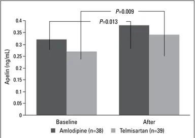
(p<0.001). Apelin concentrations were similar between treat-ment groups (p=0.150). Apelin concentration was significantly increased in whole patients taking antihypertensive therapy (from 0.29±0.15 ng/mL at baseline to 0.36±0.17 ng/mL at 1 month, p<0.001). There was also a significant increase in plasma ape-lin concentration after 1 month of treatment in both the amlo-dipine (from 0.32±0.17 ng/mL at baseline to 0.38±0.17 ng/mL at 1 month, p=0.013) and telmisartan groups (from 0.27±0.13 ng/ mL at baseline to 0.34±0.18 ng/mL at 1 month, p=0.009). The in-cremental change was similar in each of the treatment groups (p=0.922, Fig. 1 and Table 3).
After 1 month of treatment, significant reductions in SBP and DBP were observed in patients with hypertension (p<0.001 for both); the extent of the reduction was similar for both in each of the treatment groups (p=0.658 for SBP and p=0.271 for DBP, Table 3).
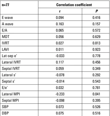
Changes in diastolic function after treatment are presented in Table 4: there were significant improvements in IVRT, lat-sep e’, lateral IVRT, septal IVRT, E/e’, left atrial volume index, lateral MPI and septal MPI in both treatment groups, and the extent of the improvements were similar in each of the groups. The proportional change in plasma apelin concentration after an-tihypertensive therapy was not significantly associated with the proportional change in blood pressure or diastolic indices, except lateral MPI (Table 5).
0.4 0.35 0.3 0.25 0.2 0.15 0.1 0.05 0 Apelin (ng/mL) Baseline P=0.013 P=0.009 After Amlodipine (n=38) Telmisartan (n=39)
Figure 1. Effects of a month of treatment with amlodipine or telmisartan on plasma apelin levels. Data are shown as means
Table 3. Comparison of apelin concentration, systolic and diastolic blood pressures before and after treatment
Group Baseline After P* P**
treatment Apelin, ng/mL Telmisartan 0.27±0.13 0.34±0.18 0.009 0.922 Amlodipine 0.32±0.17 0.38±0.17 0.013 SBP, mm Hg Telmisartan 154±7 122±8 <0.001 0.658 Amlodipine 154±10 123±10 <0.001 DBP, mm Hg Telmisartan 92±6 74±9 <0.001 0.271 Amlodipine 95±9 76±8 <0.001
Values are expressed as mean±standard deviation. DBP - diastolic blood pressure; SBP - systolic blood pressure. *P value of paired samples test; **P value of compari-son of groups in terms of after treatment values corrected for baseline values
Table 4. Echocardiographic indices for the assessment of diastolic function before and after treatment
Telmisartan group Amlodipine group
Baseline After P* Baseline After P* P**
treatment treatment E wave, cm/s 68±12 67±11 0.326 68±14 66±13 0.344 0.795 A wave, cm/s 72±16 69±13 0.070 78±13 69±13 <0.001 0.057 MDT, ms 226±25 221±16 0.122 227±20 222±25 0.217 0.922 IVRT, ms 98±14 89±10 <0.001 101±12 91±10 <0.001 0.773 E/A ratio 1.00±0.31 0.99±0.23 0.959 0.89±0.25 0.99±0.30 0.006 0.110 Lat-sep e’, cm/s 8.9±1.8 10.7±1.8 <0.001 8.3±2.3 10.0±2.6 <0.001 0.564 Lateral IVRT, ms 96±11 90±9 <0.001 99±9 91±10 <0.001 0.692 Septal IVRT, ms 95±9 88±7 <0.001 100±12 90±7 <0.001 0.785 Lateral s’, cm/s 11.5±0.5 11.8±0.6 0.286 11.1±0.8 11.6±0.7 0.156 0.324 Septal s’, cm/s 10.9±0.6 11.3±0.5 0.212 10.8±0.7 11.1±0.7 0.243 0.355 E/e’ ratio 7.8±1.5 6.3±1.1 <0.001 8.6±2.2 6.9±1.9 <0.001 0.516 LAVI, mL/m2 21.0±3.6 18.9±2.7 <0.001 21.1±3.0 19.0±2.7 <0.001 0.959 Lateral MPI 0.64±0.16 0.61±0.23 0.021 0.64±0.11 0.58±0.07 <0.001 0.270 Septal MPI 0.61±0.10 0.58±0.08 0.006 0.63±0.08 0.59±0.07 <0.001 0.837
Values are expressed as mean±standard deviation. E wave and A wave-peak early filling (E) and atrial filling (A) velocities; IVRT-isovolumic relaxation time; Lat/sep e’-mean of lateral and septal peak early diastolic velocities; LAVI-left atrial volume index; MDT-deceleration time of peak E wave; MPI-myocardial performance index; s’-peak systolic annular velocity. *P value of paired samples test; **P value of comparison of groups in terms of after treatment values corrected for baseline values
Discussion
The main findings of the present study are that plasma ape-lin concentrations increased significantly in newly diagnosed patients with primary hypertension after 1 month of effective treatment with either telmisartan or amlodipine. This is the first study demonstrating an increase in the apelin concentration to a similar extent with two anti-hypertensive drug regimens with different mechanisms of action. The change in the apelin con-centration was not associated with the observed improvements in left ventricular diastolic function as well as the decrease in blood pressure.
It has previously been reported that apelin may play a role in the pathophysiology of hypertension (4–6, 16–21). In animal mod-els and clinical studies, decreased apelin concentrations have been documented in hypertension compared with participants with normal blood pressure (4, 5, 20, 21). Papadopoulos et al. (21) measured plasma apelin concentration in 130 patients who underwent 24-hour ambulatory blood pressure monitoring; 24 were found to have masked hypertension. They found that apelin concentration was significantly lower in patients with masked hypertension compared with normotensive controls. In a recent study, even subjects with high normal blood pressure had
sig-nificantly lower apelin levels than subjects with optimal blood pressure (22).
Przewlocka-Kosmala et al. (5) enrolled 232 patients with hy-pertension, 56 of whom had never been treated. Their control group comprised 76 healthy volunteers, and patients with hyper-tension were found to have lower plasma apelin concentration than controls. In that study apelin concentration was reported to be in the range of 120–480 pg/mL, which are in a similar range as our findings (reported as ng/mL in our study). Interestingly, patients with hypertension who had been taking antihyperten-sive drugs (discontinued 2 weeks before entering the study) and those who had never been treated had similar plasma apelin concentrations, however, they also had similar SBP and DBP (in the entire cohort, mean SBP was 155±13 mm Hg and the mean DBP was 94±10 mm Hg). The finding that the comparably low plasma apelin concentrations in treatment naive patients with hypertension and those who had discontinued antihypertensive treatment had in the study by Przewlocka-Kosmala et al.(5), in-formed our decision to examine the change in apelin concentra-tion in patients newly diagnosed with hypertension after being established on an effective treatment regime, as well as to com-pare the baseline plasma apelin concentration of patients with healthy controls. In our cohort, treatment brought about a sig-nificant decrease in blood pressure and a sigsig-nificant increase in plasma apelin concentration; however, there was no correla-tion between the magnitudes of these changes. These findings are in agreement with Przewlocka-Kosmala et al. (5), who also found no significant correlation between the extent of reduction in circulating apelin concentration and blood pressure in 232 pa-tients with essential hypertension. They suggested that ambula-tory blood pressure monitoring might be a more accurate tech-nique to illuminate a possible association; we used ambulatory blood pressure monitoring to evaluate the efficiency of therapy 1 month after the initiation of treatment, but made the diagnosis of hypertension on the basis of recordings taken in our outpatients department. Most of the patients in our cohort had stage 1 hy-pertension requiring monotherapy, and patients requiring com-bination therapy were excluded from the study. The lack of a sta-tistically significant relationship between the change in plasma apelin concentration and the change in blood pressure concurs with other investigations but might also be explained by our diag-nostic methods, the lack of a wide variation in blood pressure in the treatment groups, and a relatively small sample size.
Apelin is thought to act centrally and peripherally to reduce blood pressure. In the peripheries, apelin causes arterial vaso-dilatation via the endothelial nitric oxide pathway (19, 23, 24); however, it has no effect on venous tone (25). An increase in in-tracerebral apelin causes a hypertensive response (23, 24); thus it appears that the central and peripheral components of the ape-linergic system seem to have opposing effects on arterial blood pressure. It is difficult to explain the mechanism by which plasma apelin concentration might increase after control over blood pressure has been restored, as the activity and pharmacology
Table 5. Correlation of proportional change in apelin concentration and proportional change in echocardiographic parameters and blood pressure after 1 month of therapy
n=77 Correlation coefficient r P E wave 0.094 0.416 A wave 0.163 0.157 E/A 0.065 0.572 MDT 0.056 0.629 IVRT 0.027 0.813 LAVI 0.011 0.923 Lat-sep e’ -0.033 0.778 Lateral IVRT 0.117 0.456 Septal IVRT 0.059 0.349 Lateral s’ -0.078 0.292 Septal s’ -0.014 0.543 E/e’ 0.032 0.781 Lateral MPI -0.233 0.041 Septal MPI -0.098 0.395 SBP 0.073 0.526 DBP 0.075 0.516
Data obtained from Spearman’s correlation analysis are shown in the table. A wave-peak atrial filling (A) velocity; DBP - diastolic blood pressure; E wave - wave-peak early filling velocity; IVRT - isovolumic relaxation time; Lat/sep e’ - mean of lateral and septal peak early diastolic velocities; LAVI - left atrial volume index; MDT - deceleration time of E wave; MPI - myocardial performance index; s’ - peak systolic annular velocity; SBP - systolic blood pressure
of the apelin/APJ pathway is not fully understood; our study was not designed to identify the underlying mechanism. It has been reported that apelin opposes angiotensin in the cardiovascular system, and that angiotensin converting enzyme 2 (ACE 2) acts to eliminate apelin (26). In experimental animals, lack of activity in the APJ pathway reportedly suppressed expression of the angio-tensin II type 1 receptor, which increased when APJ was activat-ed (27). In a rat model of hypertensive heart failure, down regula-tion of apelin and APJ expression was significantly increased in animals treated with olmesartan (28). These observations sup-port our findings that plasma apelin concentration is elevated in patients treated with telmisartan; nonetheless amlodipine treatment elevated circulating apelin concentration to a simi-lar extent. It is plausible; therefore, that any increase in plasma apelin concentration occurs through one or more mechanisms triggered by reduction in blood pressure, and are independent of the drug used. Further studies are needed to test this hypothesis.
We observed that diastolic function had improved after 1 month of therapy with telmisartan or amlodipine, and that the magnitude of the improvement with each drug was comparable. Zaliunas et al. (29) have also reported a measurable benefit of amlodipine on LV diastolic function in patients with arterial hy-pertension and stable angina after 4 weeks of treatment. In the longer term, Mattioli et al. (30) observed significant improve-ments in diastolic filling parameters, LA volume and LV hyper-trophy after 3, 6, 9, and 12 months of telmisartan monotherapy. We used these findings to inform our decision that 1 month of treatment was enough to control hypertension, and for improve-ment in diastolic function and changes apelin concentration to be evident. Lower levels of apelin is reportedly independently as-sociated with more profound impairment of LV diastolic function (5), but despite the beneficial effects of treatment on diastolic function and apelin concentration, we did not find any associa-tion between the change in circulating apelin concentraassocia-tion and indices of diastolic function. The pathophysiological mechanism underpinning the relationship between apelin and diastolic func-tion is not understood. Przewlocka-Kosmala et al. (5) suggested that the deleterious effect of decreased apelin concentration on LV diastolic function could be a consequence of diminished en-dothelial nitric oxide synthesis; however, further studies will be needed to examine this hypothesis. The absence of a relation-ship between follow-up apelin concentration and improvement in diastolic function in our cohort may be due to the relatively short duration of our study.
Study limitations
Our study has several limitations. First, our study sample size might not have been large enough to detect significant associa-tions between change in apelin concentration and the echocar-diographic parameters used to determine LV diastolic function. Second, using clinic measurements to diagnose hypertension may have influenced the composition of the study cohort,
al-though we used ambulatory monitoring to measure blood pres-sure at the end of the treatment period, and the protocol was the same for all participants. Third, the study design was open label. Fourth, 13 patients were excluded from the analysis for a variety of reasons, and although this is a relatively high pro-portion of the study cohort, exclusions were evenly distributed between the groups. We did not use more sensitive echocardio-graphic methods of evaluating cardiac function, such as myo-cardial strain and strain rate. These methods are able to detect subtle changes in myocardial mechanics and a relationship with change in apelin concentration could have been observed; how-ever, we elected to use well-established parameters to evaluate LV diastolic function recommended in international guidelines. Finally, even though we were able to show significant differenc-es in apelin concentration, blood prdifferenc-essure measurements, and echocardiographic indices for diastolic function, relatively short study period is another limitation and longer duration follow-up studies may strengthen the accuracy of data.
Conclusions
Plasma apelin concentration was significantly lower in pa-tients with hypertension than the control group, who had normal blood pressure. A significant increase in apelin concentration was observed after 1 month of effective treatment with either telmisartan or amlodipine. The magnitude of the change in apelin concentration was associated neither with the extent of the im-provement in diastolic function nor with the degree of change in blood pressure. Effective blood pressure control, rather than the drug choice, seems to increase apelin concentration.
Acknowledgements: There is no conflict of interest regarding any of the authors. All support for this study came from institutional and departmental resources.
Funding: This research received no grant from any funding agency in the public, commercial, or not-for-profit sectors.
Conflict of interest: None declared. Peer-review: Externally peer-reviewed.
Authorship contributions: Concept – B.P.; Design – B.P., K.O.; Super-vision – B.P., K.O., U.A.B., M.Z.U., D.Ö., H.M.; Materials – B.P., K.O., U.A.B., M.Z.U., D.Ö., H.M.; Data collection &/or processing – B.P., K.O., U.A.B., M.Z.U., D.Ö., H.M.; Analysis &/or interpretation – B.P., D.Ö.; Literature search – B.P., K.O.; Writing – S.S.B., B.P., K.O.; Critical review – B.P., K.O.; Other – D.Ö.
References
1. Tatemoto K, Hosoya M, Habata Y, Fujii R, Kakewaga T, Zou MX. Iso-lation and characterization of a novel endogenous peptide ligand for the human APJ receptor. Biochem Biophys Res Commun 1998; 251: 471-6. Crossref
2. Lee DK, Cheng R, Nguyen T, Fan T, Kariyawasam AP, Liu Y. Charac-terization of apelin, the ligand for the APJ receptor. J Neurochem 2000; 74: 34-41. Crossref
3. Barnes G, Japp AG, Newby DE. Translational promise of the apelin/ APJ system. Heart 2010; 96: 1011-6. Crossref
4. Sönmez A, Çelebi G, Erdem G, Tapan S, Genç H, Taşcı I, et al. Plasma apelin and ADMA levels in patients with essential hypertension. Clin Exp Hypertens 2010; 32: 179-83. Crossref
5. Przewlocka-Kosmala M, Kotwica T, Mysiak A, Kosmala W. Re-duced circulating apelin in essential hypertension and its associa-tion with cardiac dysfuncassocia-tion. J Hypertens 2011; 29: 971-9. Crossref
6. Charles CJ. Putative role for apelin in pressure/volume homeosta-sis and cardiovascular disease. Cardiovasc Hematol Agents Med Chem 2007; 5: 1-10. Crossref
7. Mancia G, Fagard R, Narkiewicz K, Redán J, Zanchetti A, Böhm M, et al. 2013 Practice guidelines for the management of arterial hypertension: the Task Force for the Management of Arterial Hy-pertension of the European Society of HyHy-pertension (ESH) and of the European Society of Cardiology (ESC). J Hypertens 2013; 34: 1925-38. Crossref
8. Zhou Y, Wang Y, Qiao S. Apelin: a potential marker of coronary ar-tery stenosis and atherosclerotic plaque stability in ACS patients. Int Heart J 2014; 55: 204-12. Crossref
9. Cebeci AN, Güven A, Kuru LI. Perinephric adipose tissue thickness in relation to blood pressure, plasma apelin and C -reactive protein levels in obese adolescents. Minerva Endocrinol 2016; 41: 166-74. 10. Habchi M, Duvillard L, Cottet V, Brindisi MC, Bouillet B, Beacco M,
et al. Circulating apelin is increased in patients with type 1 or type 2 diabetes and is associated with better glycaemic control. Clin En-docrinol (Oxf) 2014; 81: 696-701. Crossref
11. Peltonen T, Näpänkangas J, Vuolteenaho O, Ohtonen P, Soini Y, Ju-vonen T, et al. Apelin and its receptor APJ in human aortic valve stenosis. J Heart Valve Dis 2009; 18: 644-52.
12. Zhang DL, Liao H, Wei YY, Zhang Y, Zhang QD, Wang ZG. Elevation of serum apelin-13 is positively correlated with ADMA in patients on maintenance hemodialysis. Clin Nephrol 2009; 71: 405-12. Crossref
13. Dalzell JR, Rocchiccioli JP, Weir RA, Jackson CE, Padmanabhan N, Gardner RS, et al. The Emerging Potential of the Apelin-APJ System in Heart Failure. J Card Fail 2015; 21: 489-98. Crossref
14. Chapman CB, Ewer SM, Kelly AF, Jacobson KM, Leal MA, Rahko PS. Classification of left ventricular diastolic function using American Society of Echocardiography Guidelines: agreement among echo-cardiographers. Echocardiography 2013; 30: 1022-31. Crossref
15. Tei C, Nishimura RA, Seward JB, Tajik AJ. Noninvasive Doppler-derived myocardial performance index: correlation with simulta-neous measurements of cardiac catheterization measurements J Am Soc Echocardiogr 1997; 10: 169-78. Crossref
16. Tatemoto K, Takayama K, Zou MX, Kumaki I, Zhang W, Fujimiya M. The novel peptide apelin lowers blood pressure via nitric oxide-dependent mechanism. Regul Pept 2001; 99: 87-92. Crossref
17. Lee DK, Saldivia VR, Nguyen T, Cheng R, George SR, O’Dowd BF. Modification of the terminal residue of apelin-13 antagonizes its hypotensive action. Endocrinology 2005; 146: 231-6. Crossref
18. Ishida J, Hashimoto T, Hashimoto Y, Nishiwaki S, Iguchi T, Harada S. Regulatory roles for APJ, a seven-transmembrane receptor re-lated to angiotensin-type 1 receptor in blood pressure in vivo. J Biol Chem 2004; 279: 26274-9. Crossref
19. Kagiyama S, Fukuhara M, Matsumura K, Lin Y, Fujii K, Iida M. Cen-tral and peripheral cardiovascular actions of apelin in conscious rats. Regul Pept 2005; 125: 55-9. Crossref
20. Zhang J, Ren CX, Qi YF, Lou LX, Chen L, Zhang LK, et al. Exercise training promotes expression of apelin and APJ of cardiovascular tissues in spontaneously hypertensive rats. Life Sci 2006; 79: 1153-9. Crossref
21. Papadopoulos DP, Mourouzis I, Faselis C, Perrea D, Makris T, Tsiou-fis C. Masked hypertension and atherogenesis: the impact of apelin and relaxin plasma levels. J Clin Hypertens (Greenwich) 2013; 15: 333-6. Crossref
22. Liakos CI, Sanidas EA, Perrea DN, Grassos CA, Chantziara V, Viniou NA, et al. Apelin and visfatin plasma levels in healthy individuals with high normal blood pressure. Am J Hypertens 2016; 29: 549-52. 23. Seyedabadi M, Goodchild AK, Pilowsky PM. Site-specific effects
of apelin-13 in the rat medulla oblongata on arterial pressure and respiration. Auton Neurosci 2002; 101: 32-8. Crossref
24. Zhang Q, Yao F, Raizada MK, O’Rourke ST, Sun C. Apelin gene trans-fer into the rostral ventrolateral medulla induces chronic blood pressure elevation in normotensive rats. Circ Res 2009; 104: 1421-8. 25. Japp AG, Cruden NL, Amer DA, Li VK, Goudie EB, Johnston NR, et
al. Vascular effects of apelin in vivo in man. J Am Coll Cardiol 2008; 52: 908-13. Crossref
26. Iwanaga Y, Kihara Y, Takenaka H, Kita T. Down-regulation of cardiac apelin system in hypertrophied and failing hearts: possible role of angiotensin II-angiotensin type 1 receptor system. J Mol Cell Car-diol 2006; 41: 798-806. Crossref
27. Sun X, Iida S, Yoshikawa A, Senbonmatsu R, Imanaka K, Maruyama K, et al. Non-activated APJ suppresses the angiotensin II type 1 re-ceptor, whereas apelin-activated APJ acts conversely. Hypertens Res 2011; 34: 701-6. Crossref
28. Fukushima H, Kobayashi N, Takeshima H, Koguchi W, Ishimitsu T. Effects of olmesartan on Apelin/APJ and Akt/endothelial nitric ox-ide synthase pathway in Dahl rats with end-stage heart failure. J Cardiovasc Pharmacol 2010; 55: 83-8. Crossref
29. Zaliunas R, Jurkevicus R, Zabiela V, Brazdzionyte J. Effect of am-lodipine and lacidipine on left ventricular diastolic and long axis functions in arterial hypertension and stable angina pectoris. Acta Cardiol 2005; 60: 239-46. Crossref
30. Mattioli A, Zennaro M, Bonatti S, Bonetti L, Mattioli G. Regression of left ventricular hypertrophy and improvement of diastolic func-tion in hypertensive patients treated with telmisartan. Int J Cardiol 2004; 97: 383-8. Crossref

![Microwave supported synthesis of some novel 1,3-Diarylpyrazino[1,2-a] benzimidazole derivatives and investigation of their anticancer activities](data:image/gif;base64,R0lGODlhAQABAIAAAP///wAAACH5BAEAAAAALAAAAAABAAEAAAICRAEAOw==)