The effect of tibial dyschondroplasia on metabolic parameters in
broiler chickens
Halit İMİK1, Kübra Asena TERİM KAPAKİN2, Recep GÜMÜŞ1, Samet KAPAKİN3, Ali KURT4
1 Department of Animal Nutrition and Nutritional Disorders, Faculty of Veterinary Sciences, Ataturk University; 2 Department of
Pathology, Faculty of Veterinary Sciences, Ataturk University; 3Department of Anatomy, Faculty of Medicine, Ataturk University; 4Department of Pathology, Erzurum Regional Research and Education Hospital, Erzurum Turkey.
Summary: Tibial dyschondroplasia (TD) deteriorates the welfare of broiler chickens and causes economic loss. The present study was aimed at the investigation of the effect of TD on metabolic parameters and the demonstration of the aetiology of this disorder. For this purpose, blood serum mineral, lipid, protein, glucose, uric acid and enzyme levels, and histopathological alterations in the tissues of the tibiotarsal joint were determined. Ten broiler chickens suffering from TD and 10 healthy broiler chickens, all which were of the Ross 308 breed, constituted the material of the study. Analyses showed that calcium, phosphorus and free fatty acid blood serum levels were lower, whilst triglyceride, cholesterol, low density lipoproteins (LDL), very low density lipoproteins (VLDL) rates and uric acid and creatinine levels were higher in animals suffering from tibial dyschondroplasia, compared to healthy animals (P<0.05). No statistically significant difference existed between the glucose and amylase levels and alanine aminotransferase (ALT), aspartate aminotransferase (AST), γ-glutamyltransferase (GGT), and alkaline phosphatase (ALP), activities of broilers diagnosed with tibial dyschondroplasia and healthy broilers. The histopathological examination of the tissues of the tibiotarsal joint of the animals diagnosed with TD revealed degenerative alterations characterized by the dominance of hypertrophy and the lack of urate crystals and calcification in chondrocytes. In animals with TD, calcium and phosphorus blood serum levels were low, whilst the lipid profile and amount of uric acid were high. On the other hand, ALP enzyme activity was determined not to display any alteration.
Key words: Broiler, metabolic parameter, tibial dyschondroplasia.
Etçi piliçlerde tibial diskondoplazinin metabolik parametrelere etkisi
Özet: Tibial diskondoplazi (TD) piliçlerin refahını bozmakta ve ekonomik kayıplara neden olmaktadır. Bu çalışma TD’nin metabolik parametrelere etkisini ve etiyolojisini belirlemek amacıyla yapılmıştır. Bu nedenle hayvanların kan serumlarında mineral, lipit, protein, glikoz, ürik asit ve enzim değerleri ve tibiotarsal eklem dokusundaki histopatolojik değişimler tespit edilmiştir. Araştırmada10 adet TD’li ve 10 adet sağlıklı broiler (Ross 308) kullanılmıştır. Tibial diskondoplazili hayvanların kan serumlarındaki kalsiyum, fosfor seviyesi ve serbest yağ asitleri oranı sağlıklı hayvanlardan düşük; tigliserit, kolesterol, düşük yoğunluklu lipoprotein (LDL), çok düşük yoğunluklu lipoprotein (VLDL) oranları, ürik asit ve kreatinin miktarları ise yüksek bulunmuştur (P<0.05). Tibial diskondoplazili ve sağlıklı broilerin glikoz ve amilaz seviyeleri ile alanin aminotransferaz (ALT), aspartat aminotransferaz (AST), γ-glutamyltransferaz (GGT), ve alkalin fosfataz (ALP), aktiviteleri arasında ise istatistiksel farklılık tespit edilmemiştir. Araştırmada TD’li hayvanların tibiotarsal eklem dokusunun histopatolojik incelenmesinde dejeneratif değişiklikler, kondrositlerde hipertrofinin baskın olduğu, ürat kristalleri ve kalsifikasyonun ise olmadığı tespit edildi. Tibial diskondoplazili hayvanların kan serumlarında kalsiyum ve fosfor seviyelerinin düşük, lipit profilinin ve ürik asit miktarının yüksek, ALP enzim aktivitesinin ise değişmediği belirlenmiştir.
Anahtar sözcükler: Broyler, metabolik parameter, tibiyal diskondroplazi.
Introduction
Dyschondroplasia is a disorder of the growth plates, commonly observed in broiler chickens, ducks and turkeys. As the deformty are most frequently observed on the epiphyseal growth plate of the tibial tarsal bone, the disorder is referred to as tibial dyschondroplasia (TD) (28).
The use of fast-growing hybrids in broiler production is associated with the more frequent
observation of leg problems, mainly tibial dsychondroplasia (26). This disorder has adverse implications for both the welfare and the development of animals and causes 30% of the economic losses encountered in broiler production. Although the underlying mechanism of tibial dyschondroplasia remains unclear, it is known that predisposing factors of fast-growing poultry breeds, including genetic factors, misapplications in broiler production (14, 24, 26, 28) and factors related
to the feeding of animals (15, 17, 23, 28, 31) play a major role in the development of this disorder.
It is observed that, the lesions develop, in general, bilaterally, and sometimes unilaterally in the proximal region of the tibial tarsal bone. At early stage, the disorder clinically manifests as deformation and lameness, which later may be associated with bone fractures. Macroscopically, thickening of the epiphyseal plate and the formation of an abnormal opaque cartilaginous mass, extending from the distal end of the epiphyseal plate into the metaphysis, are observed (4, 28, 30). This abnormal cartilaginous mass is non-mineralized, devoid of blood vessels, and of soft consistency. The most pronounced histopathological lesions in such cases are the presence of hypertrophic and immature chondrocytes in the epiphyseal plate (4, 12, 20, 28, 30). Moreover, degeneration of chondrocytes and patches of necrosis may also be observed. Furthermore, reports point out to increase in the matrix with no calcification in the necrotic regions.
Previously conducted several studies have demonstrated that the development of tibial dyschondroplasia could be minimized by means of various modifications to the rations fed to animals (15, 18, 19, 34). Oso et al (23) reported that different calcium sources incorporated into broiler rations have different effects on bone mineralisation and the development of TD. Houshmand et al (11) reported that the supplementation of broiler rations with low levels of calcium increased the incidence of TD. Liu et al (19) suggested that hydrolysed soy and fish oil added to quail rations were much more beneficial for bone metabolism, compared to soy oil and poultry fat. Yalçın et al (33) demonstrated that the temperatures to which avian embryos were subjected to during their early developmental stage increased the incidence of TD with significant effects on the ash rate and no effect on the calcium rate and weight of the tibia.
In order to elucidate the aetiology of tibial dyschondroplasia, it is important to understand the impact of this disorder on metabolic parameters. With an aim to fully understand the mechanism of action of tibial dyschondroplasia, the mineral, protein, energy, lipid, enzyme, uric acid, creatinine and Blood Urea Nitrogen (BUN) blood serum levels and histopathological alterations in the tissues of the tibiotarsal joint were investigated in the present study.
Materials and Methods
Experimental animals, trial method and blood samples: This study was approved by the Local Ethics
Board for Experimental Animals of Atatürk University (Decision No: 2011/3/11). Twenty broiler chickens of the Ross 308 breed aged of 42 days, 10 of which suffered from tibial dyschondroplasia and 10 of which were healthy, constituted the material of the study. The
feed given to the animals was prepared in accordance with the recommendations of the National Research Council (22). Chemical analyses of starter, grower and finish diets were run using references of Association of Official Analytical Chemists (AOAC) (3). The nutrient composition of the feed provided to the experimental animals in the present study is shown in Table 1.
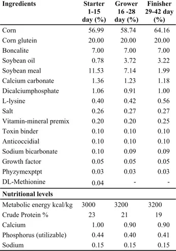
Table 1. Composition and nutrient content of diets.
Tablo 1. Karma yemlerin bileşimleri ve besin madde içerikleri. Ingredients Starter 1-15 day (%) Grower 16 -28 day (%) Finisher 29-42 day (%) Corn 56.99 58.74 64.16 Corn glutein 20.00 20.00 20.00 Boncalite 7.00 7.00 7.00 Soybean oil 0.78 3.72 3.22 Soybean meal 11.53 7.14 1.99 Calcium carbonate 1.36 1.23 1.18 Dicalciumphosphate 1.06 0.91 1.00 L-lysine 0.40 0.42 0.56 Salt 0.26 0.27 0.27 Vitamin-mineral premix 0.20 0.20 0.25 Toxin binder 0.10 0.10 0.10 Anticoccidial 0.10 0.10 0.10 Sodium bicarbonate 0.10 0.09 0.09 Growth factor 0.05 0.05 0.05 Phyzymexptpt 0.03 0.03 0.03 DL-Methionine 0.04 - - Nutritional levels
Metabolic energy kcal/kg 3000 3200 3200 Crude Protein % 23 21 19
Calcium 1.00 0.90 0.90
Phosphorus (utilizable) 0.44 0.40 0.41
Sodium 0.15 0.15 0.15
Biochemical Analyses: On the 42nd day of production, in the morning and before the animals were fed, blood samples were collected by wing vein puncture into sterile tubes, which were not coated with an anticoagulant. After clotting, the samples centrifuged at 4000 rpm for 10 mn by a cooler centrifuge at +4o C. Then sera were carefully harvested and stored at -20°C until analysis. All biochemical analyses were performed in a week.
Biochemical parameters in sera were analyzed through an automatic analyzer in commercial test kits (Cobas 6000 analyzer, Roche). The parameters were glucose, total protein, albumin, globulin, triglyceride, cholesterol, high density lipoprotein (HDL), low density lipoproteins (LDL) amylase (AMYL), aspartate aminotransferase (AST), alanine aminotransferase (ALT), γ-glutamyltransferase (GGT), alkaline phosphatase (ALP), calcium (Ca), phosphor (P), magnesium (Mg), and iron (Fe). Additionally, VLDL was calculated by dividing triglyceride by five.
Analysis of Lipid Profile: High performance
thin-layer chromatography (HPTLC) was used to separate and determine the composition of the serum lipids. To this end, 500μl n-hexane/iso-propanol (2:1 (v/v)) mixture was added to 500μl of serum. After vigorous vortexing, the tubes were centrifuged at 4°C, 5.000 x g for 10 minutes to obtain phase separation; the upper phase (n-hexane phase) was used for HPTLC. Five microlitre portions of the extracted lipids were spotted with a micropipette 2 cm from the bottom of the HPTLC plates. The lipids were developed at 6 cm with a mixture of n-hexane: diethylether: formic acid (80:20:2 (v/v/v)). After development, the entire plate was sprayed with 10% CuSO4 (/v) in 8% H3PO4 (v/v) and charred at 180°C for 10 minutes (24) to visualize the lipid classes. A standard lipid mixture comprised of L-α-phosphatidylcholine, cholesterol, palmitic acid, triolein, squalene, and the serum lipids were separated into the following classes: hydrocarbons (HC), triacylglycerol (TAG), free fatty acids (FFA), cholesterol (CH), and polar lipids (PL) (13).
Histopathological Examination: On the 42nd day, the animals were sacrificed by cervical dislocation for necropsy. Tissue samples were taken from proximal ands of the tibiotarsal joints. The samples were fixed 10% neutral buffered formalin. Then, the samples were decalcified in 36.8% formic acid and 6.8% sodium formate. Finally, the samples were post-fixed and embedded in paraffin. After routin eprocedures, 5 μm thick sections were cut and stained routinely with haematoxylin eosin, Alcian Blue, von Kossa, Masson’s Trichrome (MTC), and examined under a light microscope.
Statistical analysis: The serum parameters pertaining
to the healthy animals and animals suffering from TD were evaluated statistically using the independent-samples t-test (29).
Results
Biochemical Findings: The biochemical parameters
determined in the blood serum samples of broiler chickens diagnosed with tibial dyschondroplasia and healthy broiler chickens are presented in Table 2. Serum calcium and phosphorus levels were significantly lower in the broiler chickens diagnosed with tibial dyschondroplasia, in comparison to the healthy animals (P<0.05); whilst Mg, Fe, Na, K and Cl levels were similar in both groups. The serum triglyceride, cholesterol, LDL, VLDL levels of the animals diagnosed with tibial dyschondroplasia were significantly higher than those of the healthy broilers (P<0.05). On the other hand, HDL levels did not differ between the two groups. It was determined that the serum hydrocarbon levels of the healthy broiler chickens were significantly lower and the free fatty acid levels were higher than those of the broiler chickens suffering from TD (P<0.05). Furthermore, the serum TAG, CH and PL levels, glucose, total protein, albumin, globulin, and of liver
enzymes, AMYL, ALT, AST, GGT, ALP and BUN values did not statistically differ between the healthy broiler chickens and the broiler chickens suffering from TD. Furthermore, the serum uric acid and creatinine levels of the chickens suffering from TD were significantly higher than the levels of the healthy broiler chickens (P<0.05).
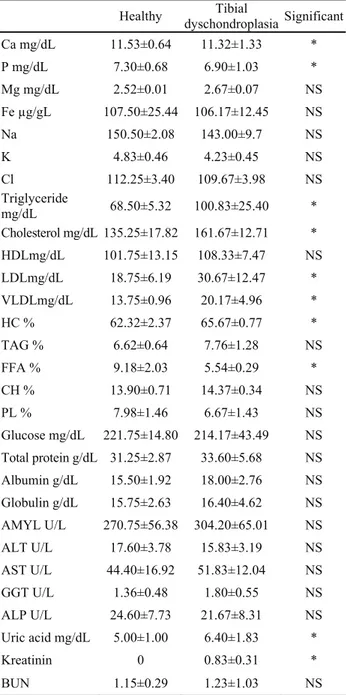
Table 2. Serum energy, lipid, enzyme, protein, uric acid and mineral levels of healthy broiler vs with tibial dyschondroplasia. Table 2. Sağlıklı ve tibial diskonroplazili broilerin serum enerji, lipit, enzim, protein, ürik asit ve mineral seviyeleri.
Healthy dyschondroplasiaTibial Significant Ca mg/dL 11.53±0.64 11.32±1.33 * P mg/dL 7.30±0.68 6.90±1.03 * Mg mg/dL 2.52±0.01 2.67±0.07 NS Fe µg/gL 107.50±25.44 106.17±12.45 NS Na 150.50±2.08 143.00±9.7 NS K 4.83±0.46 4.23±0.45 NS Cl 112.25±3.40 109.67±3.98 NS Triglyceride mg/dL 68.50±5.32 100.83±25.40 * Cholesterol mg/dL 135.25±17.82 161.67±12.71 * HDLmg/dL 101.75±13.15 108.33±7.47 NS LDLmg/dL 18.75±6.19 30.67±12.47 * VLDLmg/dL 13.75±0.96 20.17±4.96 * HC % 62.32±2.37 65.67±0.77 * TAG % 6.62±0.64 7.76±1.28 NS FFA % 9.18±2.03 5.54±0.29 * CH % 13.90±0.71 14.37±0.34 NS PL % 7.98±1.46 6.67±1.43 NS Glucose mg/dL 221.75±14.80 214.17±43.49 NS Total protein g/dL 31.25±2.87 33.60±5.68 NS Albumin g/dL 15.50±1.92 18.00±2.76 NS Globulin g/dL 15.75±2.63 16.40±4.62 NS AMYL U/L 270.75±56.38 304.20±65.01 NS ALT U/L 17.60±3.78 15.83±3.19 NS AST U/L 44.40±16.92 51.83±12.04 NS GGT U/L 1.36±0.48 1.80±0.55 NS ALP U/L 24.60±7.73 21.67±8.31 NS Uric acid mg/dL 5.00±1.00 6.40±1.83 * Kreatinin 0 0.83±0.31 * BUN 1.15±0.29 1.23±1.03 NS
*P<0.05. Different letters in the same column represent a statistical significance between the groups. Calcium (Ca), phosphor (P), magnesium (Mg), iron (Fe), High-density lipoprotein (HDL), low-density lipoprotein (LDL), Very-low-density lipoprotein (VLDL), amylase (AMYL), aspartate aminotransferase (AST), alanine aminotransferase (ALT), γ-glutamyltransferase (GGT), alkaline phosphatase (ALP), Hydrocarbons (HC), triacylglycerol (TAG), free fatty acids (FFA), cholesterol (CH), and polar lipids (PL).
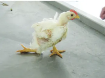
Macroscopic Findings: Macroscopically, out of 10
broiler chickens diagnosed with tibial dyschondroplasia, 6 unilateral and 4 bilateral thickening and swelling at the growth plate of the tibiotarsal joint. In addition, the dull white color cartilage mass extending from distal end of epiphyseal plate into metaphysis was observed. It was observed that the animals had difficulties in consuming feed and water associated with significant weight loss, feather loss, lameness and difficulty in maintaining standing position (Figure 1).
Microscopic Findings: Microscopic examination
revealed that the most pronounced finding in the epiphyseal growth plate of the tibiotarsal joints was extreme hypertrophy of chondrocytes, an increased number of immature chondrocytes (Figures 2a-b) and lack of blood vessels. Apart from degenerative changes and mucoid substance (Figures 2a-b), it was ascertained that patches of necrotic chondrocytes existed. Alcian blue and MTC staining demonstrated that the amount of mucoid substance (Figures 3a-b) had increased in the
Figure 1 a-b: Swollen and enlargement of the tibiotarsal joint and feather loss.
Şekil 1 a-b: Tibiotarsal eklemde kalınlaşma ve tüylerde dökülme.
Figures 2 a-b: Immature chondrocytes (arrow) and mucoid substance (arrow head) HE., Bar: 50 μm. Şekil 2a-b: Olgunlaşmamış kondrositler (ok), ve mukoid madde (ok başı). HE., Bar: 50 μm.
Figures 3 a: Mucoid substance in necrose region (arrow) Alcian blue, Bar: 50 μm.; b: Mucoid substance in necrose region (arrow) MTC., Bar: 20 μm.
Şekil 3 a. Nekrotik bölgede mukoid madde (ok). Alcian blue, Bar: 50 μm.; b. Nekrotik bölgede mukoid madde (ok). MTC., Bar: 20 μm.
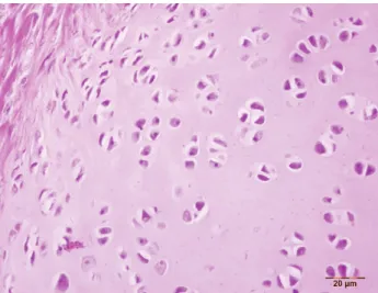
degenerated region, whilst von Kossa staining showed that there was no calcium accumulation. Furthermore, MTC staining revealed significant proliferation of connective tissue cells. In none of the cases were urate crystals determined in histopathological examination. No histopathological lesion was evidenced in control broilers (Figure 4).
Discussion and Conclusion
The use of fast-growing breeds in broiler production has led to an increased incidence of leg problems resulting from developmental disorders of bone tissue. It is known that bone formation is affected by multiple factors, including genetics, diseases, toxins, anti-nutritional feed, age, sex, diet, physical activity and the endocrine system (27). Bones are composed of inorganic salts that accumulate within the organic matrix made up of collagen fibrils and glycoproteins. The general structure of bone minerals is in the form of hydroxyapatite [Ca10(PO4)6(OH)2] crystals and contains several inorganic elements, including among others, calcium, phosphorus, magnesium, sodium, potassium, chloride and fluoride (6). Leg problems occur as a result of the incomplete development of bone and cartilage tissues, and mainly as a consequence of tibial dyschondroplasia (10, 14). Although the exact aetiology of TD remains unclear, it is known that genetics, feeding and management play a major role in such cases and lead to morphological, biochemical and molecular alterations in tissues and cells. In tibial dyschondroplasia, lesions are generally observed in the epiphyseal growth plate of the tibial tarsal bone. Furthermore, it has been suggested that such cases occur upon the enlargement of the cartilage to a level, which prevents the entirety of the cells within the matrix from receiving nutrients and oxygen (2, 6, 25). Macroscopic signs that aid in the diagnosis of TD are lameness, and the swelling and soft consistency of the joints (4, 7, 10, 28, 30), whilst definitive
diagnosis of the disorder is based on the histopathological determination of the presence of an increased number of hypertrophic and/or immature chondrocytes (4, 12, 20, 28, 30). Further histopathological signs observed in TD have been reported as increase in mucoid substance as a result of degenerative changes and lack of both mineralisation and blood vessel formation (7, 10, 27, 28, 30).
It has been reported that the lesions are generally found unilateraly in animals with TD. In present study, the lesions are located unilateraly in 6 broilers and bilateraly in 4 broilers. Macroscopically, in our cases thickening at the growth plate of the tibiotarsal joint and presence of dull cartilage mass entering from distal end of epiphyseal plate into metaphysis this findings are in accordance with previous studys. In the present study, the observation of thickening and enlargement in the tibiotarsal joint associated with the extreme hypertrophy of chondrocytes, increase in the number of immature chondrocytes, presence of patches of necrotic chondrocytes, degenerative changes, increase in the amount of mucoid substance and proliferation of connective tissue cells was in compliance with literature reports.
It is known that leg health is affected by excessive or poor levels of minerals, vitamins and trace elements in the ration. Moreover, Ca, P and Vitamin D have been reported to have major effect on the development of bone tissue (8, 21, 26, 31). In a study, in which broiler rations were supplemented with 200, 1500, 2500 and 3500 IU/kg of cholecalciferol, it was determined that in direct proportion to these supplemental doses, ash rates of the tibial and toe bones and serum calcium and phosphorus levels increased with significant decrease in the incidence of TD (15, 21) reported that, the supplementation of poultry rations with different doses of Ca and 1,25-(OH)2D3 decreased the incidence of tibial dyschondroplasia and increased significantly bone ash rates. The formation of bone tissue is directly related to serum calcium and phosphorus levels. In the present study, the serum Ca and P levels of broiler chickens diagnosed with TD being lower than that of healthy broiler chickens demonstrates that low levels increase the incidence of tibial dyschondroplasia. These findings are further supported by those obtained in previously conducted studies (8, 34). Moreover, the histopathological determination of affected regions being devoid of calcification and blood vessels in TD cases in the present study is also in compliance with literature reports.
Lipids incorporated into the ration also play a significant role in bone metabolism. When lipids containing n: 3 are added to the ration, PGE2 production in bones decreases and osteoclastic activity increases. Fleming (8) reported that, the rate of n=3 PUFA/n=6 PUFA was correlated with the leg health of broiler
Figure 4: Apperance of normal chondrocytes. HE., Bar: 20 μm. Şekil 4: Normal kondrositlerin görünümü. HE., Bar: 20 μm.
chickens. As the rate of n=3 PUFA in the ration increased, the leg health of the animals improved. Liu et al. (19) indicated that the supplementation of Japanese quail rations with soy oil and poultry fat adversely affected the metabolism and histological structure of bones when compared to fish oil, and significantly decreased bone mineral levels and significantly increased prostaglandin E2 levels in the bone marrow and tibial bone. Korotkova et al (16), in a study in which they added polyunsaturated fatty acids (PUFA) to rat feed, determined that the proportion of n=6:n=3 had a significant effect on bone metabolism. These researchers also reported that the bone growth of the rats given n=6+n=3 were better than those given n=3 and n=6. Chan et al (5) reported that, while n=3 fatty acids had a significant role in bone formation and the induction of osteoblasts, prostaglandins containing n=6 fatty acids inhibited bone development. Griel et al (9) reported that n=3 PUFA contributed to bone formation by decreasing bone resorption and positively influencing bone metabolism. Watkins et al (32) determined that, in rats, prostaglandin E2 production in bones decreased with the presence of n=3 and increased with the presence of n=6 in the rations. In the present study, the serum triglyceride, cholesterol, LDL and VLDL levels and hydrocarbon rates of animals suffering from TD being higher than those of healthy animals demonstrated the effect of lipids on leg health. It is evident that further studies are required to elucidate the effect of fatty acids on leg health.
The growth of bones involves complex metabolic processes. While osteoclasts enable the secretion of minerals required for bone formation into the extracellular fluid, osteoblasts enable the uptake of these minerals from the extracellular fluid during bone formation. Another factor, which affects bone growth, is the enzyme ALP. This enzyme adversely affects calcification by inducing osteoclast cells (1, 33). On the other hand, Rath et al. (27) reported that ALP enzyme activity in animals with TD was similar to that of healthy animals. Findings obtained in the present study, suggesting similarity between the serum ALP activity of animals with TD and healthy animals, were in agreement with the findings of Rath et al. (27).
It is known that, in animals exposed to stress, serum uric acid levels are elevated for the sustainability of vital functions and maintenance of adaptation. When analysing the serum parameters of animals diagnosed with tibial dyschondroplasia, significantly elevated uric acid and creatinine levels can be interpreted as indicators of animals having been exposed to stress. In the present study, histopathological examination revealed no urate crystals in the tibiotarsal joint.
In conclusion, the TD, based on histopathological findings, suggest that the mineral and lipid composition
of the ration fed to the animals may be influential in the aetiology of this disorder. For this reason, it is considered that the adjustment of the calcium and phosphorus levels and n=6 and n=3 rates of the lipids of the ration could be of use for the minimisation of the occurrence of leg problems. Furthermore, it is considered that further studies are required to fully understand serum calcium and phosphorus levels being low and the triglyceride, cholesterol, LDL, VLDL levels and hydrocarbon rates being high in the broiler chickens diagnosed with and elucidate the effects of lipids incorporated into the ration.
References
1. Anderson HC (2003): Matrix vesicles and calcification. Curr Rheumatol Rep, 5, 222–226.
2. Angel R (2007): Metabolic Disorders: Limitations to
Growth of and Mineral Deposition into the Broiler Skeleton after Hatch and Potential Implications for Leg Problems. J Appl Poult Res, 16, 138–149.
3. AOAC (1984): Association of Official Analytical Chemists, Official Methods of Analysis. 152-167. Arlington, USA. 4. Bains BS (1994): Broilers suffer from dyschondroplasia
and femoral necrosis. World Poult, 10, 109—111.
5. Chang DJ, Ji C, Ki KK, Casinghino S, McCarthy TL, Centrella M (1998): Reduction in Transforming Growth
Factor β Receptor I Expression and Transcription Factor CBFa1 on Bone Cells by Glucocorticoid. J Biol Chem,
273, 4892–4896.
6. Coe FI, Favus MJ (1992): Disorders of Bone and Mineral
Metabolism. Raven Press. New York, N.
7. Farquharson C, Jefferies D (2000): Chondrocytes and
longitudinal bone growth: the development of tibial dyschondroplasia. Poultry Sci, 79, 994–1004.
8. Fleming R (2008): Symposium on ‘dietand bone health’
nutritional factors affecting poultry bone health. P Nutr
Soc, 67, 177–183.
9. Griel AE, Kris-Etherton PM, Hilpert KF, Zhao G, West SG, Corwin RL (2007) An increase in dietary n-3
fatty acids decreases a marker of bone resorption in humans. Nutr J, 6, 281-288.
10. Herzog A, Genin O, Hasdai A, Shinder D, Pines M (2011): Hsp90 and angiogenesis in bone disorders lessons
from the avian growth plate. Am J Physiol Regul Integr
Comp Physiol, 301, 140–147.
11. Houshmand M, Azhar K, Zulkifli I, Bejo MH, Meimandipour A, Kamyab A (2011): Effects of
non-antibiotic feed additives on performance, tibial dyschondroplasia incidence and tibia characteristics of broilers fed low-calcium diets. J Anim Physiol An N, 95,
351–358.
12. Hüseyin Y, Metin P, Gürsel S, İlker A, Bestami Y (2009): Effects of lighting schedule and ascorbic acid on
performance and tibiotarsus bone characteristics in broilers. Turk J Vet Anim Sci. 33, 469-476.
13. Imik H, Ozkanlar S, Kaynar O, Koc M (2009): Effects
of vitamin E, C, and α-lipoic acid supplementation on the serum glucose, lipid profile, and proteins in quails under heat stress. Bull Vet Inst Pulawy, 53, 521–526.
14. Kestin SC, Su G, Sørensen P (1999) Different
commercial broiler crosses have different susceptibilitiesto leg weakness. Poultry Sci. 78, 1085–1090.
15. Khan S, Shahid R, Mian AA, Sardar R, Anjum MA (2010): Effect of the level of cholecalciferol
supplementationdiets on the performance and tibial dyschondroplasia. J Anim Physiol An N, 94, 584–593.
16. Korotkova M, Ohlsson C, Hanson LA, Strandvik B (2004) Dietary n-6:n-3 fattyacidratio in the perinatal
period affects bone parameters in adult female rats. Brit J
Nutr, 92, 643–648.
17. Leach Jr RM, Monsonego-Ornan E (2007): Tibial Dyschondroplasia 40 Years Later. Poultry Sci, 86, 2053– 2058.
18. Ledwaba MF, Roberson KD (2003): Effectiveness of
twenty-five-hydroxycholecalciferol in the prevention of tibial dyschondroplasia in ross cockerels depends on dietary calcium level. Poultry Sci, 82, 1769–1777.
19. Liu D, Veit P, Wilson J, Denbow MD (2003): Long-Term
Supplementation of Various Dietary Lipids Alters Bone Mineral Content, Mechanical Properties and Histological Characteristics of Japanese Quail. Poultry Sci, 82, 831–
839.
20. Mısırlıoglu D, Carlı KT, Sevimli A, Petek M (2001) A
pathological, bacteriological and serological approach to leg problems in broilers. Vet Bil Der, 17, 101—108.
21. Mitchell RD, Edwards JrHM, Mcdaniel GR, Rowland, III GN (1997). Dietary 1,25-dihydroxycholecalciferol has
variable effects on the incidences of leg abnormalities, plasma vitamin D metabolites, and vitamin D receptors in chickens divergently selected for tibial dyschondroplasia.
Poultry Sci, 76, 338–345.
22. NRC (1994): Nutrient Requirements of Poultry. Edited by the National Academy Press, Washington, DC.
23. Oso AO, Idowu AA, Niameh OT (2011): Growth
response, nutrient and mineral retention, bone mineralisation and walking ability of broiler chickens fed with dietary inclusion of various unconventional mineral sources. J Anim Physiol An N, 95, 461–467.
24. Petek M, Sonmez G, Yıldız H, Baspınar H (2005):
Effects of different management factors on broiler performance and incidence of tibial dyschondroplasia.
Brit. Poultry Sci, 46, 16–21.
25. Quarles LD (2008): Endocrine functions of bone in
mineral metabolism regulation. J Clin Invest. 118, 3820–
3828.
26. Rath NC, Huff GR, Huff E, Balog JM (2000): Factors
regulating bone maturityand strength in poultry. Poultry
Sci, 79, 1024–1032.
27. Rath NC, Richards MP, Huff E, Huffand GR, Balog JM (2005): Changes in the tibial growth plates of chickens
with thiram-induced dyschondroplasia. J Comp Path, 133,
41–52.
28. Saif YM, Fadly AM, Glisson JR, McDougald LR, Nolan LK, Swayne DE (2007). Diseases of poultry. 918–920. 12th edn. C Blackwell Publishing, London, UK.
29. SPSS (1996): Statistical Packages for the Social Sciences
for Windows release 10.01. SPSS Inc., Chicago.
30. Thorp BH, Whitehead CC, Rennie JS (1991): Avian
tibial dyschondroplasia: a comparison of the incidence and severity as assessed by gross examination and histopathology. Res Vet Sci, 51, 48—54.
31. Waldenstedt L (2006): Nutritional factors of importance for optimal leg health in broilers: A review. Anim Feed Sci Tech, 126, 291–307.
32. Watkins BA, Li Y, Allen KGD, Walter E, Hoffmann WE, Seifert MF (2000): Dietary Ratio of (n-6)/(n-3)
Polyunsaturated fatty acids alters the fatty acid composition of bone compartments and biomarkers of bone formation in rats. J Nutr, 130, 2274–2284.
33. Yalçın S, Molayoğlu B, Baka M, Genin O, Pines M (2007): Effect of temperature during the ıncubation period
on tibial growth plate chondrocyte differentiation and the ıncidence of tibialdyschondroplasia. Poultry Sci, 86,
1772–1783.
34. Zhao J, Shirley RB, Vazquez-Anon M, Dibner JJ, Richards JD, Fisher P, Hampton T, Christensen KD, Allard JP, Giesen AF (2010): Effects of chelated trace
minerals on growth performance, breast meat yield, and footpad health in commercial meat broilers. J Appl Poult
Res, 19, 365–372.
Geliş tarihi: 15.02.2012 / Kabul tarihi: 03.05.2012
Address for correspondence:
Dr. Halit İmik,
Department of Animal Nutrition and Nutritional Disorders, Faculty of Veterinary Sciences, Ataturk University, Erzurum, Turkey.