Radiological and ultrasonographical evaluation of lower urinary tract
diseases in cats
İrem ERGİN
1, Yusuf ŞEN
1, Oytun Okan ŞENEL
1, Deva Başak ÖZGERMEN
2, Ali BUMİN
1 1Ankara University, Faculty of Veterinary Medicine, Department of Surgery, Ankara; 2Aksaray University, Faculty of VeterinaryMedicine, Department of Surgery, Aksaray, Turkey.
Summary: The aim of this study was to evaluate lower urinary tract diseases of cats with plain radiography, ultrasonography and contrast cystography retrospectively and assess the most appropriate imaging method for each disease. In the study, 134 cats with different age, sex and breed were presented with one or more clinical signs of lower urinary tract diseases (LUTD). All cats were evaluated by radiography and ultrasonography, and contrast radiography had been performed on cats, in which plain radiography and ultrasonography were inadequate for diagnosing LUTD. LUTD included cystitis (n=20), urinary crystals (n=35), urinary crystals with cystitis (n=51), bladder calculi (n=6), bladder polyps (n=8), blood clots (n=9), bladder rupture (n=1) and idiopathic obstructions (n=4). The present study confirms that ultrasonography is an efficient imaging method for examination of lower urinary tract, but radiography and contrast cystography are still useful for diagnosing calculi, bladder ruptures, suspected urethral obstructions, and chronic cystitis which can not be evaluated by ultrasonography.
Keywords: Cat, cystography, lower urinary tract diseases, radiography, ultrasonography.
Kedilerde alt idrar yolu hastalıklarının radyolojik ve ultrasonografik değerlendirmesi
Özet: Bu çalışmanın amacı, kedilerin alt idrar yolu hastalıklarının direkt radyografi, ultrasonografi, kontrast sistografi ile retrospektif olarak değerlendirilmesi ve her hastalık için en uygun görüntüleme metodunun ortaya konmasıdır. Çalışmada farklı yaş, cinsiyet ve ırkta 134 kedi, alt idrar yolu hastalıklarının bir veya daha fazla klinik bulgusu ile getirilmiştir. Kedilerin tamamı radyografi ve ultrasonografi ile değerlendirilmiştir. Alt idrar yolu hastalığı olarak sistitis (n=20), idrar kristalleri (n=35), sistitisle birlikte seyreden idrar kristalleri (n=51), idrar taşları (n=6), idrar kesesi polipleri (n=8), idrar kesesi içindeki kan pıhtıları (n=9), idrar kesesi rupturu (n=1) ve idiopatik obstrüksiyonlar (n=4) belirlenmiştir. İdrar kesesinin opak taşları direkt abdominal radyografi ile kolaylıkla tespit edilmiştir. Kronik sistitis ve idrar kesesi rupturunun belirlenmesinde kontrast sistografi; sistitis, taş, kristal, polip ve kan pıhtılarının teşhisi için ultrasonografi kullanılmıştır. Çalışmada, alt idrar yollarının değerlendirilmesinde ultrasonografinin yeterli bir görüntüleme metodu olduğu doğrulanmış, ancak radyografi ve kontrast sistografinin de idrar taşı, idrar rupturu, şüpheli idrar yolu obstrüksiyonu ve ultrasonografi ile belirlenemeyen kronik sistitis teşhisinde yararlı olduğu görülmüştür.
Anahtar sözcükler: Alt idrar yolu hastalıkları, kedi, radyografi, sistografi, ultrasonografi.
Introduction
Lower urinary tract disease (LUTD) is a common problem affecting urethra and/or urinary bladder in cats. A number of factors are involved in etiology, which can be listed as metabolic disorders, bacterial urinary tract infections, trauma, anatomic abnormalities, neoplasia and iatrogenic causes. Idiopathic obstruction and urolithiasis can also lead to LUTD (4, 5). Regardless of the underlying etiology, clinical signs are similar and include dysuria, stranguria, hematuria, pollakiuria and periuria (6).
Various imaging methods (i.e., radiography, cystography, ultrasonography and uroendoscopy) are used in diagnosis of LUTD. Anatomical position and shape of the bladder, radiopaque calculi larger than 3 mm in diameter, bladder abnormalities, calcifications of the bladder wall, and gas accumulations can be diagnosed
using plain abdominal radiographs. Positive/negative, or
double-contrast radiography and abdominal
ultrasonography allow a detailed examination of the bladder to diagnose cystitis through measuring bladder wall thickness and attaining presence of small and/or radiolucent calculi, blood clots, bladder rupture, neoplasia, dislocation and diverticulum (3, 6, 10).
An accurate diagnosis is prerequisite of effective management of LUTD in cats. Although there is no specific clinical sign in feline LUTD, history, time of the disease, urine culture and urinary tract imaging findings are important for diagnosis. The aim of this study was to evaluate LUTD of cats with plain radiography, ultrasonography and contrast cystography retrospectively and assess the most appropriate imaging method for each disease.
Materials and Methods
One hundred and thirty-four cats of different age, sex and breed, with one or more clinical signs of LUTD (dysuria, hematuria, urinary incontinence and/or urinary retention etc.) were compiled. Plain radiography and ultrasonography of all cats and contrast radiography depending on the case were performed in the study.
Shape and anatomical position of the bladder and presence of any abnormal structures within lumen were assessed by plain abdominal radiographs in latero-lateral and ventro-dorsal positions. All radiographs were examined by the same two radiologists.
Abdominal ultrasonography had been performed with full bladder, using 5.0 or 7.5 MHz convex and linear probes in dorsal recumbency (Esaote AU5 color Doppler, Italy). For differential diagnosis of urinary crystals, the sediment was let cumulate and form a layer at the base of the bladder. Differential diagnosis of blood clots and neoplasia was done by Doppler ultrasonography.
Contrast radiography, including positive and double contrast cystography, had been performed on cats, in which plain radiography and ultrasonography were inadequate for diagnosing LUTD. For positive contrast cystography, urine had been drained through a catheter from the bladder. Contrast agent (Urografin® 76%, Schering, Germany) had been diluted to 20% by saline solution and dosed to be 6-12 ml/kg body weight (BW). Urinary bladder had been filled with contrast agent and abdominal radiographs were taken. For double contrast cystography, diluted contrast agent had been given into previously emptied bladder with a dose of 1 ml/kg BW. Then, bladder had been filled by injecting air (1-5 ml/kg BW) through the catheter.
Results
There were 47 Persian, 20 Himalayan, 17 Siamese, 12 Russian blue, 9 Manx, 8 Angora, 7 Scottish fold, 7 Burmese and 7 mix breed cats. Forty seven of female (n=60) and 63 of male (n=74) cats with LUTD were neutered. Cats aged between 1 and 5 were most affected by the disease (Table 1).
Table 1. Age groups of the cats with lower urinary tract diseases. Tablo 1. Alt idrar yolu hastalığı olan kedilerin yaş grupları.
Age groups Number of cats
Age ratios within the group (%)
Up to 1 year old 18 13.4
1-5 years old 46 34.3
6-10 years old 36 26.9
11-15 years old 25 18.7
Above 16 years old 9 6.7
Total 134 100
In 54 cats (32 Persian, 14 Himalayan, 4 Siamese, 2 Manx, 1 Russian blue, 1 Angora cat) with obstructive LUTD, 52 of them were male, 2 of them were female. Idiopathic obstructions occurred in one Manx, Persian, Himalayan and Angora cat. In 80 cats with non-obstructive LUTD, 41 of them were male, 39 of them were female (Table 2).
Owners of 85 cats had complaints of inappropriate urination or excessive time spent in the litterbox. The color of the urine ranged from reddish to dark red in 56 cats. 91 cats were reported to be fed only dry food.
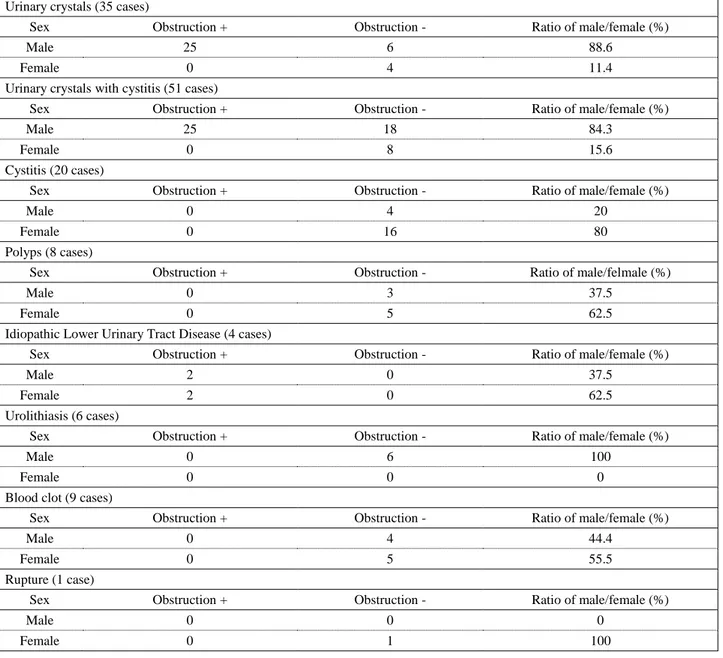
In this study; LUTD cases included urinary crystals (n=35), urinary crystals with cystitis (n=51), cystitis (n=20), bladder calculi (n=6), bladder polyps (n=8), blood clots (n=9), bladder rupture (n=1) and idiopathic LUTD (n=4). These cases were diagnosed by one of the three imaging methods (plain radiography, ultrasonography or contrast cystography) for each case. Details are shown on Table 3.
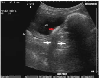
Urinary crystals appeared as hyperechoic foci of various sizes at the base of the bladder with acoustic shadow (Figure 1). Radiographic examination of these cats revealed normal bladder position, shape and sizes.
Figure 1. Ultrasonographic view of urinary crystals in a 2 years old male Persian cat (7.5 mHz). Red arrow shows the crystals which form a thick layer at the base of the bladder. White arrows show the acoustic shadow.
Şekil 1. İki yaşlı, erkek, İran ırkı bir kedide idrar kristallerinin ultrasonografik görünümü (7.5 mHz). Kırmızı ok, idrar kesesinin tabanında kalın bir tabaka oluşturan kristalleri göstermektedir. Beyaz oklar akustik gölgeyi gösterir.
In cats with chronic cystitis, bladder wall was irregular on contrast cystography. This irregularity, making indentations towards the lumen of the bladder that causes filling defects, appeared in a more prominent way. Thickening of the mucosal layer was more noticable in the trigone region.
The most typical finding in ultrasonographic examination of the cases was diffuse or focal thickening of the bladder wall, especially in chronic cystitis (Figure 2).
Table 2. Sex distribution of urinary tract obstructions in cats. Tablo 2. Kedilerde idrar yolu tıkanıklığının cinsiyet dağılımı.
Urinary crystals (35 cases)
Sex Obstruction + Obstruction - Ratio of male/female (%)
Male 25 6 88.6
Female 0 4 11.4
Urinary crystals with cystitis (51 cases)
Sex Obstruction + Obstruction - Ratio of male/female (%)
Male 25 18 84.3
Female 0 8 15.6
Cystitis (20 cases)
Sex Obstruction + Obstruction - Ratio of male/female (%)
Male 0 4 20
Female 0 16 80
Polyps (8 cases)
Sex Obstruction + Obstruction - Ratio of male/felmale (%)
Male 0 3 37.5
Female 0 5 62.5
Idiopathic Lower Urinary Tract Disease (4 cases)
Sex Obstruction + Obstruction - Ratio of male/female (%)
Male 2 0 37.5
Female 2 0 62.5
Urolithiasis (6 cases)
Sex Obstruction + Obstruction - Ratio of male/female (%)
Male 0 6 100
Female 0 0 0
Blood clot (9 cases)
Sex Obstruction + Obstruction - Ratio of male/female (%)
Male 0 4 44.4
Female 0 5 55.5
Rupture (1 case)
Sex Obstruction + Obstruction - Ratio of male/female (%)
Male 0 0 0
Female 0 1 100
Table 3. Number of diagnoses of lower urinary tract diseases for each imaging method. Tablo 3. Her bir görüntüleme yöntemi için alt idrar yolu hastalıklarının sayısı.
Imaging Method
Diagnosed Lower Urinary Tract Disease (LUTD) Plain Radiography Ultrasonography Contrast Cystography Total
Urinary crystals - 35 - 35
Urinary crystals with cystitis - 32 19 51
Cystitis - 10 10 20 Calculi 3 3 - 6 Polyps - 8 - 8 Blood clots - 9 - 9 Bladder rupture - - 1 1 Idiopathic obstruction - - - 4 Total 134
Figure 2. Ultrasonographic view of chronic cystitis in a 5 years old female Himalayan cat (7.5 mHz). Thickening of the bladder wall is shown with white arrows.
Şekil 2. Kronik sistitisin 5 yaşlı, dişi, Himalaya ırkı bir kedide ultrasonografik görünümü (7.5 mHz). İdrar kesesi duvarının kalınlaşması beyaz oklarla gösterilmektedir.
Figure 3a. Radiographic appearance of radiopaque stones in the bladder of 6 years old, male mix breed cat. V/D position (on top) and L/L position (below).
Şekil 3a. Altı yaşlı, erkek melez kedinin idrar kesesinde radyoopak taşların görünümü. V/D pozisyon (üstte) ve L/L pozisyon (altta).
Figure 3b. Ultrasonographic appearance of the same cat bladder. Hyperechoic stone formations causing acoustic shadowing (between arrows) is seen in the bladder.
Şekil 3b. Aynı kedinin idrar kesesinin ultrasonografik görünümü. İdrar kesesinde akustik gölgeye (oklar arasında) neden olan hiperekoik taş şekilleri görülmektedir.
Radiopaque stones of various numbers, shapes and sizes were seen in the bladder on plain radiographs in 3 cases (Figure 3a). Hyperechoic stone formations causing acoustic shadowing were seen in ultrasonographic examination (Figure 3b). Three radiolucent stones not determined in plain radiography appeared the same in ultrasonography as radiopaque stones.
Bladder polyps were diagnosed by ultrasonography (n=8), accompanied by wall thickening. Polypoid structures were seen to extend into the lumen mostly in trigone region (Figure 4).
In one cat with history of traffic accident, urinary bladder could not be determined on radiographic examination. Positive contrast cystography revealed rupture of the bladder and contrast media leaked into the abdominal cavity. Ultrasonographic examination of 9 cases showed large, echogenic, organized and mobile blood clots. The mobility of this mass clearly showed it was inside the bladder and not on the bladder wall (Figure 5).
Figure 4. Ultrasonography of polypoid cystitis in a 10 years old, male, Siamese cat. Different size polypoid structures are seen to extend into the lumen. The large one is in trigone region. Şekil 4. On yaşlı, erkek, Siyam ırkı bir kedide polipoid sistitis ultrasonografisi. Lümen içerisine doğru uzanmış değişik boyutlarda polipoid yapılar görülmektedir. Büyük olan trigon bölgesindedir.
Figure 5. Ultrasonographic appearance of a large, echogenic, organized blood clot inside the bladder (white arrow) in a 6 years old, male, mix breed cat.
Şekil 5. Altı yaşlı, erkek, melez kedinin idrar kesesi içinde büyük, ekojen, organize olmuş kan pıhtısının ultrasonografik görünümü (beyaz ok).
In 4 cases with obstructive idiopathic LUTD and unknown history of disease or trauma, in which plain radiographs showed that the bladders were full; there were no abnormal conditions in the bladder wall. In these cats, no abnormalities in the bladder lumen or mucosa were found on cystography and ultrasonography.
Discussion and Conclusion
LUTD describes the combination of clinical signs related to irritative voiding without identifying underlying etiology. Most cats have idiopathic or interstitial cystitis (6). Although the underlying causes of LUTD are not identified, there is a significant association between cats’ breeds and some specific types of the disease. Persian, Himalayan and Russian blue cats have great risk of developing urocystoliths. Burmese cats are less likely to have urethral obstruction than other cats. Manx cats have great risk of developing urinary incontinence (11). In this retrospective study, the most common LUTD was urinary crystals with cystitis. Idiopathic obstruction was observed in 4 cases. Persian was the most affected cat breed by LUTD. Alternatively, environmental features, especially the behavior of the owner on the cat, may play a role in development of LUTD. Buffington et al. (2006) observed that the owners of affected cats were often frustrated by the presence of a chronic disease in their cats, and appeared to feel anger toward the cats. This attitude might have activated the response of the cat (1). The present study did not identify environmental factors associated with LUTD, but this is considered to be important especially for indoor cats.
Castrated males and spayed females appear to have increased risk for LUTD (11). Although rates of males and females are quite similar, urethral obstructions due to crystals and stones were only diagnosed in male cats. Bladder stones irritate the bladder mucosal surface and provide a suitable environment for growth of bacteria. Female cats’ urethra is shorter and larger in diameter than male cats. Therefore, female cats rarely become obstructed. Male cats have a relatively long and quite narrow urethra and they are prone to congesting and obstructing the distal end of the urethra with stones.
No clinical sign is particularly diagnostic for LUTD in cats. Clinical signs, history, time of the disease, urinalysis and urinary tract imaging should be considered for diagnosis (11). In this study, cats with complaints of one or more common clinical signs had similar history. Radiographic, cystographic and ultrasonographic findings provide a major contribution for diagnosis of the disease (2). The use of ultrasound as an imaging method is important for the detection of urethral obstruction. Especially for treatment planning in cats presented with urethral obstruction, it is a valuable method (13). Radiographic examination is mostly preferred in lateral
position to avoid superposition of the urinary bladder with sacrum, vertebral column and bowels (2). Altough two different positions were used in this study, lateral position was found to be more satisfactory.
For a proper diagnostic examination, urinary bladder lumen must be full with urine. In a full bladder, localization, size, opacity and presence of radiopaque stones can easily be determined by radiography (3, 10). Radiopaque stones could be determined by radiography and radiolucent stones were identified by ultrasonography in this study.
In contrast cystography, radiopaque appearance of the thickened bladder wall having cystitis is very typical in dogs. In cats, severe cystitis can be diagnosed by double-contrast radiographs properly (7, 9). In cats with severe cystitis, the cumulation of contrast agent on inflammed mucosa was quite obvious in double-contrast radiographs in this study.
The appearance of crystalline and cellular sediments may not be differentiated during ultrasonographic examination. In cats, crystalline deposits may form a thick layer with hyperechoic appearance and acoustic shadow (12, 14). In this study, precipitate was assessed and crystals were diagnosed according to hyperechoic appearance and acoustic shadow.
Although crystalluria is often detected in cats without any consequences, crystals may adhere to bladder mucosa and cause cystitis by irritating mucosa (8, 10). In this study, the majority of cases, especially male cats had urinary crystals accompanied by cystitis. Radiography was insufficient to inspect the wall in initial stages of cystitis while radiopacity of the wall was noted in cats with chronic cystitis in contrast radiography. Increase in opacity occurs due to calcifications in mucosa (2). Double-contrast cystography was also inadequate for imaging bladder in acute cystitis. These conditions increased the need for ultrasonography. Indeed, ultrasonographic examination revealed bladder wall thickening and irregularity of mucosal layer in all cases with cystitis.
The present study confirms that ultrasonography is an efficient imaging method for examination of lower urinary tract, and radiography and contrast cystography are still useful for diagnosing bladder stones, ruptures, or suspected urethral obstructions. Anamnesis and clinical examination are very important in diagnosis and findings of imaging methods have to be compatible with clinical findings.
References
1. Buffington CAT, Westropp JL, Chew DJ, et al. (2006):
Risk factors associated with clinical signs of lower urinary tract disease in indoor-housed cats. JAVMA, 228, 722-725.
2. Burk RL, Feeney DA (2003): The abdomen. In: Burk RL, Feeney DA (Eds), Small Animal Radiology and Ultrasonography, A Diagnostic Atlas and Text. 1st edition, 249-476, Saunders, USA.
3. Farrow CS (2003): Kidney, ureteral bladder, prostatic and
urethral disease. In: Farrow CS (ed), Veterinary Diagnostic
Imaging: The Dog and Cat. Mosby Inc, Philadelphia. 4. Gerber B, Boretti FS, Kley S, et al. (2005): Evaluation of
clinical signs and causes of lower urinary tract disease in European cats. J Small Anim Pract, 46, 571-577.
5. Gunn-Moore DA (2003): Feline lower urinary tract
disease. J Feline Med Surg, 5, 133-138.
6. Hostutler RA, Chew DJ, Dibartola SP (2005): Recent
concepts in feline lower urinary tract disease. Vet Clin
Small Anim Pract, 35, 147-170.
7. Itkin RJ, Krawiec DR, Cloran JA, et al. (1994):
Ulcerative urocystitis in a dog associated with a Nocardia-like microorganism. JAVMA, 31, 296-299.
8. Johnston GR, Walter PA, Feeney DA (1986):
Radiographic and ultrasonographic features of uroliths and other urinary tract filling defects. Vet Clin North Am Small
Anim Pract, 16, 261-292.
9. Kalınbacak A, Atalay Ö, Kırmızıgül AH, et al. (2004):
Kedi ve köpeklerde sistitis’in tanısında çift kontrast sistografi tekniğinin kullanımı ve tedavide enrofloksasin’in etkinliğinin araştırılması. Ankara Üniv Vet Fak Derg, 51,
111-115.
10. Kealy JK, Mcallister H, Graham JP (2011): The
abdomen. In: Kealy JK, Mcallıster H, Graham JP (Eds),
Diagnostic Radiology and Ultrasonography of the Dog and Cat. 23-198, Saunders Elsevier, USA.
11. Lekcharoensuk C, Osborne CA, Lulich JP (2001):
Epidemiologic study of risk factors for lower urinary tract diseases in cats. JAVMA, 218, 1429-1435.
12. Leveille R (1998): Ultrasonography of urinary bladder
disorders. Vet Clin North Am Small Anim Pract, 28,
799-821.
13. Nevins J, Mai W, Thomas E (2015): Associations between
ultrasound and clinical findings in 87 cats with urethral obtruction. Vet Radiol Ultrasound, 56, 439-447.
14. Nyland TG, Mattoon JS, Wisner ER (1995):
Ultrasonography of the urinary tract and adrenal glands.
In: Nyland TG, Mattoon JS (Eds), Veterinary Diagnostic Ultrasound. 95-124, W.B. Saunders Company, Philadelphia.
Geliş tarihi: 16.08.2016 / Kabul tarihi: 01.03.2017
Address for correspondence:
Doç. Dr. Oytun Okan ŞENEL
Ankara University, Faculty of Veterinary Medicine Department of Surgery,
06110, Dışkapı, Ankara, Turkey. e-mail: [email protected]