Esra Cansever Mutlu1,2 , Özge Kaya3 , Arzu Birinci Yildirim4 and Ayhan Çetinkaya5 1Beykent University, Faculty of Engineering and Architecture, Department of Biomedical Engineering, Istanbul, Turkey. ²Scientific Industrial and Technological Application and Research Center, Bolu Abant Izzet Baysal University, Bolu, Turkey. ³Department of Biology, Faculty of Arts and Sciences, Bolu Abant Izzet Baysal University, Bolu, Turkey.
⁴Department of Field Crops, Faculty of Agricultural and Natural Sciences, Bolu Abant Izzet Baysal University, Bolu, Turkey. ⁵Department of Physiology, Faculty of Medicine, Bolu Abant İzzet Baysal University, Bolu, Turkey.
ÖZ
Ekzosomlar, kanser tedavisinde doğal, homojen, nano boyutlu, hedeflenmiş veziküller (~ 50 ila 100 nm) olarak özel özelliklerin son yıllarda kullanılmaya başlandığı doğal nanopartiküllerdir. Hedeflenen kanser hücresi yüzeylerine bağlanmak için yüksek bir aviditeye (birçok konformasyonel tutunmaya) sahiptirler. Özgün doğasında kanser hücresi membranı ile kolaylıkla etkileşebilen yapışkan pro-teinlere sahip olan biyoaktif çift-katmanlı lipit tabakalarından oluşurlar. Bu çalışmada, küçük hücreli olmayan A549 epitelyal kanser hücrelerinin ekzosomlarının doğal ya da sentetik ilaç taşıyıcı olabilme potansiyelleri araştırıldı. Öncelikle, ekzosom içermeyen or-tamlarda üretilen A549 hücre dizisinin ekzosomları üretildi. Hemen sonrasında, ekzosomlarının izolasyonu ultrasantrifüj prosedürü kullanılarak yapıldı. SEM görüntü, partikül boyutu ve zeta potansiyel ölçümleri, exosomal RNA analizleri ve Bradford yöntemi ile pro-tein içeriği analizi gerçekleştirildi. A549 hücrelerinin exozomlarının özelliklerine ilişkin bulgular (Boyut: 168 nm; zeta: -16mV), kanser hücresi terapisi için ilaç taşıyıcı olarak kullanılma potansiyellerini kanıtlamıştır.
Anahtar Kelimeler
Ekzosom, ultrasantrifüj, A549 hücre hattı, miRNA.
A B S T R AC T
E
xosomes are natural nanoparticles that their special features as a natural, homogeneous, nanosized, targeted vesicles (~ 50- 100 nm) have started to be used in the treatment of cancer very recently They have high avidity (many conformational attach-ment) to attach onto targeted cancer cell surfaces. They are composed of bioactive double-layered lipid layers in which their ori-ginal nature has the adhesive proteins interacting with the cancer cell membrane easily. In this study, the exosomes of non-small cell lung cancer, A549-epithelial carcinoma cells were investigated for their potential to be the natural or synthetic drug carrier. Firstly, exosomes of A549 cell line were produced using exosome-free media. Immediately after, isolation of their exosomes were performed by using ultracentrifugation procedure. Their SEM image, particle size and zeta potential measurements, exosomal RNA analysis and Protein Content by Bradford assays were performed. Findings (Size: 168 nm; zeta: -16mV) on the properties of A549 cell exosomes proved their potential to be used as the drug carrier for cancer cell therapy.Key Words
Exosome, ultracentrifugation, A549 cell Line, miRNA.
Article History: Received: Jan 31, 2019; Revised: Apr 20, 2019; Accepted: Jul 23, 2017; Available Online: Nov 1, 2019. DOI: https://doi.org/10.15671/hjbc.520101
Correspondence to: Cansever Mutlu, Beykent University, Faculty of Engineering and Architecture, Department of Biomedical Engineering, Istanbul,
Turkey.
E-Mail: [email protected]
Exosome Production, Isolation and Characterization from A549 Epithelial
Carcinoma Cells
A549 Epitelyal Karsinom Hücrelerinden Eksozom Üretimi, İzolasyonu ve
Karakterizasyonu
Hacettepe Journal of Biology and Chemistry
Research Article
E. Cansever Mutlu et al. / Hacettepe J. Biol. & Chem., 2019, 47 (4), 383-388
384
INTRODUCTION
E
xosomes as extracellular vehicles below 100 nm are composed of natural lipid bilayers and they are res-ponsible for cellular functions between cells and their environments. Moreover, they have got many oncolo-gical vital functions when they are especially secreted by cancer entitled oncosomes [1, 2]. Not only they can interact cellular membranes easily via adhesive prote-ins on their surface, but also avoid entrapments of mo-nonuclear phagocytes. This also represents their superi-or property fsuperi-or enhancing the delivery of incsuperi-orpsuperi-orated drugs to desired cells by altering therapeutic efficiency. Therefore, exosomes now are one of the new hot topics in the subject of nanomedicine among cancer therapy studies [3, 4].Lung cancer is one of the major health problems affec-ting too many people in all around the world. In 2012, the most common cancer type with 13% of occurren-ce frequency in the world was the lung canoccurren-cer. At the same time, lung cancer is the most common cause of death, corresponding to 19.4% out of total cancer de-aths in the world. Lung cancer is clinically based on the size and appearance of malignant cells; small cell lung (16.8%) and non-small cell lung cancer (80.4%) were di-vided into two main groups [5, 6].
Recently, it was indicated that the cancer cells secre-te their characsecre-teristic exosomes which are not present among exosomes of healthy cells. Exosomes from tu-mor cells play vital role to suppress immune system components [7]. Interestingly, they can be larger (in micron diameters) than ones from normal cells vesicles termed oncosomes. However, they are desired to be obtained in diameters below 200 nm.
In this study, we developed an isolation technique for the exosomes from A549 Epithelial carcinoma cells in order to evaluate their drug carrier potential according to their sizes and stabilities. Their miRNA, total RNA and protein content have been investigated due to the suf-ficiency of drug loading [3, 4, 8].
MATERIALS and METHODS
Materials
DMEM/F-12 (Dulbecco’s Modified Eagle Medium/Nut-rient Mixture F-12) is a widely used basal medium for supporting the growth of A549 Lung Cancer Cells, was
purchased as PAN BiotecTM . Fetal Bovine Serum (FBS)
was obtained as BioSeraTM. Penicilin-Streptomycin
(5000 U/mL), Trypsin-EDTA (0.25% w/phenol red), PBS (phosphate buffered saline w/o Calcium Magnesium Phenol Red) were used Gibco, Thermo Fisher Scientific. Ultracentrifuge tubes were purchased as HITACHI MO-DEL S303922ATM.
Pretreated Solutions
• PBS, ddH2O, FBS were subjected to ultracentrifu-gation to obtain ExoFreePBS, ExoFreeWater and ExoFreeFBS.
• After sterilization of ultracentrifuge tubes at 121oC, each has fulfilled using PBS, water and FBS. • After balance measurements, ultracentrifugation
were performed overnight at 120.000 g [9]. • Each tube had a volume of 8 mL. Supernatant was
collected gently after ultracentrifugation in order to be used during our further studies.
• Supernatant collected in another 15-mL sterile fal-con tubes and DexFreeFBS were kept -20oC again. DexFreePBS and DexFreeWater were kept +4oC.
Production of Exosomes
• A549 Epithelial Lung Carcinoma Cells were obta-ined from Bolu Abant Izzet Baysal University, De-partment of Physiology stocks.
• Cells were grown under optimum full growth con-dition (DMEM-F12 90%, 10% DexFreeFBS, Antibi-otic Solution (50 U.mL-1 penicillin, and 50 mg.mL-1
streptomycin), 0.5% Amphotericin in t25 flask). • After 3 days in a 5% CO2 humid atmosphere at 37°C,
cells were transferred to TPPTM t75 flasks for op-timal full growth condition by using Tripsin-EDTA and PBS.
• After 3 passages, 6,23x10³ cells were obtained in each t75 media. Cell Counting were performed by using Bio-Rad’s TC20™ Cell Counter.
Isolation of Exosomes
• Cells were transferred into centrifuge tubes. After centrifugation at 300g for 15 min at 4°C, cell pel-lets were removed.
• Supernatants were transferred into 8-mL ultra-centrifuge tubes placed in dry ice. Immediately after ultra-centrifugations were performed at 17.000g to eliminate cell debris completely. • Supernatants were filtered by sterile syringe
more than 200 nm [10].
• Filtered part were transferred to new sterile ultra-centrifuge tubes. Ultracentrifugation was perfor-med at 120.000 g for 60 minutes at 4°C.
• Pellet (Exosomes) were resuspended by ExoFree BPS by (3 X 50 L) to sterile cryotubes.
• Cryotubes were kept at -80 °C for exosome analysis.
SEM Analysis
Cryotubes containing exosomes were left to melt down at room temperature. A sample from each tube was dropped using sterile Pasteur pipettes onto metal grids with double sided adhesive carbon tape. After they are dried at room temperature, coated with gold to ~500×10−8 cm in thickness using sputter coater under
high vacuum, 0.1 Torr, 1.2 kV, and 50mA at 27 ± 1°C. The surface morphology of coated samples was evaluated by scanning electron microscopy (SEM), LeicaTM(2)[4].
Zeta/Size Analyses
Cryotubes were left to melt down at room temperatu-re. Each tube was diluted using 2 mL ExoFree PBS and measurements were performed by Malvern Nanosizer/ Zetasizer nano-ZS ZEN 3600. Completely disposable dip-cell cuvettes were used during measurements [4].
Extraction and Quantification of Exosomal RNA
In order to evaluate the exosomal RNA content; two co-lumn base commercial RNA isolation kits were used na-mely innuPREP RNA MinianalytikTM (Jena, Germany) for
total RNA extraction and miRNATM (Omega Bio-tek, Inc,
Guangzhou, China) kit for miRNA extraction. 30µL of exosome samples suspended in DexFree PBS were mi-xed with lysis buffer of the each kit and manufacturer’s instructions are followed. Isolated RNA molecules were eluted from the columns by centrifuge method as desc-ribed their procedures by using nuclease-free water. To-tal RNA and miRNA quantification measurements were carried out by using QuantiFluor® RNA Dye on fluoro-metry (Quantus™2 Fluorometer, Promega®, Madisson, USA) and Thermo Scientific / 2000 TMNanodrop. [4, 9]
and Nuclease free water used as the control. During the extraction whole working area was cleaned with the “RNase-ExitusPlus” from AppliChem and all the materi-als used for RNA extraction was nuclease-free in order to prevent RNase contamination.
Analysis of Protein Content
Total protein content of the A549 exosome samples sus-pended in DexFreePBS were evaluated with Bradford (1976) [11] method by using Pierce™ Coomassie (Brad-ford) Protein Assay Kit. The amount of total protein was measured colorimetrically at 595 nm using a UV/Vis spectrophotometer (Jasco V-530 UV/Vis spectropho-tometer, Jasco International Corporation, Tokyo, Japan) and was quantified as Bovine serum albumin equivalent (BSA) value.
E. Cansever Mutlu et al. / Hacettepe J. Biol. & Chem., 2019, 47 (4), 383-388
386
RESULTS and DISCUSSION
Size/zeta measurements of exosomes were performed in order to prove its drug delivery potential with respect to their dimensions and stabilities. Size distribution of exosomes displayed bimodal dispersion (Figure 1). The distribution of first peak is 168.3 ± 25.8 nm; second peak is 8.83 ± 0.9 nm. While, intensity of first peak is %87.9; intensity of the second peak is %12.1.
Zeta potential measurement showed that the exosome of A549 Lung Cancer Cell Line was -16 ± 8.72 mV (Fi-gure2).
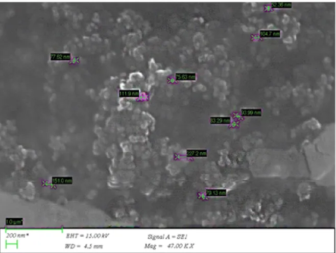
SEM images showed that the exosomes of A549 cell li-nes have the distribution between 50-150 nm (Figure 3). Images proved that coagulation has poor and the exosomes are highly durable even at 70°C. After 80°C, their spherical structures converted into more rigid structures.
RNA could not be detected in miRNA measurements. In the exosomes samples obtained from A549 cells, 4.5 ng/106 cell total RNA was detected, while RNA was not detected in the control sample with fluorometric measurements. Bradford (1976) [11] method was used to determine the exosomal protein concentration. So, exosome samples of A549 cells were measured protein content equivalent to 13.2 µg/106 cells of BSA. No pro-tein was detected in the control (DexFreePBS) samples. In this study, the potential of promising usage of exo-somes from A549 cell lines were investigated for their
drug loading capacities for probable use in cancer the-rapy as the next strategy [12, 13]. Thereby, we evalua-ted all the results to enlighten the material properties of natural nanoparticles, exosomes, of A549 cell line. According to our findings, all exosomes are quite small (≤200 nm), (-16 mV) and have lowest amount of RNA content. The prospective challenge would be that they have highly protein content before drug loading. In fact, even clinical study was performed previously [14]. Although a few studies in the literature were carri-ed out to produce exosomes of A549 cell line from mic-rovesicles (MVs), there is no spotlight exosomal study of this cancer line searching for their sizes, zeta, image analysis and molecular content such as miRNA, totalR-NA and protein contents [15].
CONCLUSION
Cancer cell exosomes has the great potential for cancer therapy, this perspective brings with many unknown questions for therapy. Steric stabilization and pH ef-fects of them are of note to change general phenomen for zeta potential measurements. Our results showed that exosomes of A549 cell line -16 mV. This circums-tance showed chemical structure of A549 cell line exo-somes should be investigated as the prospective study to enlighten protein content of their surface which may affect zeta potential value. Nonetheless, exosomes of A549 cell line has the great potential for synthetic drug loading by sonication method; for, they are spherically durable even at 70°C. These results, has been
yed the new research quide and the method article for A549 cell line exosomes in cancer therapy.
Acknowledgments
Authors thank to Bolu Abant İzzet Baysal University Research Fund through BAP Project 2018.31.01.1359.
R e f e r e n c e s
1. G. Mignot, S. Roux, C. Thery, E. Ségura, L. Zitvogel, Prospects for exosomes in immunotherapy of cancer, J. Cell. Mol. Med., 10 (2006) 376-388.
2. M. Morello, V. Minciacchi, P. De Candia, J. Yang, E. Posadas, H. Kim, D. Griffiths, N. Bhowmick, L. Chung, P. Gandellini, Large oncosomes mediate intercellular transfer of functional microRNA, Cell cycle, 12 (2013) 3526-3536.
3. P. Vader, X. O. Breakefield, M. J. Wood, Extracellular vesicles: emerging targets for cancer therapy, Trends. Mol. Med., 20 (2014) 385-393.
4. M. S. Kim, M. J. Haney, Y. Zhao, V. Mahajan, I. Deygen, N. L. Klyachko, E. Inskoe, A. Piroyan, M. Sokolsky, O. Okolie, Development of exosome-encapsulated paclitaxel to overcome MDR in cancer cells, Nanomed-Nanotechnol., 12 (2016) 655-664.
5. A. Jemal, R. Siegel, E. Ward, Y. Hao, J. Xu, T. Murray, M. J. Thun, Cancer statistics, 2008, CA: a cancer journal for clinicians, 58 (2008) 71-96.
6. R. Siegel, C. DeSantis, A. Jemal, Colorectal cancer statistics, 2014, CA: a cancer journal for clinicians, 64 (2014) 104-117. 7. C. Théry, M. Ostrowski, E. Segura, Membrane vesicles as
conveyors of immune responses, Nat. Rev. Immunol., 9 (2009) 581.
8. J. Palma, S. C. Yaddanapudi, L. Pigati, M. A. Havens, S. Jeong, G. A. Weiner, K. M. E. Weimer, B. Stern, M. L. Hastings, D. M. Duelli, MicroRNAs are exported from malignant cells in customized particles, Nucleic Acids Res., 40 (2012) 9125-9138.
9. S. El-Andaloussi, Y. Lee, S. Lakhal-Littleton, J. Li, Y. Seow, C. Gardiner, L. Alvarez-Erviti, I.L. Sargent, M.J. Wood, Exosome-mediated delivery of siRNA in vitro and in vivo, Nat. Protoc., 7 (2012) 2112.
10. C. Lässer, M. Eldh, J. Lötvall, Isolation and characterization of RNA-containing exosomes, Jove-J. Vis. Exp., (2012). 11. M.M. Bradford, A rapid and sensitive method for the
quantitation of microgram quantities of protein utilizing the principle of protein-dye binding, Anal. Biochem., 72 (1976) 248-254.
12. W. Li, D. Mu, F. Tian, Y. Hu, T. Jiang, Y. Han, J. Chen, G. Han, X. Li, Exosomes derived from Rab27a-overexpressing tumor cells elicit efficient induction of antitumor immunity, Mol. Med. Rep., 8 (2013) 1876-1882.
E. Cansever Mutlu et al. / Hacettepe J. Biol. & Chem., 2019, 47 (4), 383-388
388
13. M. Wysoczynski, M.Z. Ratajczak, Lung cancer secreted microvesicles: underappreciated modulators of microenvironment in expanding tumors, Int. J. Cancer, 125 (2009) 1595-1603.
14. M.A. Morse, J. Garst, T. Osada, S. Khan, A. Hobeika, T.M. Clay, N. Valente, R. Shreeniwas, M. A. Sutton, A. Delcayre, A phase I study of dexosome immunotherapy in patients with advanced non-small cell lung cancer, J. Transl. Med., 3 (2005) 9.
15. Y.T. Tang, Y.-Y. Huang, L. Zheng, S.-H. Qin, X.P. Xu, T.X. An, Y. Xu, Y.S. Wu, X.M. Hu, B.H. Ping, Comparison of isolation methods of exosomes and exosomal RNA from cell culture medium and serum, Int. J. Mol. Med., 40 (2017) 834-844.