Glucagon-Like Polypeptide-1 and Brain
Glukagon Benzeri Polipeptid-1 ve Beyin
Okan Sefa BAKINER, Akadlı Kürşad ÖZŞAHİN* Department of Endocrinology and Metabolism Diseases, Başkent University Faculty of Medicine, Ankara, TURKEY *Department of Family Physician, Başkent University Faculty of Medicine, Ankara, TURKEY
Turk J Endocrinol Metab. 2020;24:343-355
Introduction
Glucagon-like polypeptide-1 (GLP-1) is mainly produced in the small intestinal en-tero-endocrine cells, pancreatic alpha cells, and the nucleus tractus solitari (NTS) of the central nervous system, using pproglucagon as a precursor. It can be re-leased after conversion by prohormone convertase, and in less than 2 min, is broken down and inactivated by dipeptidyl pepti-dase-4 (DPP-4). In the circulation, GLP-1 can freely pass the blood-brain barrier (BBB), although only 20% reaches the brain
owing its short half-life in circulation (hu-moral pathway) (1,2). Industrially produced GLP-1 receptor agonists (GLP-1RA) can pass the BBB, whereas DPP-4 inhibitors cannot (3).
The important effects of GLP-1 in the brain have recently been discovered. It was ob-served that the main sources of GLP-1 in the brain were the neurons that expressed pre-proglucagon in the NTS (4). Unlike the in-testinal enteroendocrine cells, which are stimulated by food, these cells are activated with gastric distention, systemic
cholecys-Glucagon-like polypeptide-1 has specific effects on the cen-tral nervous system, including regulation of glucose meta-bolism, positive cardiovascular effects, slowing intestinal motility, immune modulation, and regulation of appetite and energy expenditure. Recently, positive effects of GLP-1 on brain energy utilization, inhibition and restoration of neuro-degeneration, response to stress, and protection against ischemic neuron damage have been demonstrated. Herein, the effects of glucagon-like polypeptide-1 on the central nervous system will be discussed.
Keywords: Glucagon-like polypeptide-1;
brain; central nervous system; neurodegeneration; stroke
Glukagon benzeri peptid 1’in glukoz bağımlı insülin sekres-yonu, beta hücre proliferassekres-yonu, glukagon sekresyon inhi-biyonu ile hepatik glukoz çıkışının azalması gibi metabolik etkilerinin yanında kardiyovasküler sistem üzerine olumlu etkiler, gastrointestinal motilitede yavaşlama, immunomo-dülasyon gibi etkileri olduğu bilinmektedir. Son zamanlarda santral sinir sistemi üzerine de belirgin etkiler gösterdiği saptanmıştır. Santral etkiler esas olarak iştah regülasyonu ve metabolizma üzerine olan etkilerin dışında beyin enerji kullanımının düzenlenmesi, nörodejenerasyonun önlenmesi ve düzeltilmesi, iskemik nöron hasarına karşı koruma ve or-ganizmanın strese olan cevabının düzenlenmesi şeklinde sı-ralanabilir. Burada glukagon benzeri peptid 1’in santral sinir sistemi üzerine olan etkileri tartışılacaktır.
Anahtar kelimeler: Glukagon benzeri peptid 1;
beyin; merkezi sinir sistemi; nörodejenerasyon; inme
Address for Correspondence: Okan Sefa BAKINER, Department of Endocrinology and Metabolism Diseases, Başkent University Adana Dr. Turgut Noyan Application and Research Center, Adana, TURKEY
Phone: +90 530 222 16 98 E-mail: [email protected] Peer review under responsibility of Turkish Journal of Endocrinology and Metabolism.
Received: 13 Jun 2020 Received in revised form: 25 Jul 2020 Accepted: 11 Aug 2020 Available online: 30 Sep 2020 1308-9846 / ® Copyright 2020 by Society of Endocrinology and Metabolism of Turkey.
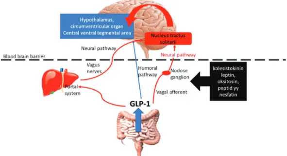
tokinin, lipopolysaccharide, or the applica-tion of lithium chloride, local leptin, or chole-cystokinin stimuli. Also, GLP-1 in the peripheral circulation has the potential to excite the neurons that produce pre-proglucagon in the NTS. Moreover, vagal ex-citation is the main source of stimulus on the preproglucagon neurons in NTS and se-cretes 1 locally (neural pathway). GLP-1 secreted from the intestine can generate vagal excitation by either local effect via in-testinal vagal afferents, or by stimulating the GLP-1 receptors on the portal venous system. Vagal afferents can also be directly stimulated by cholecystokinin, leptin, oxy-tocin, peptide yy, and nesfatin (Figure 1). Injection of GLP-1 in the portal vein in-creases insulin secretion from the pancreas, whereas this effect vanishes after hepatic vagotomy or knockdown of GLP-1 receptors in vagal afferents. This indicates that GLP-1 generates its insulinotropic effect not only by directly acting on beta cells but also by central effect (neuroendocrine effect) (5). Also, excitatory, inhibitory, or neuromodula-tory effects were observed on the feeding behavior of test animals after vagal dener-vation. Therefore, GLP-1 might play an im-portant role in long-term energy homeostasis, besides its known systemic ef-fects. This hypothesis is also supported by
the projection of neurons in the NTS that produces preproglucagon for supply to the presynaptic areas of regions such as area postrema, interpeduncular nucleus, pos-terodorsal and ventral tegmental nucleus, hypothalamic nuclei, thalamic nuclei, nu-cleus accumbens, and lateral septum, espe-cially for feeding and energy homeostasis. The presence of GLP-1 receptors in these areas also support this hypothesis (6). GLP-1 in both circulation and brain show their effect by connecting with the GLP-1 re-ceptors (GLP-1R). Excitation of GLP-1R in different centers of the central nervous sys-tem can create different responses. As a re-sult of GLP-1 activation in the brain, various physiological processes, such as appetite modulation, regulation of nausea, inhibition of reward and motivation centers, energy homeostasis, neuroprotection, increase in neural insulin sensitivity, neural plasticity, and memory formation, and immune modu-lation can occur.
Experiments conducted using GLP-1R analogs have demonstrated that they influ-ence the arcuate nucleus (ARC), paraven-tricular nucleus (PVN), and lateral hypothalamic area by acting like GLP-1R, mainly on the Proopiomelanocortin/Cocaine- and amphetamine-regulated transcript (POMC/CART) anorexigenic neurons and
directly inhibit the neuropeptide Y/Agouti-related peptide (NPY/AgRP) orexigenic neu-rons through GABAergic signals. Similarly, the stimulation of GLP-1 in the ventral tegmental area, known as the reward-moti-vation center, has been observed to control the mesolimbic dopaminergic signals, which decreases the pleasure of eating (7). It has also been demonstrated that the effect of GLP-1 analogs on the ventromedial hypo-thalamic neurons increases the sympathetic activity, which increases the thermogenesis with retention of glucose and triglyceride in the brown fat tissue, and causes the trans-formation of the white fat tissue to brown fat tissue (8).
GLP-1Rs are located in the G protein-cou-pled receptor group, and their stimulation activates some intracellular pathways (2). Stimulation of the receptor by GLP-1 acti-vates some pathways which can lead to acute or chronic responses. Examples of acute responses include insulin secretion, exocytosis, and an increase in the intracel-lular calcium ion concentration. The acute response mainly occurs with the activation of phosphoryl kinase via the cyclic adeno-sine monophosphate (cAMP) pathway. Ex-amples of chronic effects include modulation of the gene expression, cell growth, cell pro-liferation, cell differentiation, and anti-apop-totic activity. These effects occur through the phosphoinositide 3-kinase (PI3K) path-way, which is similar to the insulin down-stream pathway. Activation of PI3K triggers the activation of protein kinase B (AKT). This molecule is responsible for protein synthe-sis, an increase in anti-apoptotic factors, and inhibition of intracellular apoptotic fac-tors. Another important pathway responsi-ble for the chronic effect is the mitogen-associated protein kinase/extracel-lular signal-regulated kinase (MAPK/ERK) pathway. The activation of this pathway is responsible for important cytoprotective ef-fects, such as inhibition of apoptosis, a de-crease in oxidative stress, and a dede-crease in the inflammatory response.
Metabolic Basis of Neurodegenerative Diseases
Neurodegenerative diseases with dementia can affect memory, learning, cognitive func-tions, and behavior. There are
approxi-mately 50 million dementia patients
world-wide, and it is the 7th leading cause of
death. Alzheimer’s disease (AD) and Parkin-son’s disease (PD) account for a major pro-portion of neurodegenerative diseases. AD is characterized mainly by the extracellular accumulation of amyloid β protein, with the intracellular accumulation of hyperphospho-rylated τ protein in ligamentous form, neu-roinflammation, and decrease in the utilization of glucose by the brain. On the other hand, PD is characterized by the ac-cumulation of neuronal a-synuclein and Lewy fibrils, with the loss of dopaminergic neurons due to neuronal mitochondrial dys-function. The common features of neurode-generative diseases include the formation of neurotoxic aggregates, increased oxidative stress, activation of apoptotic pathways, neurotransmitter insufficiency, and neural differentiation failure (9). A decrease in neuronal glucose utilization due to cerebral insulin resistance is an important feature of AD (10). Initially, the brain was thought to be insensitive to insulin, but the eventual discovery of the presence of insulin recep-tors in certain parts of the brain has indi-cated that this molecule plays an important role in the metabolic, neurotropic, neuro-modulatory, and neuroendocrine regulation in the brain, as well as memory and learn-ing (11). Glucose utilization in the neurons occurs via insulin-independent glucose transporter-3 (GLUT-3) protein, whereas the forebrain, cerebral cortex, and hip-pocampus show coexpression of the insulin-dependent glucose transporter-4 (GLUT-4) protein (12). It is suggested that activities such as learning and remembering, which increase metabolic needs, can cause the ex-pression of the translocated GLUT-4. Simi-larly, expression of GLUT-4 has also been shown in the hypothalamus, which plays an important role in metabolic control. Besides the utilization of cerebral glucose, insulin plays an important role in synaptic plastic-ity, neurite multiplication, neuroprotection, memory formation, cognitive arrangement, multiplication, differentiation and myelina-tion of oligodendrocytes, and determinamyelina-tion of the levels of neurotransmitters such as
acetylcholine and norepinephrine (13,14).
It has been demonstrated that insulin is synthesized in the cerebral cortex and
hip-pocampus in the central nervous system, and that small amounts of pancreatic insulin can pass the BBB via selective transport and affect brain functions (15). Insulin levels in the cerebrospinal fluid (CSF) are correlated with those in plasma, and its transport through the BBB may be affected by obe-sity, diabetes, inflammation, and high plasma triglyceride levels. It was demon-strated that the presence of peripheral in-sulin resistance decreased the CSF/peripheral insulin ratio and flow of in-sulin from blood to the CSF (16). In diabetic animal models, the effect of insulin in the brain decreases and is associated with
de-mentia (17). The Rotterdam study is among
the pioneering research that highlighted the risk of increased dementia in type-2 dia-betic population (18). Following this, the idea of an association between AD, insulin resistance, and brain glucose utilization was
first hypothesized 22 years ago (19). The
risk of dementia increases by 65% in type-2 diabetics compared to non-diabetics (20). Also, peripheral insulin resistance is associ-ated with decreased cerebral glucose uti-lization, decreased cerebral perfusion, and brain atrophy (11). Obesity and type 2 dia-betes, which are risk factors for AD, are as-sociated with an increase in the fat tissue-originated pro-inflammatory adipokines and plasma free fatty acids.
These cytokines can pass the BBB in AD and other conditions where the cerebral circula-tion is decreased. In the neurons, it can cause the phosphorylation of insulin recep-tor substrate 1 (IRS-1) via the activation of serine kinase. Thus, the PI3K pathway is blocked, and insulin resistance occurs (21). On the other hand, insulin increases the he-patic clearance of neuronal amyloid β. In the case of peripheral insulin resistance, the ac-cumulation of cerebral amyloid β is in-creased due to diminished hepatic clearance
of amyloid β (22). However, some studies
advocate that the type of insulin resistance in the pathogenesis of AD is not the periph-eral insulin resistance, but the neuronal one, which is present in the central nervous sys-tem (23). The fact that tight glycemic con-trol cannot help dementia regression in diabetic patients, and that the regression is even more aggravated due to frequent hy-poglycemia, also indicates that intranasal
in-sulin should improve cognitive functions and memory, in doses that do not cause periph-eral hypoglycemia. These findings confirm that central insulin resistance should be dif-ferentiated from peripheral resistance. Ac-cordingly, some researchers have named the association between central insulin re-sistance and dementia as type-3 diabetes (24–27).
In cerebral insulin resistance, inhibition of the AKT pathway after phosphorylation of IRS-1 results in an increase in the levels of Glycogen Synthase Kinase-3 (GSK-3). Fur-thermore, the pathway causes an increase in Protein Kinase A (PKA), and both these conditions contribute to the pathogenesis of AD by causing the phosphorylation of τ-pro-tein. Increasing GSK-3 levels cause an in-crease in the production of amyloid-β proteins. Neurotoxic amyloid oligomers and plaques are created from amyloid-β pro-teins. Amyloid-β oligomers may further con-tribute to neuronal insulin resistance by directly causing an increase in the phospho-rylation of IRS-1, activation of TNF-α signal, or activating the glial cells to increase cy-tokine (lL-1 β, lL-6, TNF-α) secretion. Finally, decreased intracellular entry of glucose due to insulin resistance causes a decrease in neuronal ATP production and deterioration of synaptic activity. Thus, cognitive dysfunc-tion occurs (28).
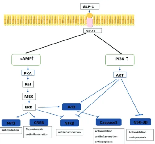
Effects of GLP-1 on Alzheimer Disease GLP-1 affects the brain through basic anti-apoptotic, antioxidant, and neurotrophic im-pact on GLP-1R. PKA and PI3K pathways are activated upon increasing cAMP, which oc-curs due to the stimulation of GLP-1R. Acti-vation of PKA actiActi-vation increases ERK, while the activation of PI3K increases AKT. The increase in both ERK and AKT triggers the transcription of genes responsible for antioxidant, anti-apoptotic, neurotropic, and anti-inflammatory effects (Figure 2) (29). GLP-1 can decrease insulin resistance in the brain and reverse it. It was demonstrated that liraglutide normalized the phosphoryla-tion of neuronal IRS-1 serine and the levels of AKT and GSK-3 β, as well as decreased the cerebral insulin levels (30). GLP-1 was also demonstrated to improve cognitive functions by increasing the utilization of glu-cose in the brain (31).
GLP-1 modulates the activation of microglia. It causes immunomodulation by inhibiting specific caspases and NF-κB (Nuclear Fac-tor-κB) with PI3K/AKT pathway activity and decreases the secretion of pro-inflammatory cytokines (32).
GLP-1 protects the neurons from oxidative stress and reduces oxidative stress. GSK-3 β is the main responsible factor for oxidative stress in neurons. An increase in levels of AKT decreases in the levels of GSK-3 β and
reduces oxidative stress (33). GLP-1
re-duces the accumulation of intracellular ROS (Reactive oxygen species) in microglia, in-creases the production of nitric oxide, and increases the levels of antioxidant glu-tathione peroxidase and superoxide dismu-tase-1. These activities protect the neurons from oxidative stress.
GLP-1, via decreased caspase 3/7 activity, also inhibits microglial apoptosis and de-creases the secretion of cytokines such as TNFα, IL1β, and IL-6, that trigger insulin
re-sistance (34). When administered intraperi-toneally, exenatide was observed to de-crease TNF-alpha levels and neuroinflammation (35).
GLP-1 enhances synaptic plasticity in the brain. Rats treated with lixisenatide were observed to show an increase in the synap-tic plassynap-ticity and learning (36). Neurites play a key role in the formation of inter-neuronal functional synapses, as well as between neurons and the surrounding microenviron-ment. Experimentally, exenatide has been shown to increase the levels of active neu-rites in cells (37). It has been observed that in rat models, lixisenatide inhibits synaptic damage due to amyloid accumulation and strengthens spatial memory (38).
GLP-1 regulates neurogenesis in the brain. It facilitates the development of new neu-rons and their differentiation from neuronal stem cells, rather than damaged neurons via the MAPK pathway. It also facilitates the maturation of neurites (39). Liraglutide has
Figure 2: The neuroprotective signaling pathways of GLP-1.
been experimentally shown to increase the differentiation of neural progenitor cells to mature neurons. It inhibits neuron apoptosis by increasing the levels of the survival fac-tors Bcl-2 (B-cell lymphoma 2) and Bcl-XL (B-cell lymphoma-extra-large) through the PI3K/AKT pathway. It increases cell prolifer-ation through the same pathways and stim-ulates differentiation of neurons and growth of neurites by increasing growth factors such as brain-derived neurotrophic factor (BDNF) and glial cell-derived neurotrophic
factor (GDNF) (31,40,41). Liraglutide has
been shown to increase memory in experi-mental animals by preventing neurodegen-eration and synaptic damage (42).
GLP-1 decreases the phosphorylation of τ-protein and accumulation of amyloid-β pro-tein. It also decreases the production of GSK-3 β in the neurons via the PI3K path-way. GSK-3 β is responsible for the phos-phorylation of neuronal τ-protein. Liraglutide has been observed to prevent the hyper-phosphorylation of τ-protein in diabetic db/db rats (43). Besides, GLP-1 reduces the levels of endogen amyloid-β protein in the brain and prevents the accumulation of
amyloid plaques (42). The formation of
amyloid plaques in experimentally created AD in female rat models has been shown to reduce by treatment with lixisenatide (44). DPP-4 (dipeptidyl peptidase) inhibitors are used as incretinergic agents for the treat-ment of diabetes, as are GLP-1 receptor analogs. These drugs inhibit the DPP-4 en-zyme and inactivate it. They cannot pass the BBB, due to which their effect on the central nervous system is associated with increas-ing endogenous GLP-1 levels. Moreover, in-hibition of the DPP-4 enzyme reduces the breakdown of stromal cell-derived factor-1 (SDF-1). SDF-1 binds to the chemokine re-ceptor-4 (CXCR-4) on the surface of the neurons and activates the PI3K/AKT path-way, which causes cell proliferation and sur-vival. DPP-4 inhibitors also positively influence vascularization in the brain by in-creasing the expression of vascular en-dothelial growth factor (VEGF) through SDF-1 (45).
To date, no randomized prospective studies have been conducted on enough humans to study the neurocognitive effect, which is dif-ferent from the antidiabetic effect, using
DPP-4 inhibitors or GLP-1 agonists. Accord-ing to a meta-analysis, seven studies have fulfilled the inclusion criteria, five of which are interventional, while two are observa-tional (46). An observational study on dia-betic patients has revealed that increased plasma DPP-4 levels are associated with mild loss in cognitive functions (47). In a study on 240 cases, DPP-4 inhibitors have been shown to improve cognitive function compared to sulfonylurea (48). A six-month-long, randomized, placebo-controlled, and double-blind study has shown liraglutide to significantly increase brain glucose metabo-lism compared to placebo (49). Similar re-sults have been reported in a two-year-long, randomized study, conducted with exenatide
(50). On the other hand, a six-month-long
randomized double-blind study comparing li-raglutide with placebo reported no differ-ence in the formation or regression of
amyloid plaque in the brain (51). Another
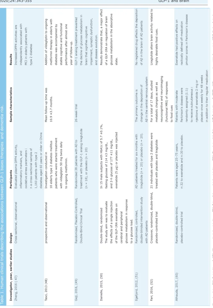
study has demonstrated that liraglutide did not show a significantly improved effect on cognitive functions over a relatively short period of 17 days, compared to the placebo (52). The clinical studies on this area are summarized in Table 1.
Therefore, the benefits of GLP-1 analogs and DPP-4 inhibitors on neurodegenerative dis-eases, independent of glycemia, have not been proven completely. The previous stud-ies have relatively small sample sizes, and some individual phenotypic differences such as apo E4 carriage may alter neurodegener-ative-diabetes and associated treatment (53).
Effects of GLP-1 on Parkinson’s Disease In preclinical studies on different experi-mental models of simulated Parkinson’s dis-ease, GLP-1 receptor agonists have been shown to reduce the loss of dopaminergic neurons, and regulate brain energy metab-olism and motor activity with neuroprotec-tive effect. These effects are similar to those described in AD by the activation of PKA and PI3K/AKT molecular pathways through GLP-1 receptors. Exenatide treatment in rats with experimentally induced dopaminergic neuron loss using toxic substances com-pletely decreased the toxicity and increased
dopaminergic neuron viability (54).
neu-Monophosphate; PKA: Protein kinase A; MEK:
Mitogen-activ
ated protein kinase; ERK: Extr
acellular regulated protein kinase; PI3K
: Phosphoinositide 3 kinase, AKT
: Protein kinase B; CREB: cAMP response element
-binding protein; NFK
β
: Nuclear factor
kappa-beta; Bcl2: B-cell lymphoma; GSK
-3
β
: Gly
cogen synthase kinase 3 beta.
Investigator, year, earlier studies
Design Participants Sample characteristics Results Zheng, 2016 ( 47) Cross-sectional, observ ational Ev
aluated plasma DPP4 activity
,
Increased DPP4 activities are
inflammatory mark
ers, and
independently associated with
oxidativ
e stress par
ameters i
MCI in elderly patients with
n a cross-sectional sample of
type 2 diabetes
1,160 patients with type 2 diabetes aged 60 y
ears or older in China
Tasci, 2013 (48) prospectiv e and observ ational In vestigation conducted in Mean follow -up time w as
Addition of vildagliptin to ong
oing
10 elderly type 2 diabetes mellitus
10.9 ±3.7 months.
metformin ther
apy in elderly with
patients who were started treatment
diabetes w
as accompanied by
with vildagliptin 50 mg twice daily
stable cognitiv
e and functional
to ongoing metformin.
performance after almost one year of follow
-up . Gejl, 2016, (49) R andomiz ed, Placebo-Controlled, R andomiz ed 38 patients with AD to 26-week trial
GLP-1 analog treatment prev
ented
Double-Blind Clinical T
rial
treatment with the GLP-1 analog lir
aglutide
the decline of glucose metabolism in
(n = 18), or placebo (n = 20)
br
ain that signifies cognitiv
e
impairment, synaptic dysfunction, and disease ev
olution.
Danielle, 2015, (50)
Double-blind, r
andomiz
ed
In 15 male subjects with HbA1c of 5.7 ±0.1%,
R
esults demonstr
ate that, a major effec
t
The study aim w
as to ev
aluate
fasting glucose of 114 ±3 mg/dL,
of a GLP-1RA on regulation of br
ain
the effects of a single injection
and 2-h glucose of 177 ±11 mg/dL,
glucose metabolism in the absorptiv
e of the GLP-1RA ex enatide on ex enatide (5 µg) or placebo w as injected state. cerebr al and peripher al
glucose metabolism in response to a glucose load.
Egefjord, 2012, (51)
R
andomiz
ed, controlled,
AD patients treated for six months with
The primary outcome is
No registered drug a
ffects the deposition
double-blinded interv
ention study
lir
aglutide (n = 20) or placebo (n = 20).
change in the deposition of
of A β in the br ain of AD patients with AD patients A β in the centr al nerv ous system Farr , 2016, (52) Crosso ve r, r andomiz ed, double-blind,
21 individuals with type 2 diabetes were
For a total of 17 da
ys, studied
Lir
aglutide alters br
ain activity related to
placebo-controlled trial
treated with placebo and lir
aglutide
metabolic changes as well as
highly desir
able food cues.
neurocognitiv
e and neuroimaging
(functional MRI) of responses to food cues
A
thauda, 2017, (60)
R
andomiz
ed, double-blind,
Patients were aged 25–75 y
ears,
Patients with moder
ate
Ex
enatide had positiv
e effe
cts on
placebo-controlled trial
n:32 to ex
enatide and n:30 to placebo
.
Parkinson's disease were
ractically defined off
-medication randomly assigned (1:1) pmotor scores in P arkinson's disease to receiv e subcutaneous i njections of ex enatide 2 mg or
placebo once weekly for 48 weeks in addition to their regular medication
Table 1. Human clinical trials describing the associations between GLP-1 bases ther
rotoxin observed higher levels of dopamine in animals administered Exendin-4. This ef-fect was associated with an increase in the viability of dopaminergic neurons, and an in-crease in the levels of tyrosine hydroxylase enzyme, which produces dopamine from its precursor, L-DOPA (55). Cell culture studies have demonstrated that liraglutide and lixisenatide also increase the activity of ty-rosine hydroxylase, and trigger the anti-apoptotic mechanism, compared to exendin (56). Long-term use of liraglutide has been shown to inhibit dopaminergic neuron loss in db-/db- diabetic mice models and prevent the impairment of motor functions and
de-velopment of PD (57). Experimentally,
li-raglutide has been shown to reduce dyskinesia, which is an important complica-tion of L-DOPA treatment (58). Semaglutide has been shown to reduce the accumulation of α-synuclein, in addition to the beneficial effects of other GLP-1R agonists (59). The first randomized double-blind placebo-con-trolled clinical study conducted on humans was published in 2017 on 62 cases with PD, wherein 2 mg/week exenatide LAR sc was compared to placebo. Exenatide was ob-served to show positive effects on clinically
monitored motor functions (60). Based on
these findings, GLP-1 agonists appear to have positive effects on the survival and functions of dopaminergic cells. The field is open for additional clinical studies for the development of an appropriate treatment strategy.
Physiological Effects pf Central GLP-1 on Stress
Recent studies have focused on the effect of GLP-1 on the stress response of the organ-ism. NTS has central GLP-1-producing cells and receives vagal stimulus from the pe-riphery, as well as a stimulus for general homeostasis. Stress activates two parallel systems in the body, hypothalamo-hy-pophysioadrenal (HPA) axis and the sympa-thetic nervous system (SNS). NTS is projected to hypothalamic and autonomic control centers, which are effective in both these systems. GLP-1 produced by NTS neu-rons can activate both the HPA axis and SNS. There is no GLP-1R in the adrenal cor-tex, and the isolated adrenal cortex cells do not release corticosterone upon GLP-1
stim-ulus. Therefore, the effect is known to have a central origin. When administered cen-trally, exendin-4 was observed to increase corticosterone levels in rodents. This in-crease is generated through CRH levels. The central blockade of the CRH receptors elim-inates this effect (61). On the other hand, the activation of GLP-1 receptors in the cen-tral autonomic system may stimulate sym-pathetic neuronal cells in the spinal cord and
adrenal medulla (62). As a result, GLP-1
may regulate the stress response of the body. Studies related to the clinical benefits of this condition and GLP-1RA practices are ongoing.
Ischemic Stroke and Glp-1
The stroke-damaged brain region can be di-vided into two parts, the ischemic core and the penumbra around the ischemic core. In the ischemic core, the damage takes place rapidly and is generally irreversible. The sur-rounding penumbra provides blood flow, and therefore, the neurons in this area can be protected by well-planned, timely interven-tions. These interventions are aimed at the chemical or surgical removal of the throm-botic clot. These treatments are generally beneficial within the first few hours after the onset of stroke, and unfortunately, may not be suitable for several patients due to late admission, difficulties in diagnosis, or con-traindications such as hypertension.
In a stroke, the mechanisms of action of GLP-1-based therapies have been studied thoroughly in animal models, but studies on human stroke patients have begun recently (ClinicalTrials.gov NCT02829502). In the LEADER (Liraglutide Effect and Action in Di-abetes) trial, a decrease in the rates of nonfatal stroke was observed in the liraglu-tide group, although the difference was non-significant (63). In the late SUSTAIN-6 (Trial to Evaluate Cardiovascular and Other Long-Term Outcomes with Semaglutide in Subjects with Type-2 Diabetes) trial, semaglutide was observed to significantly reduce the risk of the same primary com-posite endpoint as liraglutide. Both the tri-als demonstrated a decrease in major cardiovascular outcomes; however, there was no reduction in cardiovascular mortal-ity in the SUSTAIN-6 trial, as observed in the LEADER trial. There was another
im-portant difference between the studies. In the SUSTAIN-6 trial, the reduction in the major CV outcome appeared to be the re-sult of a significant (39%) decrease in the rate of nonfatal stroke (64). In large car-diovascular safety studies using DPP-4 in-hibitors, no decrease in the rates of nonfatal stroke or other major CV outcomes
was reported (65). GLP-1RA can cross the
BBB and enter the hypothalamic neurons and circumventricular organs, whereas DPP-4 inhibitors cannot. Therefore, GLP-1R agonists and DPP-4 inhibitors appear to have different effects on the ischemic brain tissue due to different pharmacological spe-cialties. In mice lacking GLP-1R, DPP-4 in-hibitors were observed to be neuroprotective (66). DPP-4 inhibitors may affect targets independent of GLP-1R. In experimental models, Rohnert et al. showed that non-selective DPP-4 inhibitors were related to a reduction in infarct vol-ume, which was not true for selective DPP-4 inhibitors (67). The mechanisms of these actions are yet to be fully clear. The DPP-4 enzyme targets more active peptides, cluding the glucose-dependent in-sulinotropic polypeptide (GIP) and the pituitary adenylate-cyclase-activating polypeptide (PACAP), than the GLP-1. Han et al. have demonstrated that a dual ago-nist targeting both GLP-1 and GIP receptors generated stronger neuroprotection against ischemic stroke than GLP-1 analog alone, indicating the neuroprotective effects of
GIP against stroke (68). Moreover, it was
reported that DPP-4 inhibition could ame-liorate endothelium-dependent relaxation, and cerebrovascular dysfunction and re-modeling, independent of glucose regula-tion (69).
Previous studies on animals have high-lighted the neuroprotective effect of GLP-1RAs in both diabetic and non-diabetic animals. Inflammation, oxidative stress, and neuronal apoptosis due to stroke are partic-ularly affected by GLP-1-based therapies. Neuroprotective Mechanisms of GLP-1-Based Therapies in Ischemic Stroke
After an ischemic stroke, GLP-1R is upregu-lated in astrocytes, GABAergic interneurons,
and microglia (70). The molecules PI3K,
cyclic adenosine monophosphate (cAMP), protein kinase-B (Akt), protein kinase-A (PKA), and cAMP response element-binding protein (CREB) are stimulated after the ad-ministration of GLP-1RAs in experimentally-induced stroke.
Inflammation, endothelial leakage, and
excitotoxicity: The effect of GLP-1-based
therapies on inflammatory response after a stroke has been studied by several searchers. Both exenatide and sitagliptin re-duced the production of the pro-inflammatory cytokine tumor necrosis factor-alpha (TNF-α) (71). Sitagliptin also reduced the expression of interleukin-6 (IL-6) and increased the production of the anti-inflammatory cytokine interleukin-10 (IL-10) (72). Therapies based on GLP-1 also influence the BBB permeability. Exendin-4 significantly reduced the expression of aquaporin-4 (AQ-4) and glial fibrillary acidic protein (GFAP), which are involved in neu-roinflammation, the formation of edema, and astrogliosis. Moreover, exendin-4 de-creases the intercellular adhesion
molecule-1 (ICAM-molecule-1) and inhibits BBB leakage (73).
Expression of endothelial NOS (eNOS), which plays a role in the maintenance of vascular homeostasis, is increased by re-combinant GLP-1, while exenatide reduces the vascular extravasation of immunoglob-ulin G (IgG), reducing endothelial leakage in the late inflammatory response to ischemia (71,74). Production of brain-derived neu-rotrophic factor (BDNF) was increased with pre-ischemic treatment with alogliptin, which reduced the release of neurotoxic glu-tamate and stabilized the neuroprotective GABAergic synapses with continuous expo-sure (75).
Oxidative stress: Both liraglutide and
exe-natide decreased the production of reactive oxygen species (ROS) after cerebral artery occlusion in diabetic mice (76). Exendin-4
decreased superoxide anion (O2) (73). In
sitagliptin-treated animals, the enzyme NADPH oxidase (NOx), which participates in the generation of ROS, was normalized (72).
Apoptosis and neuronal damage: To
iden-tify DNA damage, the “terminal deoxynu-cleotidyl transferase dUTP nick-end labeling’’ (TUNEL) assay is used. In ischemic lesions, the count of TUNEL-positive cells was signifi-cantly reduced by exenatide, liraglutide, and
modified GLP-1R agonists (77). Exenatide also reduced the activity of Caspase-3, which takes part in apoptosis (78).
Infarct volume: When administered
pre-is-chemia or post-ispre-is-chemia, GLP-1R agonists are observed to reduce infarct volume (79). High doses of exenatide were observed to be neuroprotective when administered 1.5 h and 3 h post-ischemia, and the activity di-minished upon late administration. Exe-natide administered early resulted in the greatest reduction in the infarct volume compared to later delivery (80). In contrast, the effects of DPP-4 inhibitors are more vari-able.
Functional outcome and cerebral blood
flow: Studies on GLP-1R agonists generally
reported improved functional outcomes, whereas only one out of two studies using DPP-4 inhibitors demonstrated such an ef-fect. In the first study, Ma et al. reported that linagliptin ameliorated cognitive impair-ment in diabetic mice, while the other study on alogliptin reported no beneficial effects of the compound on neurologic deficits when administered daily for three weeks before the stroke, to seven days post-ischemia (75,81). Conflicting results for the cere-brovascular effects of GLP-1RAs have been reported. In one study, treatment with ex-endin-4 was observed to increase blood flow, suggesting that GLP-1RAs impaired vascular function in ischemia, while another study reported that liraglutide did not affect the cerebral blood flow (73,82). The effects of DPP-4I administration on cerebral blood flow were also variable.
In conclusion, GLP-1-based therapies have been mainly used in diabetes and obesity, but recent studies have demonstrated their protective roles in the central nervous sys-tem. These agents, which have experimen-tally proven benefits both in neurodegenerative diseases and ischemic neuronal injuries, should be examined in this field by clinical studies. Randomized, placebo-controlled, prospective, double-blind open-ended studies and systematic meta-analyses will indicate whether GLP-based treatments have neuroprotective effects in clinical practice, regardless of the antidia-betic effect. Moreover, the new GLP-1 com-binations, developed together with GIP and glucagon agonists, also need well-planned
clinical trials, even though their benefits have been demonstrated experimentally (83). It does not appear far-fetched to use these agents as neuroprotective agents in di-abetic or non-didi-abetic patients soon.
Source of Finance
During this study, no financial or spiritual support was received neither from any phar-maceutical company that has a direct con-nection with the research subject, nor from a company that provides or produces med-ical instruments and materials which may negatively affect the evaluation process of this study.
Conflict of Interest
No conflicts of interest between the authors and / or family members of the scientific and medical committee members or members of the potential conflicts of interest, counsel-ing, expertise, working conditions, share holding and similar situations in any firm. Authorship Contributions
Idea/Concept: Okan Sefa Bakıner; Design: Okan Sefa Bakıner; Control/Supervision: Akadlı Kürşad Özşahin; Data Collection and/or Processing: Okan Sefa Bakıner; Analy-sis and/or Interpretation: Okan Sefa Bakıner; Literature Review: Okan Sefa Bakıner; Writ-ing the Article: Okan Sefa Bakıner; Critical Review: Okan Sefa Bakıner; Other (Language edition): Akadlı Kürşad Özşahin.
References
1. Campbell JE, Drucker DJ. Pharmacology, physiology, and mechanisms of incretin hormone action. Cell Metab. 2013;17:819-837. [Crossref] [PubMed]
2. Smith NK, Hackett TA, Galli A, Flynn CR. GLP-1: Molec-ular mechanisms and outcomes of a complex signaling system. Neurochem Int. 2019;128:94-105. [Crossref] [PubMed] [PMC]
3. Fuchs H, Binder R, Greischel A. Tissue distribution of the novel DPP-4 inhibitor BI 1356 is dominated by saturable binding to its target in rats. Biopharm Drug Dispos. 2009;30:229-240. [Crossref] [PubMed]
4. Holt MK, Richards JE, Cook DR, Brierley DI, Williams DL, Reimann F, Gribble FM, Trapp S. Preproglucagon neu-rons in the nucleus of the solitary tract are the main source of brain GLP-1, mediate stress-induced hy-pophagia, and limit unusually large intakes of food. Di-abetes. 2019;68:21-33. [Crossref] [PubMed] [PMC] 5. Nishizawa M, Nakabayashi H, Uehara K, Nakagawa A,
Uchida K, Koya D. Intraportal GLP-1 stimulates insulin secretion predominantly through the hepatoportal-pan-creatic vagal reflex pathways. Am J Physiol Endocrinol Metab. 2013;305:E376-E387. [Crossref] [PubMed]
6. Merchenthaler I, Lane M, Shughrue P. Distribution of pre-pro-glucagon and glucagon-like peptide-1 receptor messenger RNAs in the rat central nervous system. J Comp Neurol. 1999;403:261-280. [Crossref] [PubMed] 7. Alhadeff AL, Rupprecht LE, Hayes MR. GLP-1 neurons in
the nucleus of the solitary tract project directly to the ventral tegmental area and nucleus accumbens to con-trol for food intake. Endocrinology. 2012;153:647-658. [Crossref] [PubMed] [PMC]
8. Lockie SH, Heppner KM, Chaudhary N, Chabenne JR, Morgan DA, Veyrat-Durebex C, Ananthakrishnan G, Rohner-Jeanrenaud F, Drucker DJ, DiMarchi R, Rah-mouni K, Oldfield BJ, Tschöp MH, Perez-Tilve D. Direct control of brown adipose tissue thermogenesis by cen-tral nervous system glucagon-like peptide-1 receptor signaling. Diabetes. 2012;61:2753-2762. [Crossref] [PubMed] [PMC]
9. Ahmad K, Baig MH, Mushtaq G, Kamal MA, Greig NH, Choi I. Commonalities in biological pathways, genetics, and cellular mechanism between Alzheimer disease and other neurodegenerative diseases: an in silico-updated overview. Curr Alzheimer Res. 2017;14:1190-1197. [Crossref] [PubMed] [PMC]
10. de la Monte SM. Brain insulin resistance and deficiency as therapeutic targets in Alzheimer's disease. Curr Alzheimer Res. 2012;9:35-66. [Crossref] [PubMed] [PMC]
11. Frölich L, Blum-Degen D, Bernstein HG, Engelsberger S, Humrich J, Laufer S, Muschner D, Thalheimer A, Türk A, Hoyer S, Zöchling R, Boissl KW, Jellinger K, Riederer P. Brain insulin and insulin receptors in aging and sporadic Alzheimer's disease. J Neural Transm (Vienna). 1998;105:423-438. [Crossref] [PubMed]
12. McEwen BS, Reagan LP. Glucose transporter expression in the central nervous system: relationship to synaptic function. Eur J Pharmacol. 2004;490:13-24. [Crossref] [PubMed]
13. Stockhorst U, de Fries D, Steingrueber HJ, Scherbaum WA. Insulin and the CNS: effects on food intake, mem-ory, and endocrine parameters and the role of intranasal insulin administration in humans. Physiol Behav. 2004;83:47-54. [Crossref] [PubMed]
14. Chiu SL, Chen CM, Cline HT. Insulin receptor signaling regulates synapse number, dendritic plasticity, and cir-cuit function in vivo. Neuron. 2008;58:708-719. [Cross-ref] [PubMed] [PMC]
15. Devaskar SU, Giddings SJ, Rajakumar PA, Carnaghi LR, Menon RK, Zahm DS. Insulin gene expression and in-sulin synthesis in mammalian neuronal cells. J Biol Chem. 1994;269:8445-8454. [PubMed]
16. Banks WA. The source of cerebral insulin. Eur J Phar-macol. 2004;490:5-12. [Crossref] [PubMed] 17. Agrawal R, Zhuang Y, Cummings BP, Stanhope KL,
Gra-ham JL, Havel PJ, Gomez-Pinilla F. Deterioration of plas-ticity and metabolic homeostasis in the brain of the UCD-T2DM rat model of naturally occurring type-2 dia-betes. Biochim Biophys Acta. 2014;1842:1313-1323. [Crossref] [PubMed] [PMC]
18. Ott A, Stolk RP, Hofman A, van Harskamp F, Grobbee DE, Breteler MM. Association of diabetes mellitus and dementia: the Rotterdam Study. Diabetologia. 1996;39:1392-1397. [Crossref] [PubMed]
19. Hoyer S. Is sporadic Alzheimer disease the brain type of non-insulin dependent diabetes mellitus? A challenging hypothesis. J Neural Transm (Vienna). 1998;105:415-422. [Crossref] [PubMed]
20. Arvanitakis Z, Wilson RS, Bienias JL, Evans DA, Bennett DA. Diabetes mellitus and risk of Alzheimer disease and
decline in cognitive function. Arch Neurol. 2004;61:661-666. [Crossref] [PubMed]
21. Erickson MA, Dohi K, Banks WA. Neuroinflammation: a common pathway in CNS diseases as mediated at the blood-brain barrier. Neuroimmunomodulation. 2012;19:121-130. [Crossref] [PubMed] [PMC]
22. Tamaki C, Ohtsuki S, Terasaki T. Insulin facilitates the hepatic clearance of plasma amyloid beta-peptide (1 40) by intracellular translocation of low-density lipoprotein receptor-related protein 1 (LRP-1) to the plasma mem-brane in hepatocytes. Mol Pharmacol. 2007;72:850-855. [Crossref] [PubMed]
23. Isik AT, Bozoglu E. Acetylcholinesterase inhibition and in-sulin resistance in late onset Alzheimer's disease. Int Psy-chogeriatr. 2009;21:1127-1133. [Crossref] [PubMed] 24. Areosa Sastre A, Vernooij RW, González-Colaço
Har-mand M, Martínez G. Effect of the treatment of Type 2 diabetes mellitus on the development of cognitive im-pairment and dementia. Cochrane Database Syst Rev. 2017;6:CD003804. [Crossref] [PubMed] [PMC]
25. Yaffe K, Falvey CM, Hamilton N, Harris TB, Simonsick EM, Strotmeyer ES, Shorr RI, Metti A, Schwartz AV; Health ABC Study. Association between hypoglycemia and dementia in a biracial cohort of older adults with diabetes mellitus. JAMA Intern Med. 2013;173:1300-1306. [Crossref] [PubMed] [PMC] 26. Freiherr J, Hallschmid M, Frey WH 2nd, Brünner YF,
Chapman CD, Hölscher C, Craft S, De Felice FG, Bene-dict C. Intranasal insulin as a treatment for Alzheimer's disease: a review of basic research and clinical evi-dence. CNS Drugs. 2013;27:505-514. [Crossref] [PubMed] [PMC]
27. Chalichem NSS, Gonugunta C, Krishnamurthy PT, Du-raiswamy B. DPP4 inhibitors can be a drug of choice for type 3 diabetes: a mini review. Am J Alzheimers Dis Other Demen. 2017;32:444-451. [Crossref] [PubMed] 28. Chen Y, Deng Y, Zhang B, Gong CX. Deregulation of
brain insulin signaling in Alzheimer's disease. Neurosci Bull. 2014;30:282-294. [Crossref] [PubMed] [PMC] 29. Dong D, Xie J, Wang J. Neuroprotective effects of brain-gut peptides: a potential therapy for Parkinson's dis-ease. Neurosci Bull. 2019;35:1085-1096. [Crossref] [PubMed] [PMC]
30. Long-Smith CM, Manning S, McClean PL, Coakley MF, O'Halloran DJ, Holscher C, O'Neill C. The diabetes drug liraglutide ameliorates aberrant insulin receptor locali-sation and signalling in parallel with decreasing both amyloid-β plaque and glial pathology in a mouse model of Alzheimer's disease. Neuromolecular Med. 2013;15:102-114. [Crossref] [PubMed]
31. Parthsarathy V, Hölscher C. Chronic treatment with the GLP1 analogue liraglutide increases cell proliferation and differentiation into neurons in an AD mouse model. PLoS One. 2013;8:e58784. [Crossref] [PubMed] [PMC] 32. Athauda D, Foltynie T. The glucagon-like peptide 1 (GLP)
receptor as a therapeutic target in Parkinson's disease: mechanisms of action. Drug Discov Today. 2016;21:802-818. [Crossref] [PubMed]
33. An FM, Chen S, Xu Z, Yin L, Wang Y, Liu AR, Yao WB, Gao XD. Glucagon-like peptide-1 regulates mitochondr-ial biogenesis and tau phosphorylation against advanced glycation end product-induced neuronal insult: Studies in vivo and in vitro. Neuroscience. 2015;300:75-84. [Crossref] [PubMed]
34. Spielman LJ, Gibson DL, Klegeris A. Incretin hormones regulate microglia oxidative stress, survival and ex-pression of trophic factors. Eur J Cell Biol. 2017;96:240-253. [Crossref] [PubMed]
35. Solmaz V, Çınar BP, Yiğittürk G, Çavuşoğlu T, Taşkıran D, Erbaş O. Exenatide reduces TNF-α expression and improves hippocampal neuron numbers and memory in streptozotocin treated rats. Eur J Pharmacol. 2015;765:482-487. [Crossref] [PubMed]
36. Lennox R, Flatt PR, Gault VA. Lixisenatide improves recognition memory and exerts neuroprotective actions in high-fat fed mice. Peptides. 2014;61:38-47. [Crossref] [PubMed]
37. Luciani P, Deledda C, Benvenuti S, Cellai I, Squecco R, Monici M, Cialdai F, Luciani G, Danza G, Di Stefano C, Francini F, Peri A. Differentiating effects of the glucagon-like peptide-1 analogue exendin-4 in a human neuronal cell model. Cell Mol Life Sci. 2010;67:3711-3723. [Crossref] [PubMed]
38. Cai HY, Hölscher C, Yue XH, Zhang SX, Wang XH, Qiao F, Yang W, Qi JS. Lixisenatide rescues spatial memory and synaptic plasticity from amyloid β protein-induced impairments in rats. Neuroscience. 2014;277:6-13. [Crossref] [PubMed]
39. Salcedo I, Tweedie D, Li Y, Greig NH. Neuroprotective and neurotrophic actions of glucagon-like peptide-1: an emerging opportunity to treat neurodegenerative and cerebrovascular disorders. Br J Pharmacol. 2012;166:1586-1599. [Crossref] [PubMed] [PMC] 40. Farilla L, Bulotta A, Hirshberg B, Li Calzi S, Khoury N,
Noushmehr H, Bertolotto C, Di Mario U, Harlan DM, Per-fetti R. Glucagon-like peptide 1 inhibits cell apoptosis and improves glucose responsiveness of freshly isolated human islets. Endocrinology. 2003;144:5149-5158. [Crossref] [PubMed]
41. Li Y, Tweedie D, Mattson MP, Holloway HW, Greig NH. Enhancing the GLP-1 receptor signaling pathway leads to proliferation and neuroprotection in human neurob-lastoma cells. J Neurochem. 2010;113:1621-1631. [Crossref] [PubMed] [PMC]
42. McClean PL, Jalewa J, Hölscher C. Prophylactic liraglu-tide treatment prevents amyloid plaque deposition, chronic inflammation and memory impairment in APP/PS1 mice. Behav Brain Res. 2015;293:96-106. [Crossref] [PubMed]
43. Ma DL, Chen FQ, Xu WJ, Yue WZ, Yuan G, Yang Y. Early in-tervention with glucagon-like peptide 1 analog liraglutide prevents tau hyperphosphorylation in diabetic db/db mice. J Neurochem. 2015;135:301-308. [Crossref] [PubMed] 44. Cai HY, Yang JT, Wang ZJ, Zhang J, Yang W, Wu MN, Qi
JS. Lixisenatide reduces amyloid plaques, neurofibrillary tangles and neuroinflammation in an APP/PS1/tau mouse model of Alzheimer's disease. Biochem Biophys Res Commun. 2018;495:1034-1040. [Crossref] [PubMed]
45. Gault VA, Lennox R, Flatt PR. Sitagliptin, a dipeptidyl peptidase-4 inhibitor, improves recognition memory, ox-idative stress and hippocampal neurogenesis and up-regulates key genes involved in cognitive decline. Diabetes Obes Metab. 2015;17:403-413. [Crossref] [PubMed]
46. Dumbrill JL, Moulton CD. Effects of incretin-based ther-apies on neurocognitive function in humans: a system-atic review of the literature. Prim Care Diabetes. 2018;12:51-58. [Crossref] [PubMed]
47. Zheng T, Qin L, Chen B, Hu X, Zhang X, Liu Y, Liu H, Qin S, Li G, Li Q. Association of plasma DPP4 activity with mild cognitive impairment in elderly patients with type 2 diabetes: results from the GDMD study in China. Dia-betes Care. 2016;39:1594-1601. [Crossref] [PubMed] 48. Tasci I, Naharci MI, Bozoglu E, Safer U, Aydogdu A,
Yil-maz BF, YilYil-maz G, Doruk H. Cognitive and functional
in-fluences of vildagliptin, a DPP-4 inhibitor, added to on-going metformin therapy in elderly with type 2 diabetes. Endocr Metab Immune Disord Drug Targets. 2013;13:256-263. [Crossref] [PubMed]
49. Gejl M, Gjedde A, Egefjord L, Møller A, Hansen SB, Vang K, Rodell A, Brændgaard H, Gottrup H, Schacht A, Møller N, Brock B, Rungby J. In Alzheimer's disease, 6-month treatment with GLP-1 analog prevents decline of brain glucose metabolism: randomized, placebo-controlled, double-blind clinical trial. Front Aging Neurosci. 2016;8:108. [Crossref] [PubMed] [PMC]
50. Daniele G, Iozzo P, Molina-Carrion M, Lancaster J, Cio-ciaro D, Cersosimo E, Tripathy D, Triplitt C, Fox P, Musi N, DeFronzo R, Gastaldelli A. Exenatide regulates cere-bral glucose metabolism in brain areas associated with glucose homeostasis and reward system. Diabetes. 2015;64:3406-3412. [Crossref] [PubMed] [PMC] 51. Egefjord L, Gejl M, Møller A, Brændgaard H, Gottrup H,
Antropova O, Møller N, Poulsen HE, Gjedde A, Brock B, Rungby J. Effects of liraglutide on neurodegeneration, blood flow and cognition in Alzheimer's disease - proto-col for a controlled, randomized double-blinded trial. Dan Med J. 2012;59:A4519. [PubMed]
52. Farr OM, Sofopoulos M, Tsoukas MA, Dincer F, Thakkar B, Sahin-Efe A, Filippaios A, Bowers J, Srnka A, Gavrieli A, Ko BJ, Liakou C, Kanyuch N, Tseleni-Balafouta S, Mantzoros CS. GLP-1 receptors exist in the parietal cor-tex, hypothalamus and medulla of human brains and the GLP-1 analogue liraglutide alters brain activity related to highly desirable food cues in individuals with dia-betes: a crossover, randomised, placebo-controlled trial. Diabetologia. 2016;59:954-965. [Crossref] [PubMed] [PMC]
53. Malek-Ahmadi M, Beach T, Obradov A, Sue L, Belden C, Davis K, Walker DG, Lue L, Adem A, Sabbagh MN. In-creased Alzheimer's disease neuropathology is associ-ated with type 2 diabetes and ApoE ε.4 carrier status. Curr Alzheimer Res. 201;10:654-659. [Crossref] [PubMed] [PMC]
54. Kim S, Moon M, Park S. Exendin-4 protects dopaminer-gic neurons by inhibition of microglial activation and ma-trix metalloproteinase-3 expression in an animal model of Parkinson's disease. J Endocrinol. 2009;202:431-439. [Crossref] [PubMed]
55. Harkavyi A, Abuirmeileh A, Lever R, Kingsbury AE, Biggs CS, Whitton PS. Glucagon-like peptide 1 receptor stim-ulation reverses key deficits in distinct rodent models of Parkinson's disease. J Neuroinflammation. 2008;5:19. [Crossref] [PubMed] [PMC]
56. Liu W, Jalewa J, Sharma M, Li G, Li L, Hölscher C. Neu-roprotective effects of lixisenatide and liraglutide in the 1-methyl-4-phenyl-1,2,3,6-tetrahydropyridine mouse model of Parkinson's disease. Neuroscience. 2015;303:42-50. [Crossref] [PubMed]
57. Ma D, Liu X, Liu J, Li M, Chen L, Gao M, Xu W, Yang Y. Long-term liraglutide ameliorates nigrostriatal impair-ment via regulating AMPK/PGC-1a signaling in diabetic mice. Brain Res. 2019;1714:126-132. [Crossref] [PubMed]
58. Badawi GA, Abd El Fattah MA, Zaki HF, El Sayed MI. Sitagliptin and liraglutide modulate l-dopa effect and at-tenuate dyskinetic movements in rotenone-lesioned rats. Neurotox Res. 2019;35:635-653. [Crossref] [PubMed]
59. Zhang L, Zhang L, Li L, Hölscher C. Semaglutide is neu-roprotective and reduces α-synuclein levels in the chronic MPTP mouse model of Parkinson's disease. J Parkinsons Dis. 2019;9:157-171. [Crossref] [PubMed]
60. Athauda D, Maclagan K, Skene SS, Bajwa-Joseph M, Letchford D, Chowdhury K, Hibbert S, Budnik N, Zampedri L, Dickson J, Li Y, Aviles-Olmos I, Warner TT, Limousin P, Lees AJ, Greig NH, Tebbs S, Foltynie T. Exe-natide once weekly versus placebo in Parkinson's dis-ease: a randomised, double-blind, placebo-controlled trial. Lancet. 2017;390:1664-1675. [Crossref] [PubMed] [PMC]
61. Gil-Lozano M, Pérez-Tilve D, Alvarez-Crespo M, Martís A, Fernandez AM, Catalina PA, Gonzalez-Matias LC, Mallo F. GLP-1(7-36)-amide and Exendin-4 stimulate the HPA axis in rodents and humans. Endocrinology. 2010;151:2629-2640. [Crossref] [PubMed]
62. Yamamoto H, Lee CE, Marcus JN, Williams TD, Overton JM, Lopez ME, Hollenberg AN, Baggio L, Saper CB, Drucker DJ, Elmquist JK. Glucagon-like peptide-1 re-ceptor stimulation increases blood pressure and heart rate and activates autonomic regulatory neurons. J Clin Invest. 2002;110:43-52. [Crossref] [PubMed] [PMC] 63. Marso SP, Daniels GH, Brown-Frandsen K,
Kristensen P, Mann JF, Nauck MA, Nissen SE, Pocock S, Poulter NR, Ravn LS, Steinberg WM, Stockner M, Zinman B, Bergenstal RM, Buse JB; LEADER Steering Committee; LEADER Trial Investigators. Liraglutide and cardiovascular outcomes in type 2 diabetes. N Engl J Med. 2016;375:311-322. [Crossref] [PubMed] [PMC] 64. Marso SP, Bain SC, Consoli A, Eliaschewitz FG, Jódar E,
Leiter LA, Lingvay I, Rosenstock J, Seufert J, Warren ML, Woo V, Hansen O, Holst AG, Pettersson J, Vilsbøll T; SUSTAIN-6 Investigators. Semaglutide and cardiovas-cular outcomes in patients with type 2 diabetes. N Engl J Med. 2016;375:1834-1844. [Crossref] [PubMed] 65. Liu D, Jin B, Chen W, Yun P. Dipeptidyl peptidase 4
(DPP-4) inhibitors and cardiovascular outcomes in patients with type 2 diabetes mellitus (T2DM): a systematic re-view and meta-analysis. BMC Pharmacol Toxicol. 2019;20:15. [Crossref] [PubMed] [PMC]
66. Darsalia V, Larsson M, Lietzau G, Nathanson D, Nyström T, Klein T, Patrone C. Gliptin-mediated neuroprotection against stroke requires chronic pretreatment and is in-dependent of glucagon-like peptide-1 receptor. Diabetes Obes Metab. 2016;18:537-541. [Crossref] [PubMed] 67. Röhnert P, Schmidt W, Emmerlich P, Goihl A, Wrenger S, Bank U, Nordhoff K, Täger M, Ansorge S, Reinhold D, Striggow F. Dipeptidyl peptidase IV, aminopeptidase N and DPIV/APN-like proteases in cerebral ischemia. J Neuroinflammation. 2012;9:44. [Crossref] [PubMed] [PMC]
68. Han L, Hölscher C, Xue GF, Li G, Li D. A novel dual-glucagon-like peptide-1 and glucose-dependent in-sulinotropic polypeptide receptor agonist is neuroprotective in transient focal cerebral ischemia in the rat. Neuroreport. 2016;27:23-32. [Crossref] [PubMed] 69. Kröller-Schön S, Knorr M, Hausding M, Oelze M, Schuff A, Schell R, Sudowe S, Scholz A, Daub S, Karbach S, Kossmann S, Gori T, Wenzel P, Schulz E, Grabbe S, Klein T, Münzel T, Daiber A. Glucose-independent improve-ment of vascular dysfunction in experiimprove-mental sepsis by dipeptidyl-peptidase 4 inhibition. Cardiovasc Res. 2012;96:140-149. [Crossref] [PubMed]
70. Lee CH, Yan B, Yoo KY, Choi JH, Kwon SH, Her S, Sohn Y, Hwang IK, Cho JH, Kim YM, Won MH. Ischemia-in-duced changes in glucagon-like peptide-1 receptor and neuroprotective effect of its agonist, exendin-4, in ex-perimental transient cerebral ischemia. J Neurosci Res. 2011;89:1103-1113. [Crossref] [PubMed]
71. Kuroki T, Tanaka R, Shimada Y, Yamashiro K, Ueno Y, Shimura H, Urabe T, Hattori N. Exendin-4 inhibits matrix
Metalloproteinase-9 activation and reduces infarct growth after focal cerebral ischemia in hyperglycemic mice. Stroke. 2016;47:1328-1335. [Crossref] [PubMed]
72. El-Sahar AE, Safar MM, Zaki HF, Attia AS, Ain-Shoka AA. Sitagliptin attenuates transient cerebral ischemia/reper-fusion injury in diabetic rats: implication of the oxida-tive-inflammatory-apoptotic pathway. Life Sci. 2015;126:81-86. [Crossref] [PubMed]
73. Chien CT, Jou MJ, Cheng TY, Yang CH, Yu TY, Li PC. Ex-endin-4-loaded PLGA microspheres relieve cerebral is-chemia/reperfusion injury and neurologic deficits through long-lasting bioactivity-mediated phosphorylated Akt/eNOS signaling in rats. J Cereb Blood Flow Metab. 2015;35:1790-1803. [Crossref] [PubMed] [PMC] 74. Zhao L, Xu J, Wang Q, Qian Z, Feng W, Yin X,
Fang Y. Protective effect of rhGLP-1 (7-36) on brain ischemia/reperfusion damage in diabetic rats. Brain Res. 2015;1602:153-159. [Crossref] [PubMed] 75. Yang D, Nakajo Y, Iihara K, Kataoka H, Yanamoto H. Alogliptin, a dipeptidylpeptidase-4 inhibitor, for patients with diabetes mellitus type 2, induces tolerance to focal cerebral ischemia in non-diabetic, normal mice. Brain Res. 2013;1517:104-113. [Crossref] [PubMed] 76. Li PC, Liu LF, Jou MJ, Wang HK. The GLP-1 receptor
ag-onists exendin-4 and liraglutide alleviate oxidative stress and cognitive and micturition deficits induced by middle cerebral artery occlusion in diabetic mice. BMC Neurosci. 2016;17:37. [Crossref] [PubMed] [PMC] 77. Zhang H, Liu Y, Guan S, Qu D, Wang L, Wang X, Li X,
Zhou S, Zhou Y, Wang N, Meng J, Ma X. An orally active allosteric GLP-1 receptor agonist is neuroprotective in cellular and rodent models of stroke. PLoS One. 2016;11:e0148827. [Crossref] [PubMed] [PMC] 78. Zhang H, Meng J, Zhou S, Liu Y, Qu D, Wang L, Li X,
Wang N, Luo X, Ma X. Intranasal delivery of Exendin-4 confers neuroprotective effect against cerebral ischemia in mice. AAPS J. 2016;18:385-394. [Crossref] [PubMed] [PMC]
79. Darsalia V, Mansouri S, Ortsäter H, Olverling A, Nozadze N, Kappe C, Iverfeldt K, Tracy LM, Grankvist N, Sjöholm Å, Patrone C. Glucagon-like peptide-1 receptor activa-tion reduces ischaemic brain damage following stroke in Type 2 diabetic rats. Clin Sci (Lond). 2012;122:473-483. [Crossref] [PubMed] [PMC]
80. Teramoto S, Miyamoto N, Yatomi K, Tanaka Y, Oishi H, Arai H, Hattori N, Urabe T. Exendin-4, a glucagon-like peptide-1 receptor agonist, provides neuroprotection in mice transient focal cerebral ischemia. J Cereb Blood Flow Metab. 2011;31:1696-1705. [Crossref] [PubMed] [PMC]
81. Ma M, Hasegawa Y, Koibuchi N, Toyama K, Uekawa K, Nakagawa T, Lin B, Kim-Mitsuyama S. DPP-4 inhibition with linagliptin ameliorates cognitive impairment and brain atrophy induced by transient cerebral ischemia in type 2 diabetic mice. Cardiovasc Diabetol. 2015;14:54. [Crossref] [PubMed] [PMC]
82. Sato K, Kameda M, Yasuhara T, Agari T, Baba T, Wang F, Shinko A, Wakamori T, Toyoshima A, Takeuchi H, Sasaki T, Sasada S, Kondo A, Borlongan CV, Matsumae M, Date I. Neuroprotective effects of liraglutide for stroke model of rats. Int J Mol Sci. 2013;14:21513-21524. [Crossref] [PubMed] [PMC]
83. Tai J, Liu W, Li Y, Li L, Hölscher C. Neuroprotective ef-fects of a triple GLP-1/GIP/glucagon receptor agonist in the APP/PS1 transgenic mouse model of Alzheimer's dis-ease. Brain Res. 2018;1678:64-74. [Crossref] [PubMed]