137
Ankara Üniversitesi Tıp Fakültesi Mecmuası 2007, 60(3)
CERRAHİ BİLİMLER / SURGICAL SCIENCES Olgu Sunumu / Case Report
Penile Schwannoma
Penile Schwannoma
Halit Talas
1, Özcan Kılıç
2, Tansu Güdelci
21Ankara University Faculty of Medicine, Department of Urology 2Etimesgut Military Hospital, Department of Urology
Received: 16.02.2007 • Accepted: 03.05.2007 Corresponding author
Halit Talas
Ankara Üniversitesi Tıp Fakültesi, Üroloji Anabilim Dalı, Ankara Phone : +90 (312) 508 28 17
Fax : +90 (312) 311 21 67 E-mail address : [email protected]
A 35-year-old patient with penile schwannoma located in the shaft of penis treated by surgical excision and diagnosed by S-100 immunperoxidase staining is reported in this article.
Key Words: Penile schwannoma, treatment, diagnosis
Bu makalede penis şaftına localize olmuş penil schwannoma lezyonu bulunan 35 yaşında bir vaka sunulmaktadır, tedavisinde cerrahi eksizyon yapılmış ve tanısı S-100 immünperoksidaz boyasıyla konmuştur.
Anahtar Kelimeler: Penil schwannoma, tedavi, tanı
Schwannoma is a neoplasm, origina-ting from the Schwann cells of the neurons, which may occur in any region of the body, but is very rare in the penis (1). To our knowled-ge, penile schwannoma has been reported scarce in the literature. In this report, a case of a penile schwannoma is presented and characteristics are discussed.
Case Report
A 35-year-old white man presented for evaluation of an asymptomatic nodule on the dorsal aspect of the penile shaft (Figure 1). The patient was taken to the operating room after receiving intravenous antibi-otics for profilaxy and underwent an operation including the excisi-on of the nodule. It was well cir-cumscribed and encapsulated. It had not infiltrated the deep layer of the penis.
During microscopic examination,
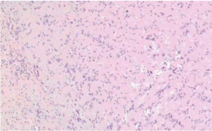
he-matoxylin-eosin staining revealed dense fasciculus formed by spind-le shaped cells (Antoni A) mixed with edematous parts consisting of the sparse tumor cells (Antoni B). Pleomorphic appearance and mitosis are rarely seen and there were no hemorrhage and necro-tic lesions. Special staining of the tumor using S-100 immunperoxi-dase was intensively positive and were negative for both actin and desmin (Figure 2). These findings are typical for the diagnosis of schwannoma. After 9 months fol-low-up period, no recurrence had been observed.
Discussion
Schwannomas are rare tumors and generally encountered in young and midlle-aged adults at the flexor surfaces of the extremities, neck, mediastinum are exceeding-ly rare in the penis (1,2). Schwan-noma may be benign or malignant
Ankara Üniversitesi Tıp Fakültesi Mecmuası 2007, 60(3)
138 Penile Schwannoma
tumors; malignancy is associated with von-Recklinghausen disease. Despite the abundant sensitive in-nervation of the penis and perine-al skin, schwannomas of the penis are rarely reported in the literatu-re (3-6).
The pathological hallmarks of schwannoma are the patterns of Antoni A and B areas. Because of
S-100 protein, expressed in the supporting cells of the nervous system, in these tumors S-100 im-munperoxidase stain should be used to establish the pathological diagnosis.
The treatment of this tumor is surgical removal. Because of schwannoma’s being a benign di-sease, recurrence rarely occurs.
In spite of rare presentation of schwannomas which would be ex-pected to occur on the dorsum of the penis, it is important to con-sider other differential diagnosis including lipoma, fibroma, athero-ma, leimyosarcoma and Peyronie’s disease.
Figure 1. Macroscopic image of the patient”s penile schwannoma Figure 2. Antoni A (Verocay bodies) and Antoni B areas in the
microscopic image of penile schwannoma (Hematoxylin Eosin reduced from x100)
REFERENCES
1. Jiang R, Chen JH, Chen M et al. Male ge-nital schwannoma, review of 5 cases. Asi J Androl 2003; 5: 251-4
2. Suzuki Y, Ishigooka M, Tomaru M et al. Schwannoma of the penis: report of a
case and review of the literature. Int J Urol 1998; 30: 197-202
3. Oberman HA, Sullenger G. Neurogenous tumors of the head and neck. Cancer 1967; 20: 1992-2001
4. Marsidi PJ, Winter CC. Schwannoma of
the penis. Urol 1980; 16: 303-4
5. Mayersak JS, Vivciano CJ, Barbiarz JW. Schwannoma of the penis. J Urol 1995; 153: 1931-2
6. Sato D, Kase T, Tajima M et al. Penile schwannoma. Int J of Urol 2001; 8: 87-89