Received 04/22/2020 Review began 05/04/2020 Review ended 05/07/2020 Published 05/15/2020 © Copyright 2020
Yurdaisik. This is an open access article distributed under the terms of the Creative Commons Attribution License CC-BY 4.0., which permits unrestricted use, distribution, and reproduction in any medium, provided the original author and source are credited.
Effectiveness of Computed Tomography in
the Diagnosis of Novel Coronavirus-2019
Isil Yurdaisik1. Radiology, Istinye University Gaziosmanpasa Medical Park Hospital, Istanbul, TUR
Corresponding author: Isil Yurdaisik, [email protected]
Abstract
Coronaviruses (CoV) belong to the coronavirus genus of the coronaviridae family. All CoVs are pleomorphic RNA viruses containing crown-like peplomers of 80-160 nm in size. This virus is a zoonotic pathogen seen with a wide range of clinical features from asymptomatic state to intensive care in humans.
So far, seven human coronaviruses have been identified with the last one being Coronavirus-2019 (COVID-19). These pathogens typically lead to mild disease, but SARS and MERS type coronaviruses have caused severe respiratory disease and even mortality within the last 20 years.
COVID-19 virus has rapidly spread worldwide after China and is continuing to cause huge economical and social impacts. Given the scarcity of resources including healthcare staff, hospital capacities, test kits, etc., timely diagnosis and treatment of this virus are of paramount importance. However, there is no vaccination or drug developed for the treatment of this disease up to today. Because the spreading rate of the virus is very high worldwide and there is no definitive treatment, diagnosis becomes even more important.
The objective of this review is to evaluate the use of chest computed tomography, one of the commonly used radiologic imaging modalities, in the diagnosis of COVID-19 in light with the current literatüre.
Categories: Radiology
Keywords: novel coronavirus, coronavirus disease, computed tomography, covid-2019
Introduction And Background
A series of pneumonia cases with unknown causes have been reported from the Wuhan state of China. A few days later, this mysterious causative agent of pneumonia was declared as a novel coronavirus [1]. After severe respiratory syndrome coronavirus (SARS-CoV) in 2002 and Middle East respiratory syndrome coronavirus (MERS-CoV) in 2012, novel coronavirus (COVID-19) was introduced in the human population as a highly pathogenic and large-scale epidemic coronavirus [2]. This novel virus was previously named as severe acute respiratory syndrome (SARS) coronavirus-2, and later the virus was named as COVID-19 by the World Health Organization (WHO). On January 20, 2020, the WHO officially declared that COVID-19 became an epidemic as an international public health emergency. On March 11, 2020, COVID-19 was characterized as a pandemic by the WHO [3]. As of April 10, 2020, 622,251 confirmed COVID-19 cases and 46,430 deaths were reported in Europe region [4].
Coronaviruses (CoV) belong to the coronavirus genus of the coronaviridae family. All CoVs are 1
Open Access Review
Article DOI: 10.7759/cureus.8134
pleomorphic RNA viruses containing crown-like peplomers of 80-160 nm in size. This virus is a zoonotic pathogen seen with a wide range of clinical features from an asymptomatic state to intensive care in humans. Although most coronaviruses affect animals, these are zoonotic pathogens that can be transmitted between animals and humans. So far seven human coronaviruses have been identified with the last one being COVID-19. These pathogens typically lead to mild disease, but SARS and MERS type coronaviruses have caused severe respiratory disease and even mortality within the last 20 years. Today, the COVID-19 virus has rapidly spread worldwide after China and is continuing to cause huge economic and social impacts. Given the scarcity of resources including healthcare staff, hospital capacities, test kits, etc., timely diagnosis and treatment of this virus are of paramount importance. There is no vaccination or drug developed for the treatment of this disease up to today. Because the spreading rate of the virus is very high worldwide and there is no definitive treatment, the diagnosis becomes even more important [5].
The objective of this review is to evaluate the use of chest computed tomography (CT), one of the commonly used radiologic imaging modalities, in the diagnosis of COVID-19 in light with the current literature.
Review
Epidemiology
COVID-19 was identified in China for the first time, but it has rapidly spreaded all over the world in a short time and currently is increasingly reported in all continents. The number of cases outside China outpaced the number and rate of new cases in China [6]. Epidemiological studies have reported that the outbreak was associated with wild animals sold in a seafood market in Wuhan province [7]. However, later confirmed cases without a history of exposure to this market indicated human-to-human transmission of COVID-19. As the epidemic
progressed, human-to-human transmission has become the main transmission route of the virus.
All ages are sensitive to COVID-19. However, the virus is more fatal in the elderly population. The infection is transmitted through droplets produced during cough and sneezing [8].
According to recent studies in the literature, viral loads are higher in the nasal cavity compared to the throat; thus, there is no significant difference between symptomatic and asymptomatic people [9]. Infected droplets can spread a few meters, accumulating on surfaces. COVID-19 can survive for days under positive atmospheric conditions, although it can be destroyed in shorter than one minute using common disinfectants such as sodium hypochlorite and hydrogen peroxide. The infection occurs by inhalation of these droplets or touching the mouth, nose, and eyes with the hands after touching contaminated surfaces.
Clinical features
The incubation period of COVID-19 is thought to be within 14 days after the exposure. However, the majority of cases have been seen within 4-5 days of exposure [10]. In a study including 1099 patients with a confirmed diagnosis of COVID-19, median incubation duration was found as four days [11]. Clinical features of COVID-19 vary from asymptomatic state to acute respiratory distress syndrome and multiorgan dysfunction. Common clinical
manifestations include fever (not in all cases), cough, sore throat, headache, and shortness of breath. In a study, fever was reported as 83%, cough as 82%, and shortness of breath as 31% of COVID-19 patients [12]. However, there are still no specific clinical features to reliably distinguish COVID-19 from other viral respiratory infections. In a part of patients, the disease may progress to pneumonia, respiratory failure, and death at the end of the first week. According to a study from China, around 15% of the patients presented with severe symptoms and 5% required intensive care [13]. In Italy and Spain, 7% to 12% of the hospitalized patients
were reported to require intensive care [14].
Diagnosis
The first approach in COVID-19 is to early detect suspected cases, immediately isolate these people, and implement infection control measures. The diagnosis of COVID-19 is established with fever, cough, and/or dyspnea together with a history of contact with an infected person in a distance shorter than approximately 2 meters and/or travel to the areas, where the disease is common within the last 14 days. However, COVID-cases may be asymptomatic and even may not have a fever. This disease is confirmed with positive molecular testing. According to the American Centers for Disease Control and Prevention (CDC), test priority criteria in suspected COVID-19 patients that were updated as of March 8, 2020 are as follows [15]:
Hospitalized patients with signs and symptoms compatible with COVID-19. Elderly people (≥ 65 years) and people with chronic medical conditions and/or
immunocompromised status (e.g. diabetes mellitus, heart disease, using immunosuppressive medications, chronic lung disease, chronic kidney disease) who can be at risk for a poor outcome.
Including healthcare personnel, people with a history of close contact with other persons with suspected or laboratory-approved COVID-19 or travel to the affected geographic areas up to 14 days before the onset of symptoms.
Molecular tests used for the diagnosis of COVID-19 are performed on respiratory samples (throat swab, nasopharyngeal swab, sputum, and endotracheal aspirates). The virus can also be identified in stool and blood in severe cases. The diagnosis of COVID-19 is mainly established with reverse transcription-polymerase chain reaction (RT-PCR) test. However, this test requires strict laboratory specifications and the results take a long time [16]. Other laboratory
investigations are usually non-specific. Indicators such as white blood cells, platelets, C-reactive protein (CRP), and erythrocyte sedimentation rate (ESR), alanine
aminotransferase/aspartate aminotransferase (ALT/AST), prothrombin time, creatinine, D-dimer, and low-density cholesterol (LDL) are also studied. Nevertheless, these markers have been associated with disease severity and are not specific to COVID-19 [17].
Chest X-ray is one of the diagnostic modalities used to help the diagnosis of COVID-19. This image usually shows bilateral infiltrates. However, the results of this method can be normal at the beginning of the disease. Chest CT is more sensitive and specific in the diagnosis of COVID-19 compared to X-ray. CT images usually show infiltrates, ground-glass opacities (GGO), and sub-segmental consolidation. In addition, CT is abnormal also in asymptomatic patients without clinical findings related to the involvement of the lower respiratory tract. CT scans have been used also in suspected COVID-19 patients with a negative molecular diagnosis, and repeated molecular tests were positive in most of these patients [17].
CT in the diagnosis of COVID-19
Imaging plays an important role in the diagnosis and management of COVID-19. Chest CT is used as the first-line imaging method in suspected cases and is a useful tool for monitoring the changes during treatment. Correct diagnosis of viral pneumonia based on chest CT indicates isolation and plays an important role in the management of patients suspected to have an infection, especially in the absence of scientifically proven treatment methods. Findings on CT may reflect the severity of the disease. Therefore, CT is an important imaging technique in helping the diagnosis and management of COVID-19 patients, and publications about the radiologic appearance of COVID-19 pneumonia are increasing in the literature. As this disease
increasingly becomes a global concern, it is more crucial for radiologists to be familiar with CT images of COVID-19 and to have basic knowledge about the clinical course and size of this infection. Radiologists to have sufficient knowledge about the clinic and chest CT imaging of COVID-19 will help early detection of the infection and evaluation of the disease course. So far reported common CT features regarding COVID-19 are as follows [18]:
● GGO
● Consolidation
● Mixed GGO and consolidation ● Centrilobular nodules ● Architectural distortion ● Cavitation
● Tree-in-bud
● Bronchial wall thickening ● Reticulation
● Subpleural bands ● Traction bronchiectasis
● Intrathoracic lymph node enlargement ● Vascular growth in the lesion
● Pleural effusions
CT images are usually examined in four distributions as craniocaudal, transverse, lung region, and scattered. Thin slice chest CT is more effective in the early detection of COVID-19 [19]. Some case series and case reports investigating chest CT imaging features of COVID-19 have been published [20-23]. It has been reported that CT abnormalities are more likely to be bilateral, to have a peripheral distribution, and to involve the lower lobes [24]. In a study comparing 219 COVID-19 patients in China and 205 patients with other viral pneumonia in the USA, peripheral distribution, GGOs, thin reticular opacities, vascular thickening, and reverse halo sign were more common, while central and peripheral distribution, air bronchogram, pleural thickening, pleural effusion, and lymphadenopathy were more rare in COVID-19 cases [25].
In a study from Wuhan, China, it was reported that COVID-19 had abnormal findings on chest CT even in asymptomatic patients [26]. In that study, chest CT findings of 81 patients with COVID-19 were examined. Examinations were performed by two radiologists who were blind to RT-PCR testing results. According to the results of the study, CT images showed bilateral lung involvement in 79%, peripheral distribution in 54%, and diffuse distribution in 44% of the
patients. The most common chest CT findings included GGO together with poorly defined margins, smooth or irregular interlobular septal thickening, air bronchogram, and thickening of adjacent pleura [26]. Remarkably, in a study by Shi et al., early CT changes were seen in the asymptomatic patient group, and these findings supported the observations in a familial cluster with COVID-19 pneumonia [19]. However, some studies have reported positive RT-PCR results in the absence of CT changes or abnormal CT results in the presence of initial false-negative RT-PCR results [18]. Predominant detection of GGOs on CT in COVID-19 makes this method more sensitive. In a study by Song et al., the most common chest CT findings were reported as GGO appearance in COVID-19 patients [21]. In a case series of 21 patients by Chung et al., GGO was the most common CT finding by 57%, and bilateral disease was found in 76% of COVID-19 patients [27]. However, CT findings may be normal also in confirmed cases [28].
In a study by radiologists from Wuhan, China, it was found that chest CT had a low misdiagnosis rate in COVID-19 and this method can help standardization of imaging and a rapid diagnosis [10]. On the other hand, it has been reported that the use of CT is limited in the detection of specific viruses and making a distinction between viruses [29]. In a study from Wuhan including 1014 patients who underwent RT-PCR testing and chest CT, the sensitivity of positive results by the consensus of two radiologists was found as 97% when PCR tests were used as a reference. However, in the same study, the specificity of CT was only 25% [30]. It was reported that this low specificity may be associated with other etiologies leading to CT findings. In general, chest findings of chest CT in COVID-19 overlap with the findings of other infections such as H1N1, SARS, and MERS, and this limits the specificity of CT.
Effectiveness of CT in COVID-19 should be carefully evaluated because the majority of the existing data come from the studies conducted in the far east. According to the studies in the literature, the preference of CT in China is resulted from the wide availability of this method, because access to CT is relatively easier in China [31]. In most of these studies, it was reported that chest CT was positive in the presence of negative test results [17]. RT-PCR results can be affected by sampling errors and low viral load [32]. In previous studies about SARS, the sensitivity of RT-PCR has been shown to be insufficient within the first five days of the disease [33]. In addition, findings of this rapidly spreading conditions have not been completely published or are not updated. CDC currently does not recommend the use of CT for the diagnosis of COVID-19. According to the CDC, viral testing is the only specific diagnosis method, and even in the case of COVID-19 radiologic findings with CT, the results must be confirmed with viral testing [34]. According to Lee et al. who are radiologists in Hong Kong university, the variability of COVID-19 presentations leads to difficulties in establishing the diagnosis. The authors emphasized that there are more things to learn from this contagious viral pneumonia, and further studies are needed on the relationship between CT findings and clinical progression [31].
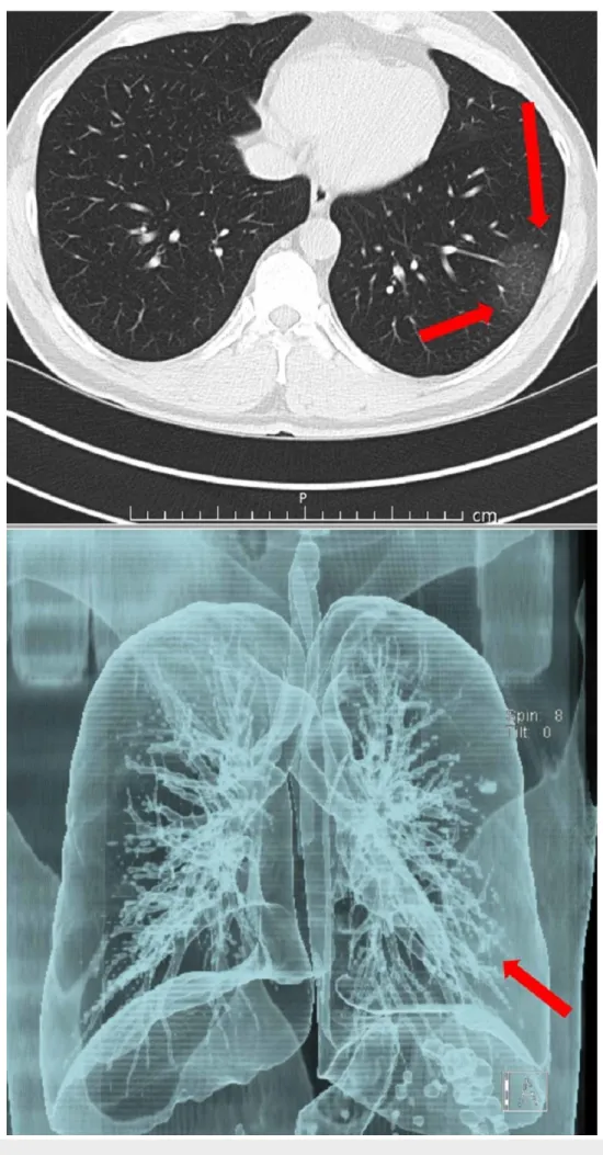
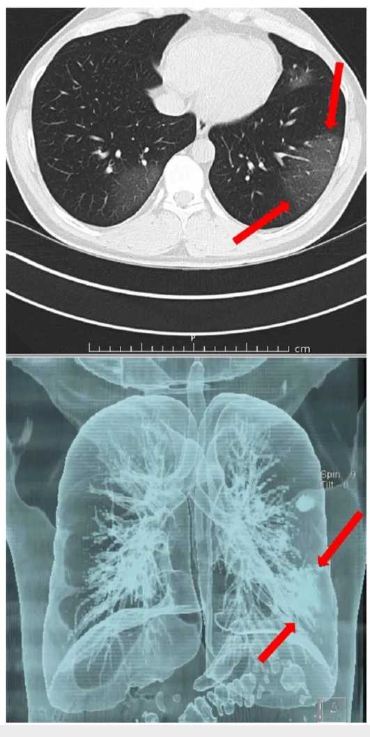
Upper left and peripheral localized lesion of ground-glass density, which is hardly noticeable on cross-sectional CT is clearly seen on 3D reconstruction.
density in the lower left lobe is well visualized on the sectional
view, but it cannot be distinguished in the
The upper left lesion is enlarged and the lower left lesion is visible. CT, computed tomography
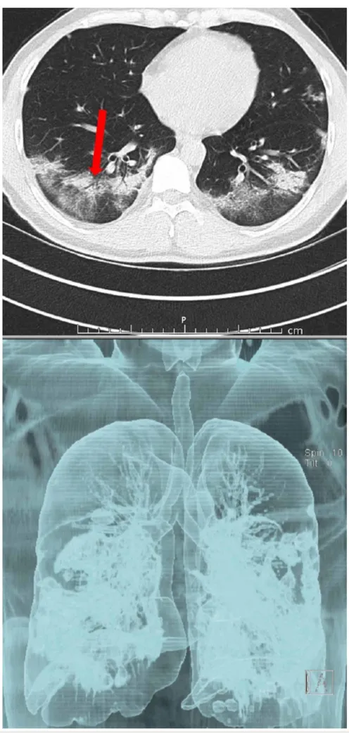
Air bronchograms and fibrosis were developed. Bilateral milimetric central aeration areas on 3D image. Sectional routine image better shows lesion details, and 3D images better
depict involvement area in the lung. CT, computed tomography
Conclusions
There is debate in the literature on the use of chest CT for the diagnosis of COVID-19. As this epidemic progresses, various presentations of COVID-19 will be increasingly observed, and the correlation between CT and RT-PCR findings will be more commonly studied. As the picture becomes clearer, the radiologic-pathologic correlation will be better understood, potential imaging predictors will be determined in more detail, and the role of radiology in the management of COVID-19 will be increased.
Additional Information
Disclosures
Conflicts of interest: In compliance with the ICMJE uniform disclosure form, all authors declare the following: Payment/services info: All authors have declared that no financial support was received from any organization for the submitted work. Financial relationships: All authors have declared that they have no financial relationships at present or within the previous three years with any organizations that might have an interest in the submitted work. Other relationships: All authors have declared that there are no other relationships or activities that could appear to have influenced the submitted work.
Acknowledgements
Thank physicians and all other healthstaff worldwide for their incredible efforts to fight this outbreak.
References
1. Novel Coronavirus - China . (2020). Accessed: May 15, 2020: https://www.who.int/csr/don/12-january-2020-novel-coronavirus-china.
2. Guo Y, Cao Q, Hong Z, et al.: The origin, transmission and clinical therapies on coronavirus disease 2019 (COVID-19) outbreak - an update on the status. Mil Med Res. 2020, 13:11.
10.1186/s40779-020-00240-0
3. Rolling updates on coronavirus disease (COVID-19) . (2020). Accessed: May 15, 2020: https://www.who.int/emergencies/diseases/novel-coronavirus-2019/events-as-they-happen. 4. WHO announces COVID-19 outbreak a pandemic . (2020). Accessed: May 15, 2020:
http://www.euro.who.int/en/health-topics/health-emergencies/coronavirus-covid-19/news/news/2020/3/who-announces-covid....
5. What you should know about COVID-19 to protect yourself and others . (2020). Accessed: May 15, 2020: https://www.cdc.gov/coronavirus/2019-ncov/downloads/2019-ncov-factsheet.pdf. 6. Novel Coronavirus - Republic of Korea (ex-China) . (2020). Accessed: May 15, 2020:
https://www.who.int/csr/don/21-january-2020-novel-coronavirus-republic-of-korea-ex-china/en/.
7. Novel Coronavirus (2019-nCoV) Situation Report - 2 . (2020). Accessed: May 15, 2020: https://www.who.int/docs/default-source/coronaviruse/situation-reports/20200122-sitrep-2-2019-ncov.pdf.
8. Rothe C, Schunk M, Sothmann P: Transmission of 2019-nCoV Infection from an
9. Zou L, Ruan F, Huang M, et al.: SARS-CoV-2 viral load in upper respiratory specimens of infected patients. N Engl J Med. 2020, 10.1056/NEJMc2001737
10. Li and Xia L: Coronavirus Disease 2019 (COVID-19): role of chest CT in diagnosis and management. AJR Am J Roentgenol. 2020, 4:1-7. 10.2214/AJR.20.22954
11. Guan WJ, Ni ZY, Hu Y, et al.: Clinical characteristics of Coronavirus disease 2019 in China . N Engl J Med. 2020, 382:1708-1720. 10.1056/NEJMoa2002032
12. Chen N, Zhou M, Dong X, et al.: Epidemiological and clinical characteristics of 99 cases of 2019 novel coronavirus pneumonia in Wuhan, China: a descriptive study. Lancet. 2020, 15:507-513. 10.1016/S0140-6736(20)30211-7
13. Wu Z, McGoogan JM: Characteristics of and important lessons from the coronavirus disease 2019 (COVID-19) outbreak in China: Summary of a report of 72 314 cases from the Chinese Center for Disease Control and Prevention. JAMA. 2020, 10.1001/jama.2020.2648
14. Lazzerini M, Putoto G: COVID-19 in Italy: momentous decisions and many uncertainties . Lancet Glob Health. 2020, 109:30110-8. 10.1016/S2214-109X(20)30110-8
15. Updated Guidance on Evaluating and Testing Persons for Coronavirus Disease 2019 (COVID-19). (2020). Accessed: May 15, 2020: https://emergency.cdc.gov/han/2020/HAN00429.asp (Accessed on April 10, 2020).
16. Huang C, Wang Y, Li X, et al.: Clinical features of patients infected with 2019 novel coronavirus in Wuhan, China. Lancet. 2020, 15:497-506. 10.1016/S0140-6736(20)30183-5
17. Huang P, Liu T, Huang L: Use of chest CT in combination with negative RT-PCR assay for the 2019 novel coronavirus but high clinical suspicion. Radiology. 2020, 295:22-23.
10.1148/radiol.2020200330
18. Xie X, Zhong Z, Zhao W, Zheng C, Wang F, Liu J: Chest CT for typical 2019-nCoV pneumonia: relationship to negative RT-PCR testing. Radiology. 2020, 10.1148/radiol.2020200343
19. Jasper Fuk-Woo Chan, Shuo Feng Yuan, Kin-Hang Kok: A familial cluster of pneumonia associated with the 2019 novel coronavirus indicating person-to-person transmission: a study of a family cluster. Lancet. 2020, 395:514-23. https://doi.org/10.1016/ S0140-6736(20)30154-9.
20. Pan FP, Ye T, Sun P, et al.: Time course of lung changes on chest CT during recovery from 2019 novel coronavirus (COVID-19) pneumonia. Radiology. 2020, 10.1148/radiol.2020200370
21. Song F, Shi N, Shan F, et al.: Emerging coronavirus 2019-nCoV pneumonia . Radiology. 2020,
10.1148/radiol.2020200274
22. Qian L, Yu J, Shi H: Severe acute respiratory disease in a Huanan seafood market worker: Images of an early casualty. Radiology. 2020, 10.1148/ryct.2020200033
23. Kim H: Outbreak of novel coronavirus (COVID- 19): What is the role of radiologists? . Eur Radiol. 2020, 10.1007/s00330-020-06748-2
24. Wei Zhao, Zheng Zhong, Xingzhi Xie, Qizhi Yu, Jun Liu: Relation between chest CT findings and clinical conditions of coronavirus disease (COVID-19) pneumonia: a multicenter study. AJR Am J. 2020, 214:1072-1077. 10.2214/AJR.20.22976
25. Bai HX, Hsieh B, Xiong Z, et al.: Performance of radiologists in differentiating COVID-19 from viral pneumonia on chest CT. Radiology. Published Online: Mar. 10:10-1148.
https://doi.org/10.1148/radiol.2020200823.
26. Shi H, Han X, Jiang N, et al.: Radiological findings from 81 patients with COVID-19 pneumonia in Wuhan, China: a descriptive study. Lancet. 2020, 10.1016/S1473-3099
27. Chung M, Bernheim A, Mei X, et al.: CT imaging features of 2019 novel coronavirus (2019-nCoV). Radiology. 2020, 10.1148/radiol.2020200230: 200230
28. Lei J, Li J, Qi X: CT Imaging of the 2019 Novel Coronavirus (2019-nCoV) Pneumonia . Radiology. 2020, 10.1148/radiol.2020200236
29. Wuhan CT scans reliable for coronavirus (COVID-19) diagnosis, limited for differentiation . (2020). Accessed: May 15, 2020: https://www.eurekalert.org/pub_releases/2020-03/arrs-wcs030520.php.
30. Ai T, Yang Z, Hou H, et al.: Correlation of chest CT and RT-PCR testing in coronavirus disease 2019 (COVID-19) in China: a report of 1014 cases. Radiology. 2020, 26:200642.
10.1148/radiol.2020200642
31. Lee KS: Pneumonia associated with 2019 novel coronavirus: can computed tomographic findings help predict the prognosis of the disease?. Korean J Radiol. 2020 21,
32. Peiris JS, Chu CM, Cheng VC, et al.: Clinical progression and viral load in a community outbreak of coronavirus-associated SARS pneumonia: a prospective study. Lancet. 2003,
361:1767-1772. 10.1016/s0140-6736(03)13412-5
33. Poon LL, Chan KH, Wong OK, et al.: Early diagnosis of SARS coronavirus infection by real time RT-PCR. J Clin Virol. 2003, 28:233-238. 10.1016/j.jcv.2003.08.004
34. ACR Recommendations for the use of Chest Radiography and Computed Tomography (CT) for Suspected COVID-19 Infection. (2020). Accessed: May 15, 2020:
https://www.acr.org/Advocacy-and-Economics/ACR-Position-Statements/Recommendations-for-Chest-Radiography-and-CT-for-S....