Original Research Article
Correlation between histopathologic and radiological changes in
articular cartilage of the knee joint with degenerative
joint disease
Hakan Cift
1, Ali Seker
1, Bulent Kilic
3, Murat Demiroglu
4*, Asli Erdogan Cakir
2,
Mehmet Isyar
1, Murat Bulbul
1, Mahir Mahirogullari
1INTRODUCTION
Osteoarthritis (OA) of the knee is among the most common disabling diseases which may cause pain and decrease in functional status. It is the commonest form of arthritis and is most prevalent in the elderly, with 50% of
adults aged 65-75 years and almost 70% of those 75+ years suffer from this disease.1-3
Aging of the population and the increasing trends of obesity will probably cause the prevalence of knee OA to raise even more in the nearby future.4
ABSTRACT
Background: Osteoarthritis (OA) of the knee is among the most common disabling diseases which may cause pain
and decrease in functional status. It is the commonest form of arthritis and is most prevalent in the elderly, with 50% of adults aged 65-75 years and almost 70% of those 75+ years suffer from this disease. The aim of this study was to investigate consistency of radiologic findings with histomorphologic structure of bone in patients with severe gonarthrosis.
Methods: 62 knees of 57 patients over 60 years old who had stage 3-4 gonarthrosis according to Kellgren-Lawrence
classification were included in the study. Patients were separated into two groups as having stage 3 or stage 4 gonarthrosis. All the patients underwent total knee replacement procedure. During the operation distal femoral medial/lateral condyle and proximal tibial medial/lateral plateau were removed and sent to histologic examination for the measurement of thickness of cartilage layer and subchondral bone, number and thickness of trabeculae, space between two trabeculae.
Results: Average thickness of subchondral bone was measured at stage 3 gonarthrosis and at stage 4 gonarthrosis.
Only the difference between medial tibial condyle values of two groups was statistically significant. Average trabecula thickness was measured both at stage 3 and at stage 4 gonarthrosis. Only the difference between lateral tibial condyle values of two groups was statistically significant. Furthermore, as for the number of trabeculas and cavity between trabeculae, a significant difference couldn’t be found.
Conclusions: Despite having radiological differences two groups can be said to show similar histopathological
characteristics.
Keywords: Gonarthrosis, Kellgren-Lawrence classification, Subchondral bone
1Department ofOrthopeadic and Traumatology, 2Department of Pathology, Istanbul Medipol University, Istanbul 3Department ofOrthopeadic and Traumatology, Gelisim University, Istanbul
4Department ofOrthopeadic and Traumatology, Medeniyet University Goztepe EAH, Istanbul Received: 16 December 2016
Accepted: 20 December 2016 *Correspondence:
Dr. Murat Demiroglu,
E-mail: [email protected]
Copyright: © the author(s), publisher and licensee Medip Academy. This is an open-access article distributed under
the terms of the Creative Commons Attribution Non-Commercial License, which permits unrestricted non-commercial use, distribution, and reproduction in any medium, provided the original work is properly cited.
There is various treatment options according to the plain radiograph findings of osteoarthrosis combined with physical examination of the patient. In this study we aimed to investigate the consistency of radiologic findings with histomorphologic structure of bone in patients with severe gonarthrosis (Kellgren-Lawrence stage 3 and 4) and try to display if there is histologic difference between two stages of the disease.
METHODS
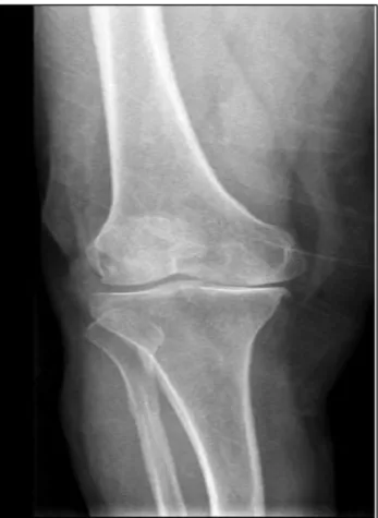
The patients with intense knee pain even during resting and had a Kellgren-Lawrence stage 3 and 4 osteoarthritis was planned for the total knee arthroplasty between 2013 and 2014 and they had constituted our study cohort. Knee roentgenographies of the patients were graded by two orthopaedic surgeons who were blind to the study (Figure 1-2). In this context, 62 knees of the 57 patients were involved in the study. The specimans including medial and lateral femoral condyle, medial and lateral tibial plateaus were obtained from the patients undergoing total knee arthroplasty.
Figure 1: The knee roentgenographie of Kellgren-Lawrence stage 3.
After surgery, the specimens underwent fixation in 10% formalin for 2 days. Then all specimens decalcified in the formic acid (10%) for 5-7 days. After decalcification, 4 samples were taken from medial-lateral tibial plateaus, medial-lateral condyles of distal femur. Both normal
cartilage and destroyed cartilage were sampled in the same section. After tissue process (graded alcohols, cleared in xylene and embedded in paraffin wax), 5 micrometer sections were stained with haematoxylin and eosin.
Figure 2: The knee roentgenographie of Kellgren-Lawrence stage 4.
In histological assessment thickness of cartilage, subchondral bone, trabecula and number of trabecula and space between trabeculas were measured. Three measurements from the most severely affected locations were measured histopathologically with light microscopy (under Leica microscope camera) for each sample. Osteoarthritis cartilage histopathology grade and stage were assessed according to the Pritzer et al article.5 All
the histological assesments were done by an expert pathologist in musculoskeletal subdivision (Figure 3, 4).
Figure 3: The histopathologically appearance with light microscopy of OARSI 4
Figure 4: The histopathologically appearance with light microscopy of OARSI 5.
Statistical analysis
The statistical analysis was performed by using SPSS 15 (SPSS Inc., Chicago, IL). The chi-square and Fisher’s exact tests were used for descriptive statistics. Pearson chi-square test was used for correlation analysis. A p value of less than 0.05 was accepted as statistically significant.
RESULTS
Average cartilage thickness was measured as 1.85, 2.38, 2.34 and 2.44 mm at medial tibial condyle, lateral tibial condyle, medial femoral condyle and lateral femoral condyle respectively at stage 3 gonarthrosis. These values were 2.32, 2.51, 2.64 and 2.97 mm at stage 4 gonarthrosis. However, the differences between same anatomic regions of stage 3 and 4 groups were not statistically significant.
Average thickness of subchondral bone was measured as 378.6, 335.2, 361.9 and 371.3 mm at medial tibial condyle, lateral tibial condyle, medial femoral condyle and lateral femoral condyle respectively at stage 3 gonarthrosis. These values were 310, 339.8, 333.9 and 308.4 mm at stage 4 gonarthrosis. The difference between medial tibial condyle values of two groups was statistically significant (p = 0.037). However, the differences were not statistically significant regarding lateral tibial condyle, medial femoral condyle and lateral femoral condyle.
Average trabecular thickness was measured as 227.4, 216.9, 207.7 and 207.9 mm at medial tibial condyle, lateral tibial condyle, medial femoral condyle and lateral femoral condyle respectively at stage 3 gonarthrosis. These values were 200.8, 191.5, 210.7 and 216.7 mm at stage 4 gonarthrosis. The difference between lateral tibial condyle values of two groups was statistically significant (p = 0.048). However, the differences were not
statistically significant regarding medial tibial condyle, medial femoral condyle and lateral femoral condyle. Furthermore, as for the number of trabeculae and cavity between trabeculae, a statistically significant difference couldn’t be found. A significant relationship between parameters at correlation analysis couldn’t be set between stage 3 and 4 gonarthrosis.
DISCUSSION
OA is regarded as a whole joint disease with a multifactorial etiology, including increased mechanical stress, ligament derangements, cartilage degradation, subchondral bone changes and muscular impairments.1-3
Supporting the recognition that OA is a disease of the whole joint, there is mounting evidence that subchondral bone plays an important role in OA. Bone remodeling in the OA joint occurs preferentially in the subchondral plate. Surgical specimens from persons with OA have demonstrated that subchondral bone changes, including subchondral bone attrition which is a flattening or depression of the subchondral bony surface unrelated to gross fracture, are common.4,6
As life getting longer with improvements in health care, osteoarthritis has been seen more frequently. This problem makes patients life quality worser. Patient related complaints usually starts with walking, but as the time passes the pain make the patient suffer even during resting. As this problem is getting important, great attempts to prevent and treat this condition has been displayed by the clinicians.
Diagnosis of the osteoarthritis is very easy and a standing knee X- ray is usually enough. There are many staging methods for the grading of osteoarthritis and in 1957 Kellgren and Lawrence introduced one of the widely used grading systems. Kellgren and Lawrence grading system were adapted at the World Healh Organization meeting in 1961. This grading system helps the orthopaedic surgeons to decide which measures to take for the treatment of this progressive condition.8,9
The most obvious histopathologic features of an osteoarthritic joint are damaged cartilage, subarticular cysts, alterations in the shape of the articular surfaces and osteophytes. Especially in load bearing areas, the cartilage may be entirely absent and have a polished marble like appearance (eburnation). Microscopic examination of the cartilage reveals a spectrum of changes. In the early stages the cartilage is thicker; with the progression of disease, the joint surface thins and vertically oriented clefts occur. Deep cartilage ulcers, extending to bone, can be seen. There are several macroscopic and microscopic grading systems for histopathologic assessment of osteoarthritis.5 These
systems are based on the cartilage surface changes in advanced osteoarthritis.
There are many treatment options in knee osteoarthritis as injections, medical treatment, and arthroscopy, osteotomies around the knee, unicondylar arthroplasty and total knee arthroplasty. Patients age, degree of osteoarthritis, complaints makes us to choose our treatment options. In severe gonarthrosis (stage 4 according to Kellgren-Lawrence classification), there is no doubt for the optimal treatment option with arthroplasty. However, in moderate gonarthrosis (stage 3 osteoarthrosis) there is still an uncertainty in the treatment strategy. All options listed above can be done for the treatment. In a study Razak BA et al compares the arthroscopic findings and Kellgren- Lawrence classification in osteoarthritic knees and found poor correlation.10 In that point, we aimed to search for the
histopathological difference between stage 3 and stage 4 osteoarthrosis if it can help us to guide our treatment strategy.
It has been hypothesized that cartilage loss is a mechanically mediated process that is more likely to occur in regions subjected to high stress; such areas of high stress are influenced by bone shape.11 Understanding
the implications of bony alterations on the biomechanical environment of the joint is critical as biomechanics are known to play an important role in knee OA. The association of static malalignment and higher dynamic knee adduction moments with knee OA progression supports the importance of mechanical influences in knee OA pathogenesis.12,13
Specifically, subchondral bone in OA has increased thickness and volume, but is weaker and less mineralized than normal bone.14-18 With alterations in its properties,
subchondral bone may be less able to absorb and dissipate energy, thereby increasing forces transmitted through the joint and predisposing the articular surface to deformation.4,7
In a study by Hunter et al, histomorphometric evaluation has also demonstrated that BMLs to have many of the same features noted to occur in OA bone, but to a greater extent; these changes include increased bone volume fraction, decreased tissue mineral density, increased trabecular thickness and spacing but decreased number, and to be more plate-like than bone in other areas of the same (osteoarthritic) knee.19 Overall, these
histomorphometric changes constitute the basic changes in osteoarthritis.20
In this study subchondral bone thickness values were compared in four different area of the knee joint. Except medial tibial plateau no difference was observed between stage 3 and 4 gonarthrosis. In medial tibial condyle we detected decreased subchondral bone thickness.
To our knowledge, our study was the first one to compare the relationship between histopathological and radiological changes in knee osteoarthritis. However, Brandt et al compare the radiographic grading with the
arthroscopic articular cartilage in the knee osteoarthritis and they found no difference.21
Despite, extensive use of Kellgren- Lawrence classification for the detection of severity of knee osteoarthritis by the clinicians, patient symptom’s and signs should be the priority for the optimal treatment modality selection for gonartrosis.
CONCLUSION
There was not a statistically significant difference between stage 3 and stage 4 gonarthrosis patients in terms of cartilage thicknesss, number of trabeculae and cavity between trabeculae which mean that radiological staging was not consistent with histopathological evaluation. Treatment of patients with gonarthrosis should not rely on radiological staging but patient’s solely symptoms and signs. Further studies might be needed to find out modalities for the better deliniation of radiological staging methods.
Funding: No funding sources Conflict of interest: None declared
Ethical approval: The study was approved by the Istanbul Medipol University with number assigned 10840098-34
REFERENCES
1. Hunter DJ, Felson DT. Osteoarthritis. BMJ. 2006;332(7542):639-642.
2. Hinton R, Moody RL, Davis AW, Thomas SF. Osteoarthritis: diagnosis and therapeutic considerations. Am Fam Physician. 2002;65(5):841-8.
3. Lawrence RC, Helmick CG, Arnett FC, Deyo RA, Felson DT, Giannini EH, et al. Estimates of the prevalence of arthritis and selected musculoskeletal disorders in the United States. Arthritis Rheum. 1998;41(5):778-99.
4. Neogi T. Clinical significance of bone changes in osteoarthritis. Ther Adv Musculoskelet Dis. 2012;4(4):259-67.
5. Pritzker KPH, Gay S, Jimenez SA, Ostergaard K, Pelletier JP, Revell PA, et al. Osteoarthritis cartilage histopathology: grading and staging. Osteoarthritis and Cartilage. 2006;14(1):13-29.
6. Bullough PG. Osteoarthritis and related disorders: pathology. IN: Klippel, J.H. and Dieppe, p. (eds), Rheumatology, 2nd ed. London: Mosby; 1998:8.8.1-8.8.8.
7. Uygur E, Kilic B, Demiroglu M, Ozkan K, Cift H. Subchondral bone and its role in osteoarthritis. Open J Orthopaed. 2015;5:355-60.
8. Kellgren JH, Lawrence JS. Radiographic assessment of osteoarthritis. Ann Rheumatol Dis. 1957;16:494. 9. Kellgren JH, Jeffrey M, Ball J. The epidemiolog of
chronic rheumatism. Atlas of standart radiographs. Vol 2. Oxford: Blackwell Scientific; 1963.
10. Bin Abd Razak HR, Heng HY, Cheng KY, Mitra AK. Correlation between radiographic and arthroscopic findings in Asian osteoarthritic knees. J Orthop Surg (Hong Kong). 2014;22(2):155-7. 11. Williams TG, Holmes AP, Waterton JC, Maciewicz
RA, Hutchinson CE, Moots RJ, Nash AF, Taylor CJ. Anatomically corresponded regional analysis of cartilage in asymptomatic and osteoarthritic knees by statistical shape modelling of the bone. IEEE Trans Med Imaging. 2010;29(8):1541-59.
12. Miyazaki T, Wada M, Kawahara H, Sato M, Baba H, Shimada S. Dynamic load at baseline can predict radiographic disease progression in medial compartment knee osteoarthritis. Ann Rheum Dis 2002;61:617-22.
13. Sharma L, Song J, Felson DT, Cahue S, Shamiyeh E, Dunlop DD. The role of knee alignment in disease progression and functional decline in knee osteoarthritis. JAMA. 2001;286:188-95.
14. Buckland-Wright C. Subchondral bone changes in hand and knee osteoarthritis detected by radiography. Osteoarthritis Cartilage. 2004;12(Suppl A):S10-19.
15. Grynpas MD, Alpert B, Katz I, Lieberman I, Pritzker KP. Subchondral bone in osteoarthritis. Calcif Tissue Int. 1991;49:20-6.
16. Li B, Aspden RM. Composition and mechanical properties of cancellous bone from the femoral head of patients with osteoporosis or osteoarthritis. J Bone Miner Res. 1997;12:641-51.
17. Li B, Aspden RM. Mechanical and material properties of the subchondral bone plate from the femoral head of patients with osteoarthritis or osteoporosis. Ann Rheum Dis. 1997;56:247-54. 18. Radin EL, Rose RM. Role of subchondral bone in
the initiation and progression of cartilage damage. Clin Orthop Relat Res. 1986;213:34-40.
19. Hunter DJ, Gerstenfeld L, Bishop G, Davis AD, Mason ZD, Einhorn TA. Bone marrow lesions from osteoarthritis knees are characterized by sclerotic bone that is less well mineralized. Arthritis Res Ther. 2009;11(1):R11.
20. Cubukcu D, Sarsan A, Alkan H. Relationships between pain, function and radiographic findings in osteoarthritis of the knee: a cross-sectional study arthritis. 2012:Article ID 984060.
21. Brandt KD, Fife RS, Braunstein EM, Katz B. Radiographic grading of the severity of knee osteoarthritis: relation of the Kellgren and Lawrence grade to a grade based on joint space narrowing, and correlation with arthroscopic evidence of articular cartilage degeneration. Arthritis Rheum. 1991;34(11):1381-6.
Cite this article as: Cift H, Seker A, Kilic B,
Demiroglu M, Cakir AE, Isyar M, et al. Correlation between histopathologic and radiological changes in articular cartilage of the knee joint with degenerative joint disease. Int Surg J 2017;4:450-4.