07-07-2017 Accepted 28-11-2017
Doi
10.16984/saufenbilder.327153
Cloning, Expression and Characterization of Xylanase (xyn-akky1) from Bacillus subtilis in
Escherichia coli
Sema BİLGİN*1, Yakup ULUSU2, Hülya KUDUĞ3, İsa GÖKÇE3
In this study, Bacillus subtilis akky1 strain was isolated from the soil of beech forest in Akkuş City, Ordu
Province, Turkey. akky1 strain was identified by 16S rRNA analysis. The full-length 16S rRNA sequence
of akky1 strain showed the 100% similarity with Bacillus subtilis strain B7 (KC310823.1) . A 642 bp DNA
fragment was obtained from genomic DNA using primers designed based on the gene sequence of Bacillus
subtilis xylanase given in GenBank. The gene encoding xylanase was cloned into pET28b (+) plasmid
vector, sequenced and expressed in Escherichia coli BL21 (DE3). The hexahistidine (6xHis) tagged fusion
protein was purified using nickel affinity chromatography and the xylanase activity was measured. The
molecular mass of the purified xylanase was approximately 26 kDa as estimated by SDS-PAGE. The
xylanase had optimal activity at pH 6.0 and 60
°C. The K
m
values of the recombinant enzyme towards
beechwood was 3.33 mg/ml.
Keywords: Bacillus subtilis, Xylanase, Recombinant Protein, Industrial Enzymes, Escherichia coli
1. INTRODUCTION
Hemicellulose is a heterogeneous polymer
composed of pentose (such as xylose,
arabinose) and hexose sugars (such as
mannose, glucose, galactose) and sugar acids.
Hemicelluloses collectively are classified
into three groups as xylan, glucomannan,
arabinogalactan (1). An essential component
of the xylan is 5-carbon sugar, D-xylose,
which can be converted into chemical fuel by
microbial cells (3). Complete degradation of
plant xylanes requires the collaboration of
several hydrolytic enzymes because of the
complex
chemical
structure
and
heterogeneity of the xylan. Therefore, it is not
surprising that producing a multitude number
of polymer disintegrate enzymes by the xylan
digesting microbial cells. The xylanolytic
enzyme system which is performed xylan
hydrolysis usually consists of several
hydrolytic enzymes: 1,4-endoxylanase,
β-xylosidase,
α-L-arabinofuranosidase,
α-glucuronidase, acetyl xylan esterase and
phenolic acid esterase (ferulic acid and
p-coumaric acid). Among them, endo-1,4-
β-xylanase (1,4-β-D-xylan-xylan hydrolase
E.C. 3.2.1.8) is the key enzyme. This enzyme
breaks down the glycosidic bonds in xylan
structure. Initially, the product of hydrolysis
is β-D-xylopyranosyl oligomers and at the
later stages small molecules such as mono-,
di- and trisaccharides of β-D-xylopyranosyl
(16). Endo-1,4- β-xylanase is produced by
various microorganisms such as funguses (3;
2), actinomycetes (6) and bacteria (4).
Xylanases derived from microorganisms
caught major attention due to their expended
industrial applications including textile
industry
(
5
),
production
of
xylo-oligosaccharides (
14
), clarification of juices
(
3
),
waste-water
treatment
(17),
bioconversion of lignocellulosic wastes into
useful economical products (ethanol, sugar
syrups, gaseous fuels etc.) (
5
), biobleaching
of pulp (
11
,
18
).
In this study, xylanase-producing Bacillus
subtilis strain akky1 was isolated from the
soil of beech forest in Akkuş City, Ordu
Province, Turkey. The identification of the
strain akky1 was performed with PCR
amplification of 16S rRNA. Xylanase gene
was amplified from the genomic DNA of
akky1 strain by polymerase chain reaction
using two oligonucleotides. After Xyn-akky1
gene had been sequenced, it was cloned into
the pET28b vector and expressed in
Escherichia coli. The recombinant xylanase
was characterized by biochemical methods.
2. MATERIALS AND METHODS
2.1. Microorganism
Isolation
and
Screening Xylanase Activity
The soil of beech forest was collected from
Akkuş City, Ordu Province, Turkey. The
growth medium contained 0.25% yeast
extract, 0.5% peptone, 0.1% glucose and
adjusted to pH 4.8 using HCl. Culter was
incubated at 37°C and 250 rpm for 30 h. The
diluted cultures were spread on agar plates
containing 0.5% peptone, 0.25% yeast
extract, 1.0% beechwood xylan and 2.0%
agar (pH 4.8). Congo red method has been
used to screen xylanase-producing strains (1)
The strains identified as xylanase producers
were inoculated into 5 ml of PCA medium pH
5.5 and incubated overnight at 37°C at 250
rpm agitation. The isolation of genomic DNA
from overnight culture after incubation was
carried
out
in
accordance
with
the
manufacturer's recommendation using the kit
‘Fermantes
.
The isolated genomic DNA and
two oligonucleotides were utilized in order to
amplify the 16S rRNA (Table 1).
Sequence
analysis was performed by Refgen Company.
So that choosen xylanase-producing strains
was identified by 16S rRNA analysis.
Table 1. Two oligonucleotides were utilized in order to amplify the 16S rRNA
Primer Sequence (5’→3’) Accession Number Unv-Bac-27F agagtttgatcmtggctcag AB579660– 765 Unv-Bac-1525R aaggaggtgwtccarcc
2.2. Cloning and Expression of the
xylanase Gene in Escherichia coli
Two different oligonucleotides were used to
amplify DNA fragment encoding the
xylanase gene for Bacillus subtilis akky1
(Table 2).
Genomic DNA used as template DNA for
PCR, isolated from Bacillus subtilis akky1
using Fermantes genomic DNA prufication
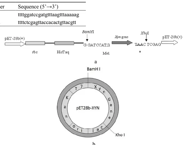
kit. xylanase gene was cloned to construct the
pET28b-xyn recombinant vector DNA using
XhoI and BamHI restriction enzymes (Figure
1). The positive clones for recombinant
xylanase were identified using the Congo
Red.
Table 2. Two oligonucleotides were utilized in order to amplify the xylanase gene
a
b
Figure 1. a. Schematic diagram of the gene region where the Xyn DNA sequence transferred to pET-28b (+) vector. b. Circular the pET28b-xyn map of construct which used to produce Bacillus subtilis xyn-akky1 xylanase.
2.3. Purification of Recombinant Xylanase
The E. coli BL21 (DE3) was transformed
with the pET28b-xyn construct and growth
on Luria Bertani agar containing kanamycin
(50 mg/ml). Briefly, the transformant was
inoculated into 3 ml culture tube containing
LB brorth and incubated overnight at 37 °C
and 200 rpm. Then this culture was inoculated
into 500 ml of LB containing kanamycin and
grown at 37 °C with shaking at 200 rpm. The
Primer
Sequence (5’→3’)
xyn1
ttttggatccgatgtttaagtttaaaaag
xyn2
ttttctcgagttaccacactgttacgtt
culture was induced for 3 hours with a final
concentration 1mM IPTG when the OD
600reached 06-07.
Next, the cells were harvested by using
centrifugation
(at +4°C, 8000 rpm 5 min),
followed by re-suspended the pellet using
RNAse (20 µg/ml) and DNAse (20 µg/ml)
with 20 mM phosphate buffer (pH 8.0) and
protease inhibitors (0.5 mM Phenyl methyl
sulfonyl fluoride (PMSF) and 2 mM
Benzamidine). Cells were first lysed using a
sonicator (Sonics VCX 130), then high-speed
(30,000 rpm) centrifugation was performed
for 1 hour. Qiagen Ni-NTA affinity column
was
used
to
prufication
of
soluble
recombinant protein carrying N-terminal 6x
histidine. The column was washed first with
50 mM phosphate buffer (pH 8.0) and then 50
mM phosphate buffer (pH 8.0) containing 30
mM imidazole. The protein was eluted from
column with 300 mM imidazole in 50 mM
phosphate buffer (pH 8.0). Purity of this
isolated protein was checked by SDS-PAGE.
Concentration of protein was determined by
UV absorption at 280 nm (19).
2.4. Plate Assay
E. coli strain BL21 (DE3) containing the
recombinant plasmid pET28b-xyn was
inoculated on LB agar plate containing 1%
beechwood xylan, 50 mg/ml kanamycin and
100 mg/ml IPTG. Following overnight
incubation at 37ºC, staining of the plates done
using 1% Congo-red solution and destained
by three washes using 1 M NaCl followed by
0.1 N NaOH. The enzyme activity was
examined by a clear zone formation around
the colony (Wood et al., 1998).
2.5. Biochemical Characterization
3,5-dinitrosalicylic acid (DNS) method was
used to determine the recombinant xylanase
activity (10). The optimal pH for purified 6x
His tagged enzyme was determined at 37ºC.
Beechwood xylane was used as a substrate in
wide pH which ranging from 4.0 to 10.0. pH
range of substrate was adjusted by Mcllvaine
buffer for pH 4-7, Tris-HCl buffer for pH 8,
and glycine-NaOH buffer for pH 9-10. To
determine the optimal temperature for
enzymatic activities the enzyme was
incubated between 30ºC to 70ºC in precense
of Mcllvaine buffer (pH 6.0). The
thermo-stability of the xylanase was tested by pre
incubating the enzyme in Mcllvaine buffer
(pH 6.0) at 50ºC, 55ºC, 60ºC without
substrate. Km and V
maxvalues for purified
enzyme were calculated in Mcllvaine buffer
(pH 6.0 at 60ºC using 1-10 mg/ml beechwood
xylan as a substrate). The data were plotted
by Lineweaver-Burk method (13).
2.7.
Nucleotide
sequence
accession
numbers
Bacillus subtilis strain akky1 16S rRNA
nucleotide sequences and xylanase gene were
deposited in the GenBank (accession
numbers KJ540929.1 and KJ540928.1).
3. RESULTS AND DISCUSSION
3.1. Microorganism Identification Using
PCR
Six strains isolated from soil samples
collected from Ordu province, Turkey
demonstrated xylanolytic activity. New
strains were identified using 16S rRNA
sequences. The xylanase activity was
revealed by strain akky1 which produce
highest zone clearance on agar plate
containing xylan (Figure 2a,b).
Figure 2. a. Hydrolysis zones of the xylanase-producing microorganisms. b. The result of agarose gel (%1) electrophoresis shows PCR products for the 16S rRNA of the xylanase-producing microorganisms. 1. λ-EcoR I /Hind III DNA marker, 2-8 16S rRNA PCR product of xylanase-producing microorganisms.
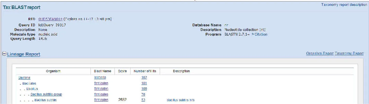
The species that have a similarity of 16S
rRNA sequences was analysed by BLAST
server at the NCBI public database. A
taxonomy report was established by using the
Taxonomy Report tool within BLAST.
According to taxonomy report, 16S rRNA
sequence of Bacillus subtilis strain akky1
(KJ540929) exhibited 100% nucleotide
identity with Bacillus subtilis strain therefore
new species were classified under the genus
Bacillus subtilis (Figure 3).
Figure 3. Taxonomy BLAST report of Bacillus subtilis strain akky1 16S ribosomal RNA gene, partial sequence (KJ540929)
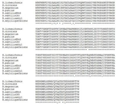
Figure 4. The multiple alignment of amino acid sequence of the xylanase enzymes of different species of Bacillus with the amino acid sequence isolated from Bacillus and used for cloning and expression studies of xylanase enzyme, by using ClustalW2 program. The accession numbers are: B. subtilis strain akky1, KJ540928.1; Bacillus pumilus, AAZ17390.1; Bacillus subtilis subsp. subtilis str. 168, NP_389765.1; Bacillus subtilis BSn5, YP_004203820.1;
Bacillus amyloliquefaciens, AAZ17388.1; Bacillus megaterium, ACT21830.1; Bacillus licheniformis, AAZ17387.1; Bacillus circulans,AAM08360.1
3.2. Cloning of the Xylanase gene in
Escherichia coli
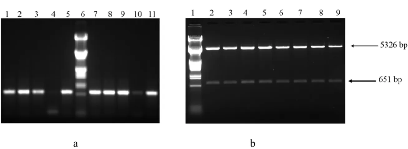
DNA fragment encoding-xylanase from
Bacillus subtilis strain akky1 xylanase was
amplified and this fragment was cloned to
pET 28b (+) vector using XhoI and BamHI
restriction enzymes. Final plasmid was
named as a pET28b-xyn. The results obtained
from colony PCR ( template: the E.coli DH5α
strains harboring pET28b-xyn; primers:
xyn1-xyn2) (Figure 5 a), restriction
fragment analysis (Figure 5 b), confirm the
success of cloning. Furthermore DNA
sequencing was done to verify correct
insertion
of
xylanase-encoding
DNA
fragment. E. coli DH5α cells transformed by
constructed pET28b-xyn were selected with
kanamycin selection. Consecutive plasmid
preparation
method
was
utilized
for
sequencing DNA samples and transformation
of competent E. coli BL21 (DE3) cells.
Figure 1b. demonstrates the circular plasmid
map of the construct.
a b
Figure 5. a. Agarose gel (1%) electrophoresis result showing PCR verification of recombinant plasmid pET28b-xyn after the cloning. Columns 1-5 and 7-11 indicates PCR products from the colonies and column 6 indicate, λ-EcoR I /Hind III DNA marker. b. 1% Agarose gel demonstrating digesting of recombinant plasmid pET28b-xyn with NcoI restriction enzyme after the cloning. 1, λ-EcoR I /Hind III DNA marker 2-9, DNA fragments obtained after the digestion.
3.3. Qualitative Analysis of the Purified
Protein
The 6xHis tagged recombinant xylanase was
purified from E. coli cell lysate by Ni-NTA
chromatography as described above. Eluted
samples were analysed on SDS-PAGE and
the purified enzyme migrated on the gel as a
single band with a molecular mass of around
26.0 kDa (Figure 6 a). The calculated
molecular mass of protein using the “ExPASy
ProtParam Tool” was 26970.6 Da which is
very close to the experimental molecular
mass.
Figure 6.a. Purification of xylanase enzyme was confirmed with SDS-PAGE (%12). Samples from induced E. coli BL21 pLysE cell lysate carrying pET28b plasmid (1). Samples from induced E. coli BL21 pLysE cell lysate carrying pET28b-xyn plasmid (2). Collected supernatant after centrifugation of the lysate (3). BioRad dual colour precision plus protein marker (4). The eluate collected from Ni-NTA agarose affinity column (imidazole concentrations are respectively 10, 25, 300 mM) (5-7). b. The image of zone formation at the periphery of the recombinant colony by Congo-red plate containing beechwood xylan. A. Recombinant colony B. E. coli BL21 without plasmid
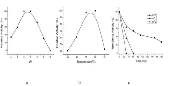
3.4.Biochemical Characterization
Activity of recombinant xylanase was
observed for various pH values. The optimum
condition for activity of recombinant
xylanase predicted as follows:
pH 6.0 (Figure 7a) and at 60°C (Figure 7b).
35% of the enzyme activity was able to
maintain stability for 200 minutes at 55°C
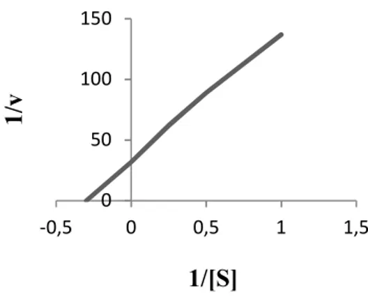
(Figure 7c). Km value for xylanase was 3.33
mg/ml when beechwood xylan was used as
substrate(Figure 8).
a b c
Figure 7. Characterization of recombinant xylanase. a. Effect of pH. b. Effect of temperature. c.Thermostability of recombinant xylanase
Figure 8. Lineweaver-Burk curve for the purified xylanase enzyme