Research Notes Dros. Inf. Serv. 92 (2009) 114
mouth parts, which are advantageous to the female beings to develop as normal. So as in the other metric traits. Perusal of literature reveals, the species under study are closely related. The present study also showed there is a similarity among the species with respect to metric traits.
References: Church, R.B., and F.W. Robertson 1996, Ecology 37: 544; Cosins, A.R., and K. Bowler 1987, Temperature Biology of Animals. London: Chapman & Hall; Coyne, J.A., and E. Beecham 1987, Genetics 177: 737; David, J.R., P. Gibert, E. Gravot, G. Petavy, J.P. Morin, and D. Karan et al., 1997, J. Therm. Biol. 22: 441-451; Delcour, L., 1969, Dros. Inf. Serv. 44: 133-134; Gibert, P., B. Moreteau, J.C. Moreteau, and J.R. David 1998, Heredity 80: 326-335; Hallgrimsson, B., and B.K. Hall 2005, Variation. Academic Press, San Diego; Hegde, S.N., and M.S. Krishna 1999, Current Science 77: 1346-1347; Hoffmann, A.A., and P.A. Parsons 1997, Extreme
Environmental Change and Evolution. Cambridge: Cambridge University Press; Hoffmann, A.A.,
and M.J. Hercus 2000, Bioscience 50: 217-226; Kitagawa, O., K.I. Wakahama, Y. Fuyama, Y. Shimada, E. Takanashi, M. Htsumi, M. Umabo, and Y. Miitay 1982, Jpn. J. Genet. 57: 141; Leather, S., K. Walters, and J. Bale 1993, The Ecology of Insects Overwintering. Cambridge: Cambridge University Press; Precht, H., J. Christophersen, H. Hensel, and W. Larcher 1973, Temperature and
Life. Berlin: Springer Verlag; Sokoloff, A., 1966, Evolution 20: 49-71.
The effects of dibutyl phthalate on the development of Drosophila melanogaster. Memmi, Burcu Koçak, and Emel Atlı. Hacettepe University, Faculty of Science, Department of Biology, Genetics Section, 06800, Beytepe, Ankara, Turkey; e-mail: [email protected]
Abstract
In this study, the developmental effects of a well-known plasticizer dibutyl phthalate (DBP) were determined in wild type Oregon strain of Drosophila melanogaster. The 72 h larvae of D.
melanogaster were exposed to four concentrations of DBP-acetone solution. After 72 hours, the
pupae were counted. The developmental times of DBP exposure groups were found extended comparing to the control group. The reductions were found statistically significant compared to the control (P < 0.05).
Key-words: Dibutyl phthalate, plasticizer, Drosophila melanogaster, development, mean pupation time.
Introduction
In recent years, the contamination with phthalate esters (1,2-benzendicarboxylates) (PAEs) turned into ubiquitous in the soil, water, and air (Steiner et al., 1998). The increased contamination with phthalate is a common problem worldwide due to its widespread utilization areas. Therefore, the concerns about possible harmful health effects have been raised (Swan et al., 2005; Hauser et al., 2006).
PAEs including dibutyl phthalate (DBP) are widely used in PVC industry and to a lesser extent in the non-polymer industry products (children’s toys, sealants, paints, printing inks, cosmetics, varnishes, shampoos, cables or fabrics, insect repellents etc.) (Heudorf et al., 2007). PAEs
Dros. Inf. Serv. 92 (2009) Research Notes 115 are oily substances and slightly soluble in water. They are not bound to plastics covalently (Mikula
et al., 2005). So, they can be leached from these products to the environment (Steiner et al., 1998).
PAEs are known to disrupt the endocrine system of animals (Fisher, 2004) and hence its exposure may cause potential health problems (Prasanth et al., 2009). DBP is a component of PAEs and has been reported a potential for endocrine disrupting effects on vertebrates (Toppari et al., 1996; Keith, 1997; Wine et al., 1997).
The toxic effects of DBP were investigated in many organisms including rat, abalone, freshwater rotifer etc. The researchers found that DBP can cause reproductive tract malformations in male rats (Wölfle et al., 2009), fertility reductions and ovarian malfunctions in female rats (Gray et
al., 2006). The developmental effects of DBP were found significantly toxic (malformation of
embryos, low larval settlements) in the exposed larvae of abalone Haliotis diversicolor supertexta (Zhihui et al., 2009). In addition, it is shown that DBP significantly prolonged the generation time of freshwater rotifer Brachionus calyciflorus (Zhao et al., 2009).
To the best of our knowledge, there are no reports in the open literature on the effects of DBP on D. melanogaster. Therefore, the major aim of the present study is to assess the effects of DBP on development of the fruit fly D. melanogaster.
Materials and Methods
In order to determine the mortality of DBP (CAS No: 84-74-2), wild type Oregon strain of D.
melanogaster was used. First, Oregon adult flies were crossed in culture bottles containing Standard Drosophila medium at 25 ± 1°C culture room. After 72 ± 2 hours, individuals were removed from
bottles and the 3rd instar larvae were collected. Ten larvae were placed into each vial. The DBP was firstly diluted in 0.1 ml acetone and after 100 ml sucrose solution was added. The solution and 1.5 gr
Drosophila medium were added in the vials. The treatment doses were 1 ml/L, 0.1 ml/L, 0.01 ml/L
and 0.001 ml/L. Each dose had two replicates. Statistical differences between the transition period of control and treated larvae were examined with two-variable t test using SPSS 11.5. The homogeneity of variances of data was tested with Levene statistics, and the variances were found homogeneous (P > 0.05).
Results
The developmental and mortality effects of DBP were investigated in 72 h larvae of D. melanogaster. After the exposure with DBP, pupation was recorded at 24 hours, 48 hours, and 72 hours. After 24 hours, pupation percentages were found to be 23.3% in control group, 5% in 0.001 ml/L dose, 3.33% in 0.01 ml/L dose, 5% in 0.1 ml/L dose, and 1.67 in 1 ml/L dose. After 48 hours, pupation percentages were found to be 66.67% in control group, 61.6% in 0.001 ml/L dose, 51.6% in 0.01 ml/L dose, 60% in 0.1 ml/L dose, and 58.33 in 1 ml/L dose. After 72 hours, pupation percentages were found to be 96.67% in control group, 88.33% in 0.001 ml/L dose, 98.33% in 0.01 ml/L dose, 95% in 0.1 ml/L dose, and 96.67% in 1 ml/L dose (Table1).
Table 1. The pupation percentages of the 3rd instar larvae of Drosophila melanogaster after treated with DBP.
No Doses (ml/L) n 24 h 48 h 72 h 1 Control 60 23.3 66.67 96.67 2 0.001 60 5 61.6 88.33 3 0.01 60 3.33 51.6 98.33 4 0.1 60 5 60 95 5 1 60 1.67 58.33 96.67 n: Number of larvae.
Research Notes Dros. Inf. Serv. 92 (2009) 116
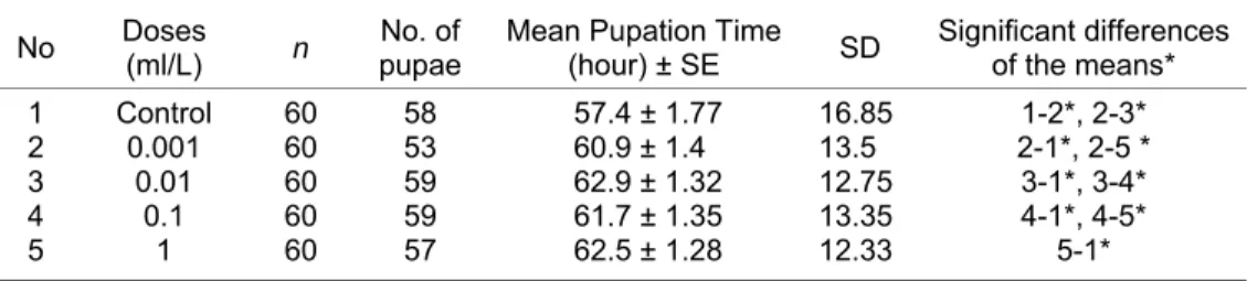
Table 2 shows mean pupation times (hour) of the control and exposure groups. The mean pupation time of control group was found to be 57.4 of DBP on development of D. melanogaster. The mean pupation times were found to be 57.4 in control group, 60.9 in 0.001 ml/L dose, 62.9 in 0.01 ml/L dose, 61.7 in 0.1 ml/L dose, and 62.5 in 1 ml/L dose. As seen in Table 2, there was a statistically significant reduction in the mean pupation times in the treated groups (P < 0.05) compared to the control group.
Table 2. The effect of DBP treatment on the mean pupae numbers of Drosophila
melanogaster in 72 hours.
No Doses (ml/L) n No. of pupae Mean Pupation Time (hour) ± SE SD Significant differences of the means*
1 Control 60 58 57.4 ± 1.77 16.85 1-2*, 2-3*
2 0.001 60 53 60.9 ± 1.4 13.5 2-1*, 2-5 *
3 0.01 60 59 62.9 ± 1.32 12.75 3-1*, 3-4*
4 0.1 60 59 61.7 ± 1.35 13.35 4-1*, 4-5*
5 1 60 57 62.5 ± 1.28 12.33 5-1*
n: Number of Larvae, SE: Standard Error, SD: Standard deviation; *p < 0.05
Discussion
In the present study, the developmental effects of plasticizer DBP were determined in wild type Oregon strain of Drosophila melanogaster. The pupation percentages of four treatment groups and control group were determined and compared. As seen in Table 1, the pupation percentages of treatment groups (5%, 3.33%, 5%, 1.67% ) in first 24 hours were found to be dramatically delayed when compared to that of control group (23.3%). After 48 hours, the differences between control and treatment groups were found stable.
The mean pupation time (hour) of the four treatment groups were calculated and compared with control group. As seen in Table 2, the mean pupation times of the treatment groups (60.9, 62.9, 61.7, 62.5, respectively) were markedly prolonged when compared to the that of control group (57.4). The results showed significant reductions among the treatment groups and control group (P < 0.05). These significant delays show that the pupation of D. melanogaster was markedly affected by DBP. Similarly to our findings, Zhao et al. (2009) determined a significantly prolonged generation time in freshwater rotifer Brachionus calyciflorus with DBP exposure.
It is reported that DBP treatment can cause fertility reductions, reproductive malformations in rats and low larval settlements in H. diversicolor supertexta (Gray et al., 2006; Wölfle et al., 2009; Zhihui et al., 2009). Besides, the researchers found that several phthalates including DBP or their metabolites, respectively, can cross the placenta barrier and reach the human fetus (Wittassek et al., 2009).
In conclusion, the present study has revealed that DBP can cause statistically significant reductions in pupation time of D. melanogaster. Concerning the potential estrogenic activity of DBP, further research is required.
References: Fisher, J.S., 2004, Reproduction 127: 305-315; Gray, L.E., Jr., J. Laskey, and J. Ostby 2006, Toxicological Sciences 93(1): 189-195; Hauser, R., J.D. Meeker, S. Duty, M.J. Silva, and A.M. Calafat 2006, Epidemiology 17: 682-691; Heudorf, U., V. Mersch-Sundermann, and J. Angerer 2007, Int. J. Hyg. Environ. Health 210: 623-634; Keith, L.H., 1997, Environmental
Dros. Inf. Serv. 92 (2009) Research Notes 117 Z. Svobodova, and M. Smutna 2005, Czech. J. Food Sci. 23: 217–223; Prasanth, G.K., L.M. Divya, and C. Sadasivan 2009, Toxicology 262: 38-42; Steiner, I., L. Scharf, F. Fiala, and J. Washuttl 1998, Food Addit. Contam. 15: 812; Swan, S.H., K.M. Main, F. Liu, S.L. Stewart, R.L. Kruse et al., 2005, Environ. Health Perspect. 113: 1056–1061; Toppari, J., J.C. Larsen, P. Christiansen, A. Giwercman, and P. Grandjean 1996, Environ. Health Perspect. 104: 741; Wine, R.N., L.H. Li, L.H. Barnes, D.K. Gulati, and R.E. Chapin 1997, Environ. Health Perspect. 105: 102-107; Wittassek, M., J. Angerer, M. Kolossa-Gehring, SD. Schafer, W. Klockenbusch et al., 2009, Int. J. Hyg. Environ. Health 212: 492–498; Wölfle, D., K. Pfaff, T. Platzek, and A. Luch 2009, Toxicology Letters 189S: S57-S273; Zhao, L.L., Y.L. Xi, L. Huang, and C.W. Zha 2009, Aquat. Ecol. 43 (2): 395-402; Zhihui, Y., Z. Xiangjing, and C. Zhonghua 2009, Chinese Journal of Oceanology and Limnology 27 (2): 395-399.
Vitamin A deprivation does not decrease fluorescence of ARF72-RFP, a label for Golgi apparatus, in Drosophila visual receptors.
Denny, George, and William S. Stark. Department of Biology, Saint Louis University, St. Louis, MO 63103. e-mail: [email protected]
Each ommatidium of the Drosophila compound eye has 8 photoreceptors (retinula cells); the rhabdomere of each is the specialized organelle that houses rhodopsin and the visual transduction molecules. One of the primary functions of the retinula cell is maintenance of the rhabdomere, including turnover of membrane and protein (Lee et al., 1996). Vitamin A deprivation reduces or eliminates rhodopsin in Drosophila rhabdomeres (Harris et al., 1977). Vitamin A replacement synchronizes de novo synthesis and export of rhodopsin (Sapp et al., 1991).
Although the quality of fixation of Drosophila photoreceptors has always been variable, electron micrographs from our lab archives showed that vitamin A deprived flies are more likely to be plagued by what we refer to as “ghosty cytoplasm” (marked with asterisks [*], Figure 1, Top left) than vitamin A replete controls (Figure 1, Middle left). (Electron micrographs are labeled thus: R is rhabdomere, G is Golgi, > is desmosome, N is nucleus, and PG is pigment granule.) We hypothesized that cytoplasmic organelles dedicated to biosynthesis, rough endoplasmic reticulum and Golgi apparatus, might be reduced by vitamin A deprivation. We tested our hypothesis using a fly stock we had been using to visualize Golgi apparatus, ARF72-RFP (ADP ribosylation factor tagged with red fluorescent protein).
Flies were lightly etherized and fixed to a glass slide for visualization of the deep pseudopupil. A typical fluorescence micrograph is presented (Figure 1, Top right); the blurry appearance compared with rhabdomere fluorescence (Stark and Thomas, 2004) is explained since Golgi apparatus is distributed throughout retinula cells. Fluorescence was quantified using a fluorescence microscope with a photometer system (Stark et al., 1985). The pseudopupil image was delimited by the photometer and fed to the photomultiplier tube. Rhabdomeres were excited with a calibrated amount of 488 nm light, and a voltage response proportional to the level of fluorescence being emitted was recorded by a computer. Flies were raised at room temperature either on our yellow cornmeal food (supplemented with beta-carotene, vitamin A replete) or on Sang’s medium lacking vitamin A. A strong correlation between age and level of fluorescence was noted (Figure 1, Middle right). Thus, for control, measurements were performed on flies within 12 hr of eclosion.
RFP fluorescence of vitamin A deprived flies (n = 28) was the same as that of flies reared on vitamin A replete medium (n = 27), as witnessed, since the error bars (95% confidence intervals)