RESEARCH ARTICLE
50
THE INVESTIGATION OF THE ACTION MECHANISM OF AMMONIUM PYRROLIDINE DITHIOCARBAMATE ON RAT AORTA SMOOTH MUSCLE
CONTRACTION-RELAXATION RESPONSES Hayri DAYIOĞLU1
, Ayhan YILMAZ2, Zeynep KELEŞ3, Fatih ALAN4 and Sinan DARCAN5
1Kütahya Dumlupınar University, Faculty of Science and Literature, Department of Biology, 43270, Kütahya,
[email protected], ORCID: 0000-0002-9270-8561
2
Kütahya Dumlupınar University, Faculty of Science and Literature, Department of Biology, 43270, Kütahya, [email protected], ORCID: 0000-0003-0410-8687
3Fevzi Çakmak Mah., Muhsin Yazıcıoğlu Cad. No:10, 34899, Pendik, İstanbul, [email protected],
ORCID: 0000-0003-3440-0785
4General Directorate of Presidential Protection Services, Presidential Complex, 06530, Beştepe, Ankara,
[email protected],ORCID: 0000-0002-0561-6192
5Kütahya Health Science University, Evliya Çelebi Campus, Tavşanlı Yolu 10. Km, 43100, Kütahya,
[email protected], ORCID: 0000-0002-2135-4807
Received Date: 20.04.2020 Accepted Date:09.10.2020
ABSTRACT
Ammonium Pyrrolidine Dithiocarbamate (APDTC) is an inhibitor of nuclear factor kappa b (NF-κB). Beside of these function, its antitumoral, antioxidant, anticarcinogenic and antiviral properties and also apoptosis inhibiting effect in smooth muscle cells were determined. Our experiments aimed to investigate the mechanism of action of APDTC on rat aortic smooth muscle. Some adrenergic and cholinergic receptors, l-type Ca2+ channels and K+ channels were blocked in 7 different groups, and therefore the action mechanism of APDTC whether is used or not on which channels and receptors and the extent to which they are effective were aimed to be determined. The contraction-relaxation responses after the administration of APDTC, atropine, phentolamine, propranolol, nifedipine, tetraethylammonium (TEA) and mix (atropine+phentolamine+propranolol) on living state controlled with potassium chloride (KCl) and blocked nitric oxide (NO) synthesis with l-ng-nitro arginine methyl ester (L-NAME) in the pre-contraction-induced aortic preparations with phenylephrine were investigated. The obtained data were evaluated by kruskal wallis and mann-whitney u tests. APDTC created the relaxation response in the aortic smooth muscle. Cholinergic receptor blocker atropine, α-adrenergic receptor blocker fentolamine, β-α-adrenergic receptor blocker propranolol, l-type calcium channel blocker nifedipine and potassium channel blocker TEA did not alter the relaxation response of APDTC. In the mix group consisting of atropine+phentolamine+propranolol, APDTC created a significant contraction response. It has been determined that APDTC can be effective on these systems via different mechanisms.
Keywords: APDTC, aorta, non‐ adrenergic non‐ cholinergic (NANC) system, L-NAME, phenylephrine, adrenergic, cholinergic, calcium channels, potassium channels.
Dayıoğlu et all., Journal of Scientific Reports-A, Number 45, 50-69, December 2020.
51 1. INTRODUCTION
The aorta is the most functional vessel that transmits blood to all parts of the body. The thickness of the aorta is approximately 2.5 cm. The blood cycle in the body lasts 60 seconds. The pressure in the aorta, brachial and other large arteries in a young adult human increases to a peak of approximately 120 mm Hg (systolic pressure) during each heart cycle and decreases to a minimum (diastolic pressure) of about 70 mm Hg [1].
APDTC is a NF-κB inhibitor. APDTC has antioxidant effects. It inhibits apoptosis in vascular endothelial cells, lymphocytes and neurons. It inhibits apoptosis in leukemia cells (HL-60) [2]. A significant increase in TAK was detected in rats treated with APDTC (0.70±0.01 μmol/mg protein). APDTC has been found to increase anti-oxidant enzyme activity in cholestasis dependent hepatic injury model. This result with APDTC is actually not surprising [3]. After showing that aldesterone was effective in 1992, it was revealed that there were aldesterone receptors in the corpus cavernosum. After investigating the penile tissues obtained from mature men, it was revealed that aldesterone did not have a direct relaxant effect, but increased the effect of neurarenaline [4] . APDTC inhibits DON-induced mitochondrial dysfunction and apaptosis via the NF-κB/iNOS pathway [5]. In models with inflammatory disease, the therapeutic effect of antioxidants as potential NF-κB inhibitors has been determined [6]. In the collagen-induced joint inflammation model, the anti-oxidant effect of APDTC in brain, stomach, lung, myocardia, organ injuries, renal ischemia/reperfusion was investigated. APDTC protects against NF-κB through pathological effects induced by some different stimuli, including lipopolysaccharide and cytokines[7]. AMTB and APDTC administered intrathecally affect TRPM8 and NF-κB in the spinal dorsal horn, even the brain [8]. It revealed the fact that inactivation of autophagy using APDTC reduces damage to the intestinal mucosal barrier. The effects of autophagy inactivation were largely dependent on the dosage of APDTC. Overactive autophagy may be weakened in accordance with physiology when using low or medium dose of APDTC. Using high-dose APDTC can completely inhibit autophagy and lead to decreased survival of instestinal mucosal cells. In this study, 10 mg/kg APDTC was shown to inhibit autophagy and perform best in maintaining inestinal mucosal integrity. The bi-directional efficacy of APDTC also requires the therapeutic window of APDTC, or other autophagy inhibitors, to be carefully investigated in humans [9]. The effects of Klotho were determined to be comparable to NF-κB inhibition, with the inhibition of NF-κB nuclear translocation and inhibition of APDTC administration, which is reported to inhibit DNA binding activity. Accumulating evidence suggests that the anti-aging protein Klotho plays an important role in inhibiting NF-κB and preventing nuclear translocation [10]. After injecting the APDTC, the pH of the gastric juice did not change significantly, but the promotional effect of NaHS on gastric acid secretion can be inhibited by APDTC. The pH value of the gastric juice changed values from 5.41±0.32 before injection, to 5.34±0.36 (p> 0.05) after injection. There is no significant difference after enterocele is injected with APDTC+NaHS. These results show that NaHS is involved in the control of gastric acid secretion by activating the NF-kB pathway [11].
Muscle fibers are structures that convert chemical energy into mechanical energy by spending ATP energy [12]. According to their microscopy, they are classified as striped and smooth muscles. The reason is due to the different physical properties of contractile proteins [13]. Muscle tissues can be stimulated, contracted, stretched and relaxed [14]. Smooth muscle cells are found on the walls of various organs and pipes of the body along with blood vessels, stomach, intestine, bladder, respiratory tract, penis, uterus and clitoral carenosal sinuses. Smooth muscle usually consists of smaller fibers, 1.5 μm in diameter and 20-500 μm in length [15]. The concentration of myosin in smooth muscle is only
Dayıoğlu et all., Journal of Scientific Reports-A, Number 45, 50-69, December 2020.
52
one third of myosin in striated muscle, while the actin concentration can be twice as high. Despite these differences, the maximal tension per sectional unit developed from smooth muscles and the maximal tension developed in skeletal muscle are similar [16]. Smooth muscle activities are different. In some organs, smooth muscle cells are active by constantly maintaining a tonus level [12]. Smooth muscle cells cause tonic and phasic contractions in response to load or length changes. Smooth muscle cells, completely separate from the stimulant, use the cross-bridge between actin and myosin strands to produce strength, and Ca2+ ions trigger contraction [17]. Similar to skeleton and heart muscles, Ca2+ plays a significant role in initiating smooth muscle contraction. However, the source of Ca2+ increase in unitary smooth muscle may be very different [18]. In order to relax contracted smooth muscle, myosin needs to be dephosphorylated because dephosphorylated myosin can not bind to actin. Myosin light chain phosphotase enzyme, which is constantly active in the smooth muscle during rest and contraction periods, mediates dephosphorilation. When cytosolic calcium rises, the myosin phosphorylation rate of activated kinase exceeds the phosphotase dephosphorylation rate, and therefore the amount of phosphorylated myosin in the cell increases and the tension increases. When the cytosolic calcium concentration decreases, the dephosphorylation rate exceeds the phosphorylation rate, and the amount of phosphorylated myosin is low and relaxation occurs [16]. However, dephosphorylation of myosin light chain kinase does not lead to smooth muscle relaxation. There are several mechanisms. One is the latch bridge mechanism, that is, the myosin cross bridges continue to hold on to actin for a while after the cytoplasmic calcium concentration has decreased. This little energy produces continuous contraction and is especially important in vascular smooth muscles [18]. In addition to the cellular mechanisms that increase smooth muscle contraction, there are also cellular mechanisms that cause relaxation. This feature is important in the smooth muscles surrounding the blood vessels in order to increase blood circulation. It has long been known that endothelial cells, which line the inside of blood cells, secrete a substance NO that relaxes smooth muscle. NO produced in endothelial cells can spread into smooth muscle to act. Once becoming intramuscular, NO directly activates the soluable guanylate cyclase to produce another second messenger molecule, cyclic guanosine monophosphate (cGMP). This molecule activates ion channels, Ca homeostasis, phosphatases, or cGMP-specific protein kinases that may affect them all, leading to smooth muscle relaxation [18]
.
Atropine or l-hyocyanimine is an alkaloid obtained from the leaves of Atropa bellodona and Datura
stramonium plants of solanacea family and effective in the peripheral system [19]. Atropine is a
non-selective muscarinic receptor antagonist. The binding strength of muscarinic receptor antagonists to the receptor may overlap among various subtypes [20]. It reduces parasympathetic tone to keep sympathetic activity high. Atropine has the ability to change the cardiac rhythm [19]. Atropine and bomatropine and similar drugs such as scopolamine inhibit the effect of acetylcholine on the muscarinic types of cholinergic effector organs [21]. Imidazoline-derived phentolamine is a non-selective α-adrenergic receptor antagonist. Phentolamine effects are nonnon-selective. Phentolamine have equal binding points on alpha-1 and alpha-2 receptors [22]. Nifedipine is a predominant calcium antagonist with vascular effects. Nifedipine inhibits voltage-dependent L-type Ca2+ channels in cardiac-related smooth muscle cell membranes. Nifedipine is a short-acting Ca2+ antagonist [19]. TEA has K+ channel blocker effect. It is the first substance showing ganglion blocker effect [23]. Taurine is blocked by TEA. However, 4-AP facilitates K+ flow by a mechanism which is not affected by glibenclamide, iberiotoxin or barium chloride (BaCl2). TEA has the effect of blocking the potassium
channel, which may be contrary to the effect of taurine. The vasorelaxan effect of taurine in rat isolated arteries is reduced by TEA [24]. Most K+ channels are blocked by externally supplied TEA. However, sensitivity to TEA is different in various K+ channel types [25]. The system that is not
Dayıoğlu et all., Journal of Scientific Reports-A, Number 45, 50-69, December 2020.
53
related to neither the cholinergic nor adrenergic part of the autonomic nervous system is called the NANC system.
Both neuronal NO and endothelial NO have vasodilation effects. NO is thought to be the major neuromediatory of NANC in most parts of the body. NO is synthesized from an amino acid (l-arginine) at the nerve endings by NO synthetase [19]. In addition to adrenergic and cholinergic conduction in the autonomic nervous system, it provides conduction in dopaminergic, peptidergic and purinergic nerves [23]. NANC fibers were defined in 3 groups. The neurotransmitter of the purinergic fibers is ATP and cause vasoconstriction. The neurotransmitter of nitrosidergic fibers is NO. It causes vasodilatation. The neurotransmitter of the peptidergic nerves is vasoactive intestinal peptide (VIP) or peptide related to the calcitonin gene (CGRP) [26]. Phenylephrine is a synthetic alpha antagonist that is partially selective to alpha-1 receptors. It increases systolic and diastolic blood pressures by causing vasoconstriction [23].
Isolated tissue and isolated organ preparations have been in use for over one hundred years, providing researchers with convenient biological models that exist without the systemic influences of the intact animal [27]. Isolated preparations can permit molecular biologists to quantitate the physiological impact of the expression of these altered genetic sequences at the tissue and organ level [28]. Some isolated organ baths are ADInstruments, DMT, Orchid Scientific, Ugo Basile and Campden Instruments Ltd. [29].
Our experiments aimed to investigate the mechanism of action of APDTC on rat aortic smooth muscle. The mechanism of action of APDTC is not fully elucidated and, therefore, it is possible to inhibit the effects of α-adrenergic and β-adrenergic receptors, cholinergic receptors, l-type Ca2+
channels and K+ channels in aortic smooth muscle contraction and dilatation responses with the aim of investigating whether these factors are effective or not in the mechanism of action of APDTC. Some adrenergic and cholinergic receptors, l-type Ca2+ channels and K+ channels were blocked in 7 different groups, and therefore the action mechanism of APDTC whether is used or not on which channels and receptors and the extent to which they are effective were aimed to be determined. The contraction-relaxation responses after the administration of APDTC, atropine, phentolamine, propranolol, nifedipine, TEA and mix (atropine+phentolamine+propranolol) on living state controlled with potassium chloride (KCl) and blocked nitric oxide (NO) synthesis with L-NAME in the pre-contraction-induced aortic preparations with phenylephrine were investigated.
2. MATERIALS AND METHODS 2.1. Laboratory Animal Supply
This study was carried out at Kütahya Dumlupınar University (KDPÜ) Experimental Animals Breeding Application and Research Center in the Faculty of Science and Art, Biology Department, Physiology Laboratory. The study was approved by KDPÜ Animal Experiments Local Ethics Committee (HAYDEK) with the decree of 2015.12.05. In the experiments, 70 male wistar albino rats, produced in Saki Yenilli Experimental Animals Production Center, were used. The rats were fed ad libitum in well ventilated rooms, at the normal day and night cycles, with standard industrial feed and tap water. The studies were carried out immediately after obtaining the permission of the Animal Ethics Committee. 70 male rats weighing 300-350 g with ages of 6 and 7 months held by Experimental Animal Breeding Application and Research Center Laboratory were placed in cages as
Dayıoğlu et all., Journal of Scientific Reports-A, Number 45, 50-69, December 2020.
54
following groups presented in 2b and 2c sections with all the experimental groups and procedures were explained.
2.2. Designing Test Groups
Test groups with used chemicals were as follows: 1st group is APDTC group (control), 2nd group is alpha adrenoreceptor antagonist (phentolamine)+APDTC, 3rd group is beta adrenoceptor antagonist (propranolol)+APDTC, 4th group is cholinergic receptor antagonist (atropine)+APDTC, 5th group is l-type calcium channel blocker (nifedipine)+APDTC, 6th group is potassium channel blocker (TEA)+APDTC and finally 7th group is both adrenergic and cholinegic receptor antagonist (phentolamine+propranolol+atropine)+APDTC. 10 animals were used per group.
2.3. Experimental Protocol
Experimental protocol for rat aorta smooth muscle contraction and relaxation response measurements is in detail as follows: After the animals were killed by cervical dislocation method, the thoracic aorta was removed by cutting from the neck to the abdomen (Figure 2.1). The reason that we use the cervical dislocation method is to avoid hormonal changes. Then, it was left into krebs-henseleit solution and was seperated from the surrounding connective tissue and then 3 mm circular sections were obtained. The weight of each organ was determined by precision scales. Isolated organs were placed in an organ bath. The temperature of the isolated organ bath was brought to 37oC and the a gas mixture of 95% O2-% CO2 was applied to the bath to oxygenate tissues. Tissue preparations under 1 g
strain in the isolated organ bath were equilibrated by washing them with a krebs-henseleit solution for 45 minutes in every 15 minutes. The viability of the organs was tested with 6×10⁻² M KCl. The organ
was washed three times with krebs-henseleit solution by fill-and-empty technique to remove KCl from the bath. After 5 minutes, 10-2 M L-NAME was given to stop the NO effects. 10-4 M phenylephrine was added to the bath for precontraction. After 10 minutes, the antagonist or blocking agent was added to the designated groups in the bath. As the 1st group was the control group, only APDTC was administered, 10-5 M phentolamine in the 2nd group, 10-6 M propranolol in the 3rd group, 10-6 M atropine in the 4th group, 10-6 M nifedipine in the 5th group, 10-3 M TEA in the 6th group and finally 7th group with phentolamine+propranolol+atropine were used. 5.5×10⁻⁵ M APDTC was added 15 minutes after the antagonist administered to the other groups except the first group. We waited 10 minutes and the relaxation responses of the organ to the given chemicals were recorded in the data acquisition analysis system via the isometric transducer. All solutions during the experiment were prepared with distilled water. We used the isolated organ bath from Commat, Turkey with data acquisition analysis system from MP36, USA and isometric transducers from Biopac, USA.
2.4. Statistical Analysis
Bath recording lasted 1 hour 43 minutes. To compare, the phenylephrine and antagonist agent range was used, and then the antagonist PDTC range. The calculation was performed as follows. When phenylephrine is 100, % value was calculated by establishing the ratio of what would be the antagonist and what would be APDTC. Simultaneous experiments were carried out in four baths. SPSS program was used for statistical analysis. When statistical analysis was made in SPSS, both standard error and standard deviation were calculated and used in statistical processing. We drew the graphics according to our SPSS results in excel files. We also drew according to aortic phenylephrine values in the 100 file. We created p-p values according to this file. First we found the p-p values. Then we calculated how the drugs affected when the contraction was 100%. Then, we set the proportion to compare them with phenylephrine, such as what is the value of atropine when phenylephrine is 100. It also gave us statistical results if there were any significant results. The exact sig. or asymp. sig. from
Dayıoğlu et all., Journal of Scientific Reports-A, Number 45, 50-69, December 2020.
55
the mann-whitney test results made us decide whether the difference is meaningful or meaningless. The data obtained from the experiments were evaluated by applying mann-whitney u and kruskal wallis tests. p<0.05 values were considered statistically significant.
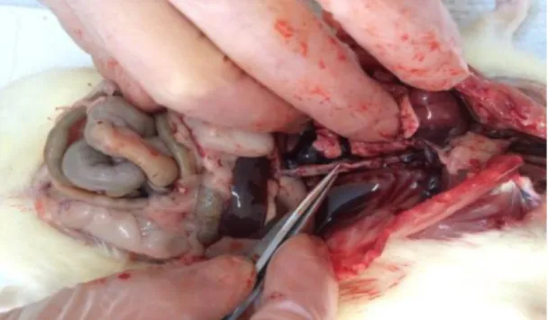
Figure 1. Rat Aorta. 3. RESULTS
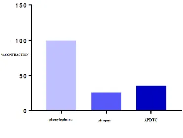
In our study, 7 different groups were studied as 1st group as the control group, 2nd group as atropine group, 3rd group as phentolamine group, 4th group as propranolol group, 5th group as nifedipine group, 6th group as TEA group and 7th group as mix group (phentolamine+propranolol+atropine). In our study, contraction-dilatation responses of the tissue to APDTC were investigated by applying various antagonists or channel blockers to pre-contracted aortic tissue with phenylephrine. There was a significant difference between nifedipine administered group and phentolamine group, propranolol group and mix groups (p<0.05). There was a significant difference between phentolamine+propranolol+atropine-treated mix group and control group, atropine group and nifedipine group (p<0.05) (Table 1 and Figure 2). APDTC on y axis in Figure 2 shows contraction percentage of smooth muscle of aorta by adding APDTC on pre-contracted aorta with phenylephrine in the presence and absence of antagonists or blockers in all 7 groups. Figure 2 is the comparison of all groups in one graph.
Figure 2. Effects of APDTC on pre-contracted aorta with phenylephrine in the presence and absence of antagonists or blockers.
Dayıoğlu et all., Journal of Scientific Reports-A, Number 45, 50-69, December 2020.
56
Table 1. Comparison of APDTC among groups. There was a significant difference between nifedipine administered group and phentolamine group, propranolol group and mix groups (p <0.05). There was a significant difference between phentolamine+propranolol+atropine-treated mix group and control group, atropine group and nifedipine group (p <0.05). Values that are smaller than 0.05 are in bold.
Groups Control Atropine Phentolamine Propranolol Nifedipine TEA Mix
Control - 0.450 0.070 0.174 0.450 0.450 0.041 Atropine 0.450 - 0.131 0.326 0.070 0.940 0.023 Phentolamine 0.070 0.131 - 0.734 0.008 0.199 0.880 Propranolol 0.174 0.326 0.734 - 0.023 0.364 0.940 Nifedipine 0.450 0.070 0.008 0.023 - 0.131 0.000 TEA 0.450 0.940 0.199 0.364 0.131 - 0.112 Mix 0.041 0.023 0.880 0.940 0.000 0.112 -
There was a significant difference between precontraction responses induced by phenylephrine in the aortic smooth muscle and APDTC responses in the presence of atropine (p <0.05). APDTC showed a contraction response in the presence of atropine on the contraction induced by phenylephrine in aortic tissue. However, there was no significant difference between the control and atropine group. Phenylephrine-contracted aortic smooth muscle relaxation responses of APDTC did not affect atropine, a nonselective muscarinic receptor antagonist (p> 0.05) (Table 2 and Figure 3).
Figure 3. The effect of APDTC in the presence of atropine and atropine substance on aortic contraction.
Dayıoğlu et all., Journal of Scientific Reports-A, Number 45, 50-69, December 2020.
57
Table 2. Comparison of APDTC in the presence of atropine and by atropine.There was a significant difference between precontraction responses induced by phenylephrine in the aortic smooth muscle and APDTC responses in the presence of atropine (p <0.05). Values that are smaller than 0.05 are in bold.
Atropine Phenylephrine Antagonist APDTC
Phenylephrine - 0.001 0.001
Antagonist 0.001 - 0.257
APDTC 0.001 0.257 -
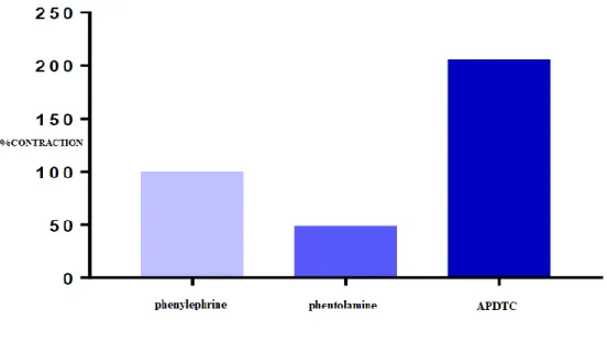
There was a significant difference between the contraction responses of phenylephrine and phentolamine in aortic smooth muscle (p<0.05). APDTC showed contraction response in the presence of phenylephrine. There was no significant difference between the control group and phentolamine group. Phenylephrine-contracted aortic smooth muscle relaxation responses of APDTC were not affected by (p>0.05) (Table 3 and Figure 4).
Figure 4. The effect of APDTC on aortic contraction in the presecence of phenilephrine and phentolamine.
Table 3. Comparison of APDTC in the presence of phentolamine and phenylephrine. There was a significant difference between the contraction responses of phenylephrine and phentolamine in aortic smooth muscle (p<0.05). Values that are smaller than 0.05 are in bold.
Phentolamine Phenylephrine Antagonist APDTC
Phenylephrine - 0.000 0.419
Antagonist 0.000 - 0.762
Dayıoğlu et all., Journal of Scientific Reports-A, Number 45, 50-69, December 2020.
58
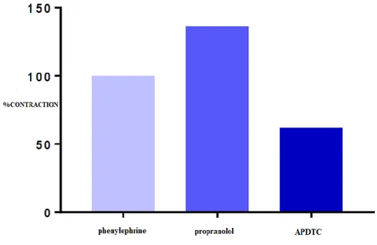
There was no significant difference between precontraction responses in the aortic smooth muscle with phenylephrine and APDTC responses in the presence of propranolol (p> 0.05). APDTC showed a relaxing effect in the presence of propranolol on contraction of aortic tissue with phenylephrine. At the same time, the contraction effect of propranolol turned into a relaxing effect with the administration of APDTC. There was no significant difference between the control group and the propranolol administered group (p> 0.05) (Table 4 and Figure 5).
Figure 5. Effect of APDTC on aortic contraction with and presence of propranolol. Table 4. Comparison of APDTC with and in the presence of propranolol.
Propranolol Phenylephrine Antagonist APDTC
Phenylephrine - 0.419 0.106
Antagonist 0.419 - 0.307
APDTC 0.106 0.307 -
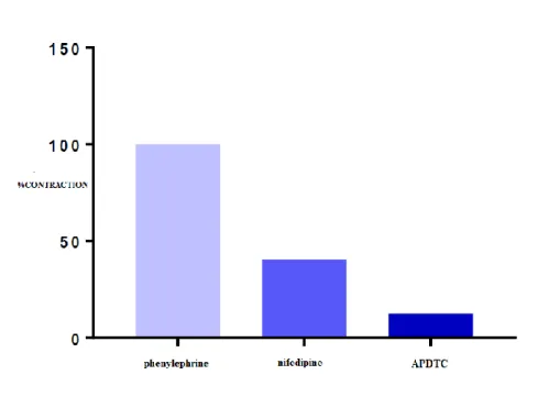
There is a significant difference in aortic smooth muscle contractile responses between APDTC responses in the presence of nifedipine and phenylephrine (p <0.05). There was no significant difference between the control group and nifedipine administered group (p> 0.05). APDTC showed a relaxing effect on contraction of aortic tissue with phenylephrine in the presence of nifedipine. In addition, the relaxant effect of nifedipine continued increasing gradually with the administration of APDTC (p <0.05) (Table 5 and Figure 6).
Dayıoğlu et all., Journal of Scientific Reports-A, Number 45, 50-69, December 2020.
59
Figure 6. The effect of APDTC on aortic contraction with and in the presence of nifedipine. Table 5. Comparison of APDTC with and in the presence of nifedipine. There is a significant difference in aortic smooth muscle contractile responses between APDTC responses in the presence of nifedipine and phenylephrine (p <0.05). The relaxant effect of nifedipine continued to increase gradually with the administration of APDTC (p <0.05). Values that are smaller than 0.05 are in bold.
Nifedipine Phenylephrine Antagonist APDTC
Phenylephrine - 0.001 0.000
Antagonist 0.001 - 0.016
APDTC 0.000 0.016 -
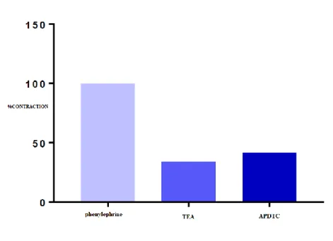
There was a statistically significant difference between contraction responses with phenylephrine and potassium channel blocker TEA in aortic smooth muscle (p <0.05). TEA, a potassium channel blocker in the aortic tissue, produced a relaxation response. However, there was no significant difference between the control group and the TEA administered group (p> 0.05) (Table 6 and Figure 7).
Dayıoğlu et all., Journal of Scientific Reports-A, Number 45, 50-69, December 2020.
60
Figure 7. The effect of APDTC on aortic contraction with and in the presence of TEA substance. Table 6. Comparison of APDTC in the presence of TEA and TEA. There was a statistically significant difference between contraction responses with phenylephrine and potassium channel blocker TEA in aortic smooth muscle (p <0.05). Values that are smaller than 0.05 are in bold.
TEA Phenylephrine Antagonist APDTC
Phenylephrine - 0.001 0.001
Antagonist 0.001 - 0.791
APDTC 0.001 0.791 -
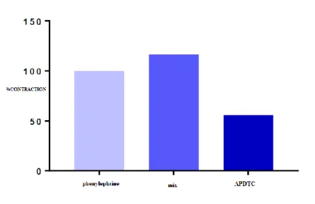
There is a significant difference in contractile responses of aortic smooth muscle induced between APDTC responses in the presence of phentolamine+propranolol+atropine and phenylephrine (p <0.05). APDTC showed a relaxation effect on contraction of phenylephrine-induced aortic tissue in the presence of adrenergic and cholinergic receptor antagonists as phentolamine+propranolol+ atropine. In addition, the contraction effect of fentolamine+propranolol+atropine, which are adrenergic and cholinergic receptor antagonists, turned into a statistically relaxing effect with the application of APDTC (p<0.05). There was a significant difference between the control group and the mix group. Phentolamine+propranolol+atropine changed APDTC's phenylephrine-contracted aortic smooth muscle relaxation responses (p<0.05) (Table 7 and Figure 8).
Dayıoğlu et all., Journal of Scientific Reports-A, Number 45, 50-69, December 2020.
61
Figure 8. The effect of APDTC in aortic contraction with and in the presence of phentolamine+ propranolol+atropine.
Table 7. Comparison of APDTC with and in the presence of mix. There is a significant difference in contractile responses of aortic smooth muscle induced between APDTC responses in the presence of phentolamine+propranolol+atropine and phenylephrine (p<0.05). The contraction effect of fentolamine+propranolol+atropine turned into a statistically relaxing effect with the application of APDTC (p<0.05). There was a significant difference between the control group and the mix group. Phentolamine+propranolol+atropine changed APDTC's phenylephrine-contracted aortic smooth muscle relaxation responses (p<0.05).Values that are smaller than 0.05 are in bold.
Mix Phenylephrine Antagonist APDTC
Phenylephrine - 0.419 0.001
Antagonist 0.419 - 0.001
APDTC 0.001 0.001 -
4. DISCUSSION AND CONCLUSION
APDTC, a specific inhibitor of low molecular weight NF-κB exibiting antioxidant ability to eliminate free toxic radicals, interferes with the production of pro-inflammatory cytokines [30]. All substances capable of inducing NF-κB can be blocked by antioxidants. It reduces the ability to bind to DNA, interferes with the activation signal pathway for NF-κB, and stabilizes or increases the IκBa synthesis mechanism to inhibit NF-κB activity [31]. Investigating the contraction-relaxation responses of APDTC, a dose-dependent relaxation response against precontraction of KCl after contraction was obtained [32]. We have investigated the mechanism of action of APDTC in vitro in aortic smooth muscle as a result of the fact that the mechanism of action of APDTC, which is determined to relax the aortic smooth muscle, has not been revealed yet.
Dayıoğlu et all., Journal of Scientific Reports-A, Number 45, 50-69, December 2020.
62
In order to determine the mechanism of action of APDTC, our experiments were carried out in seven groups ( control, atropine, phentolamine, propranolol, nifedipine, TEA, and fentolamine+propranolol +atropine) to determine which receptors and channels were used in the aortic smooth muscle contracted by phenylephrine whose NO synthesis was stopped by L-NAME. We used L-NAME to prevent the relaxation effect of NO and for the pre-contraction to run smoothly.5.5×10-5 M APDTC produced a relaxation response in the aortic smooth muscle. In a study, the cellular mechanism of nitric oxide (NO)-induced relaxation in corporeal smooth muscle (CSM) of the guinea-pig was investigated. Changes in the intracellular concentration of calcium ions ([Ca2+]i), membrane potential
and isometric tension were measured. CSM cells exhibited spontaneous depolarizations and transient increases in [Ca2+]i (Ca2+ transients) which were accompanied by contractions. This spontaneous
activity was abolished by nifedipine (10 μM). NO released by 3-morpholino-sydnonimine (SIN-1, 10 μM) hyperpolarized the membrane and prevented the generation of spontaneous depolarizations. SIN-1 also abolished Ca2+ transients and associated contractions. These effects of SIN-1 were blocked by 1H-[1,2,4]oxadiazole[4,3-a]quinoxalin-1-one (ODQ, 10 μM), an inhibitor of guanylate cyclase. Noradrenaline (NA, 1 μM) increased [Ca2+]i to levels similar to those produced by high
potassium-containing solution (high K+ solution, [K+]o = 40 mM), however, NA-induced contractions were three
times greater in amplitude than those induced by high K+ solution. In NA precontracted preparations, SIN-1 inhibited 80% of the contraction and decreased [Ca2+]i by 20%. In contrast, nifedipine reduced
[Ca2+]i by 80%, while the level of contraction was decreased by only 20%. SIN-1-induced reduction in
[Ca2+]i but not the tension effect, was abolished by pretreatment with cyclopiazonic acid (CPA, 10
μM). In high K+ precontracted preparations, SIN-1 inhibited 80% of the contraction and reduced
[Ca2+]i by 20%. Nifedipine, however, largely abolished increases in both [Ca2+]i and tension under
these circumstances. These results suggest that decreasing the sensitivity of contractile proteins to Ca2+ is probably the key mechanism of NO-induced relaxation in CSM of the guinea-pig [33].
In the atropine group, there was no statistically significant difference in phenylephrine-contracted aortic smooth muscle contraction responses between the administration of 5.5×10⁻⁵ M APDTC in the presence of atropine and the administration of 5.5×10⁻⁵
M APDTC to the control group. In the presence of 10-6 M atropine, APDTC caused contraction of the aortic smooth muscle. Previous studies have shown that atropine causes relaxation in rat aortic smooth muscle [34]. Atropine has a double effect on rat aortic contraction [34]. It causes a contraction effect at lower concentrations (10 nM-1 μM) and relaxation effect at higher concentrations (1-100 μM) on smooth muscle [34, 35]. Atropine administration reversed the relaxation response of APDTC, but was not statistically significant. This indicates that the APDTC dose of 5.5×10⁻⁵ M does not use muscarinic-cholinergic pathways in the
aortic smooth muscle. A comparative study explain some details as follow: 1. In the rabbit isolated aorta, atropine (3 x 10(-6) M-10(-4) M) inhibited contractile response to noradrenaline without affecting contraction to KCl. 2. In the presence of contraction to noradrenaline, atropine (3 x 10(-7) M-10(-4) M) caused concentration-dependent relaxation. Pretreatment with theophylline (10(-3) M) potentiated the relaxant action of atropine. Relaxation to atropine was not affected by the specific guanosine 3':5'-cyclic monophosphate phosphodiesterase inhibitor, M & B 22,948 (10(-4) M), tetraethylammonium (10 mM), indomethacin (10(-5) M), propranolol (10(-7) M), nifedipine (10(-6) M) or removal of the endothelium. 3. Relaxation to either atropine or prazosin was not affected by preincubation with prazosin and atropine, respectively. 4. In Ca(2+)-free medium containing EGTA and nifedipine, atropine (10(-7) M-10(-4) M) inhibited the residual noradrenaline response more than the subsequent Ca(2+)-induced contraction. Pretreatment with either theophylline (10(-3) M), forskolin (3 x 10(-7) M) or a low concentration of prazosin (3 x 10(-9) M) also inhibited the residual contraction to noradrenaline and Ca2+. The effect of combined treatment of atropine and any of these agents was much greater than
Dayıoğlu et all., Journal of Scientific Reports-A, Number 45, 50-69, December 2020.
63
with each individual agent. 5. Atropine (10(-6) M-10(-4) M) also inhibited increases in the level of inositol monophosphates (IP) in response to noradrenaline. Theophylline (10(-3) M) and a low concentration of prazosin (3 x 10(-9) M) also inhibited IP formation. Combined with atropine, the effect was much greater than with each of these agents individually. 6. Atropine did not affect adenosine 3':5'-cyclic monophosphate (cyclic AMP) levels in the aorta and also failed to displace specific [3H]-prazosin binding.7. These results suggest the possibility that smooth muscle relaxation to atropine may be due to the inhibition of phosphoinositide metabolism. The relaxation is not apparently due to an action of atropine on ax-adrenoceptors, or a change in the level of cyclic AMP [36].
In the phentolamine group results, administration of 5.5×10⁻⁵ M dose of APDTC in the presence of
phentolamine in the bath did not cause a statistically significant difference compared to 5.5×10⁻⁵
M APDTC applied to the control group. There was a statistically insignificant increase in APDTC aortic smooth muscle contractions in the presence of a 10-5 M dose of phentolamine. Electrical field stimulation (EFS)-induced contractions were found to be significantly reduced in the presence of phentolamine [37]. APDTC reversed the relaxant effect of phentolamine, but APDTC maintains its relaxation response. For this reason, we thought that APDTC did not use α-adrenergic receptors. A comparative study explain some details as follow: Endothelium is the main source of catecholamine release in the electrical-field stimulation (EFS)–induced aortic contractions of the non- venomous snake Panterophis guttatus. However, adrenergic vasomotor control in venomous snakes such
as Crotalus durissus terrificus and Bothrops jararaca has not yet been
investigated. Crotalus and Bothrops aortic rings were mounted in an organ bath system. EFS-induced aortae contractions were performed in the presence and absence of guanethidine (30 μM), phentolamine (10 μM) or tetrodotoxin (1 μM). Frequency-induced contractions were also performed in aortae with endothelium removed. Immunohistochemical localization of both tyrosine hydroxylase (TH) and S-100 protein in snake aortic rings and brains, as well as in human tissue (paraganglioma tumour) were carried out. EFS (4 to 16 Hz) induced frequency-dependent aortic contractions in both Crotalus and Bothrops. The EFS-induced contractions were significantly reduced in the presence of either guanethidine or phentolamine in both snakes (p<0.05), whereas tetrodotoxin had no effect in either. Removal of the endothelium abolished the EFS-induced contractions in both snakes aortae (p<0.05). Immunohistochemistry revealed TH localization in endothelium of both snake aortae and human vessels. Nerve fibers were not observed in either snake aortae. In contrast, both TH and S100 proteins were observed in snake brains and human tissue. Vascular endothelium is the main source of catecholamine release in EFS-induced contractions in Crotalus and Bothrops aortae. Human endothelial cells also expressed TH, indicating that endothelium- derived catecholamines possibly occur in mammalian vessels [38].
There was no statistically significant difference between 5.5×10⁻⁵ M APDTC in the presence of a
propranolol dose of 10-6 M and 5.5×10⁻⁵ M APDTC administered to the control group in aortic smooth muscle. In phenylephrine-contracted aortic smooth muscle, 10-6 M propranolol produced contraction. Adrenaline-induced relaxation in rat thoracic aorta has been shown to be suppressed by propranolol [39]. However, APDTC continued its relaxation responses. Blocking β-adrenergic receptors did not alter the relaxation effect of APDTC and thus we cannot conclude that it acts through these receptors. A comparative study explain some details as follow: AD-induced relaxation was completely suppressed by propranolol (10−7 M) or by ICI-118,551 (10−8 M) plus atenolol (10−6 M), and was also very strongly inhibited by ICI-118,551 (10−8 M) alone. AD (10−5 M) increased tissue cAMP levels by approximately 1.9-fold compared with that in non-stimulated aortic tissue, but did not significantly
Dayıoğlu et all., Journal of Scientific Reports-A, Number 45, 50-69, December 2020.
64
increase cAMP levels in the presence of ICI-118,551 (10−8 M) or SQ 22,536 (10−4 M). AD-induced relaxation was strongly suppressed by SQ 22,536 (10−4 M). NA-induced relaxation was almost completely suppressed by atenolol (10−6 M) plus ICI-118,551 (10−8 M) although it was hardly affected by ICI-118,551 (10−8 M) alone. NA (10−5 M) increased tissue cAMP levels by approximately 2.2-fold compared with that in non-stimulated aortic tissue, but did not significantly increase cAMP levels in the presence of atenolol (10−6 M) or SQ 22,536 (10−4 M). NA-induced relaxation was strongly suppressed by SQ 22,536 (10−4 M). In rat thoracic aorta, AD- and NA-induced relaxations, which are both strongly dependent on increased tissue cAMP levels, are mainly mediated through β2- and β1
-adrenoceptors respectively [40].
In the nifedipine treated group, there was no significant difference between the 5.5×10⁻⁵ M APDTC
control group and the phenylephrine-contracted APDTC administered group. Nifedipine inhibited phenylephrine contraction responses in the aorta and caused relaxation. In aortic preparations, it inhibits KCI-induced contractions in a concentration-dependent manner [41]. It has been found that leptin-induced vasoconstriction in the thoracic aorta with endothelium is completely destroyed in the presence of an l-type calcium blocker nifedipine [42]. The relaxation response caused by nifedipine was continued with APDTC in statistically significant manner. Nifedipine increased the relaxation effect of APDTC even more, and blocking the channel did not change this effect. L-type calcium channels do not play a role in the mechanism of action of APDTC. A comparative study explain some details as follow: Circulating leptin concentrations were increased in SHR. Serum metabolic parameters, including glucose, insulin, total cholesterol and triglyceride levels, were also elevated in SHR. Leptin did not modify the angiotensin II-induced vasoconstriction in SHR either in intact or endothelium-denuded aortic rings. In addition, leptin was not able either to diminish the angiotensin II-induced the peak rise of [Ca2+]i or to accelerate the recovery rate to basal calcium levels in VSMCs from SHR. However, OB-Ra and OB-Rb mRNA and protein expression were increased in SHR VSMCs. The lack of effect of leptin on angiotensin II-induced contraction in the aorta of SHR is due to an impaired handling of [Ca2+]i in VSMCs. Hyperleptinemia and overexpression of OB-R in VSMCs could be compensatory mechanisms against VSMC leptin resistance in genetically hypertensive rats [43].
No statistically significant difference was found between the control group and the group of 5.5×10⁻⁵
M APDTC administered organ bath containing 10-3 M TEA. The aorta showed relaxation response with TEA. The administration of APDTC to the relaxed aorta with TEA caused contraction. In rat isolated aortic rings contracted with phenylephrine, the vasorelaxation with taurine is blocked by TEA [24]. The relaxant activity of T. fagifolia aqueous fraction (Tf-AQF) has been shown to decrease in the presence of TEA (a non-selective inhibitor of K+ channels) and the pharmacological potential is weakened [44]. Contractions with APDTC are not important enough to alter the relaxation response and therefore suggesting that APDTC does not use potassium channels. A comparative study explain some details as follow: The acute toxicity, the antioxidant activity, and the pharmacological activity on the gastrointestinal tract of rodents of the ethanolic extract (TFEE) from the bark of Terminalia fagifolia Mart. & Zucc. (Combretaceae) and of its aqueous (TFAqF), hydroalcoholic (TFHAF), and hexanic (TFHEXF) partition fractions have been evaluated. TFEE presented low acute toxicity, antioxidant, and antiulcerogenic activity against ethanol-induced ulcers, which was partially blocked by pretreatment with L-NAME and indomethacin. It reduced the total acidity and raised the pH of gastric secretion. Additionally, TFEE delayed gastric emptying and slightly inhibited the small intestinal transit and also presented a weakly antidiarrheal activity. The antiulcerogenic and antioxidant activity were also detected in TFAqF and TFHAF but not in TFHEXF. The antisecretory
Dayıoğlu et all., Journal of Scientific Reports-A, Number 45, 50-69, December 2020.
65
and gastroprotective activity of TFEE partially involve the nitric oxide and prostaglandin participation. Nevertheless, TFEE, TFAqF, and TFHAF drastically reduced the mucus layer adhered to the gastric wall of rats treated with ethanol or indomethacin. Complementary studies are required in order to clarify the paradox of the presence of a gastroprotector activity in this plant that, at the same time, reduces the mucus layer adhered to the gastric Wall [45].
In the mix group, we tried to determine whether the NANC system plays a role on the mechanism of action of APDTC. 5.5×10⁻⁵ M APDTC was applied to the organ bath containing the mix and a significant difference between the control group was founded. Mix group in aortic smooth muscle produced contraction response. APDTC inhibited the contraction by producing an antagonistic effect with the mix group and produced a relaxation response. Significant decrease in relaxation in the mix group compared to the APDTC relaxant effect in the control group showed that the mechanism of action of APDTC could be either adrenergic or cholinergic.
In our investigative mechanism of action of APDTC study, with proven anti-oxidant, anti-viral and anti-inflammatory effects in aortic smooth muscle, we determined that APDTC showed relaxation response in rat aortic smooth muscle contracted with phenylephrine. This response was altered by the mix group, suggesting that APDTC may be effective through adrenergic and cholinergic receptors. We also think that the NANC system, which plays an important role in aortic smooth muscle contraction-relaxation, or different pathways, will help to elucidate the mechanism of APDTC by further studies.
REFERENCES
[1] Barrett, K.E., Barman, S.M., Boitano,S., Brooks, H., (2010), Ganong's Review of Medical Physiology, 23. Baskı, New York: McGraw-Hill Higher Education, s.110, 111 , 112, 544, 563, 564.
[2] https://www.sigmaaldrich.com
[3] Parlak, N., (2009), Pirolizidin Alkaloidinin Neden Olduğu Hepatotoksisitede Nükleer Faktör Kappa B İnhibitörü Pirolidin Ditiyokarbamat’ın Koruyucu Etkinliğinin Değerlendirilmesi, Uzmanlık Tezi, Abant İzzet Baysal Üniversitesi Tıp Fakültesi, Fizyoloji Anabilim Dalı, Bolu. [4] Parlaktaş, S.B., Fırat, F., Erdemir, F., (2010), Erektil Disfonksiyonunun Medikal Tedavisindeki
Yeni Gelişmeler, Türk Üroloji Seminerleri, s. 1, 92-98.
[5] Wan, D., Wu, Q., Qu, W., Liu, G., Wang, X., (2018), Pyrrolidine Dithiocarbamate (PDTC) Inhibits DON-Induced Mitochondrial Dysfunction and Apoptosis via the NF-κB/iNOS Pathway, Oxidative Medicine and Cellular Longevity, 1324173:8.
[6] Kovacich, J.C., Boyle, E.M.Jr., Morgan, E.N., Canty, T.G.Jr., Farr, A.L., Caps, M.Y., Frank, N., Pohlman, T.H., Verrier, E.D., (1999), Inhibition Of The Transcriptional Activator Protein Nuclear Factor Kappa B Prevents Hemodynamic Instability Associated With The Wholebody İnflammatory Response Syndrome. J. Thorac, Cardiovasc, Surg., 118, s.154–162.
Dayıoğlu et all., Journal of Scientific Reports-A, Number 45, 50-69, December 2020.
66
[7] Hagar, H.H., Medany, A.E., Eter, E.E., Arafa, M., (2007), Ameliorative Effect Of Pyrrolidinedithiocarbamate On Acetic Acid–Induced Colitis In Rats, European Journal Of Pharmacology, 554, s.69–77.
[8] Cao, S., Li. Q., Hou. J., Li, Z., Cao, X., Liu, X., Qin, B., ( 2019), Intrathecal TRPM8 blocking attenuates cold hyperalgesia via PKC and NF-κB signaling in the dorsal root ganglion of rats with neuropathic pain, Journal of Pain Research, 18;12:1287-1296.
[9] Yang, H., Ma. S., , Guo. Y., Cui. D., Yao. J.,(2019), Bidirectional Effects of Pyrrolidine Dithiocarbamate on Severe Acute Pancreatitis in a Rat Model, Dose Response, 31;17(1):1559325819825905.
[10] Zhang, B., Xu, J., Quan, Z., Qian, M., Liu, W., Zheng, W., Yin, F., Du, J., Zhi, Y., Song, N., (2018), Klotho Protein Protects Human Keratinocytes from UVB-Induced Damage Possibly by Reducing Expression and Nuclear Translocation of NF-κB, Medical Sciense Monitor, 24: 8583– 8591.
[11] Sun, H, Z., Gong, X, Y., Wu, L., Wang, X, X., Nie , Y, N., Shang, R., Wang, H., Li. Y, C., Sun ,QF., Gao, PF., Bi .JX., (2018), Journal of Physıology and Pharmacology, 69, 3, 419-422. [12] Berne, R.M., Levy, M.N., Koeppen, B.M., Stanton, B.A., (2008), Fizyoloji (Çeviri. Türk
Fizyolojik Bilimler Derneği ), Beşinci baskı, Güneş Kitabevi, İstanbul, s. 223-358.
[13] Ackermann, U., (2006), Fizyoloji, (çev. Alican, İ.), İstanbul Medikal Yayıncılık, s,36, 199, 191. [14] Akçay , M., (1971), Kas Fizyolojisi ders Kitabı, Güven matbaası, Ankara, s. 1-10.
[15] Guyton, A.C., Hall, J. E., (2007), Tıbbi Fizyoloji, ( çev. Yeğen B. Çavuşoğlu, H), Onbirinci baskı, Nobel Tıp Kitabevi, İstanbul, s. 72, 92, 94.
[16] Widmaier, E, P. , Raff, H. , Strong, K.T., (2004), Vander’s Human Psikology: the Mescanisms of Body Function, 10. Baskı, San Fransisco: Mc Graw- Hill Higher Education, s. 304.
[17] Webb, R, C., (2003), Smooth Muscle contraction and Relaxion, American physiological society, 27 (1-4):201-6.
[18] Barrett, K.E., Barman, S.M., Boitano,S., Brooks, H., (2010), Ganong's Review of Medical Physiology, 23.Baskı, New York: McGraw-Hill Higher Education, s.110,111,112, 544, 563, 564. [19] Dökmeci, Ġ., (2000), Kısaltılmış Bilgiler ve Sınav Hazırlık Soruları, Nobel Tıp Kitabevleri, s.81,
75, 96, 248.
[20] Altınkurt, O., (1981), Farmakoloji I, Ankara üniversitesi Basımevi, Ankara, s. 35, 105.
[21] Guyton A. C., Hall, J.E., (1996) Tıbbi Fizyoloji (çev. ÇavuĢoğlu, H.), 9. Baskı, Nobel Tıp Kitabevleri, İstanbul, s. 780, 781.
Dayıoğlu et all., Journal of Scientific Reports-A, Number 45, 50-69, December 2020.
67
[22] Bökesoy, T. A., Çakıvı, İ., Melli, M., (2000), Farmakoloji Ders Kitabı, Ankara: Gazi Kitabevi Yayınları, s. 46, 165, 166.
[23] Süzer, Ö., (2005), Premium Süzer Farmakoloji, 3. Baskı Klinisyen Kitabevleri, S.78, 65, 72. [24] Niu, L.G., Zhang, M. S., Liu, Y., Xue, W. X., Liu, D. B., Zhang, J., Liang, Y. Q., (2008),
Vasorelaxant effect of taurine is diminished by tetraethylammonium in rat isolated arteries, European Journal of Pharmacology, 580: 169–174.
[25] Bisset, D., Chung, S.H., (2008), Efficacy of external tetraethylammonium block of the KcsA potassium channel: Molecular and Brownian dynamics studies, Biochimica et Biophysica Acta (BBA) - Biomembranes, 1778(10): 2273-82.
[26] Ackermann, U., (2006), Fizyoloji, İstanbul Medikal Yayıncılık, s.36, 199, 191.
[27] Chien, K.R., (1996), Genes and physiology: molecular physiology in genetically engineered animals, J. Clin. Investigation 97:901-909.
[28] Blinks, J.R., (1965), Convenient apparatus for recording contractions of isolated muscle. J. Appl. Physiol. 20:755-7.
[29] https://www.adinstruments.com
[30] Yin, J., Wu, M., Duan, J., Liu, G., Cui, Z., Zheng, J., Chen, S., Ren, W., Deng, J., Tan, X., Al-Dhabi, N. A., Duraipandiyan, V., Liao, P., Li, T., Yulong, Y., (2015), Pyrrolidine Dithiocarbamate Inhibits NF-KappaB Activation and Upregulates the Expression of Gpx1, Gpx4, Occludin, and ZO-1 in DSS-Induced Colitis, Applied Biochemistry and Biotechnology, 177(8): 1716-1728.
[31] Kan, S., Zhou, H., Jin, C., Yang, H., (2015), Effects of PDTC on NF-κB expression and apoptosis in rats with severe acute pancreatitis-associated lung injury, International journal of clinical & experimental medicine, 8(3): 3258–3270.
[32] Akca, S. D., (2015), L-NAME Uygulanmış Sıçan Aortunda Atorvastatin Ca, Kafeik Asit Fenil Ester, Ammonium Pyrrolidine Dithiocarbamate ve SG-Benz’ in Kasılma-Gevşeme Üzerine Etkisi, Yüksek Lisans Tezi, Dumlupınar Üniveristesi Fen Bilimleri Enstitüsü, Kütahya, s. 45. [33] Hikaru Hashitani, Hiroyasu Fukuta, Emma J Dickens, and Hikaru Suzuki, (2002), Cellular
mechanisms of nitric oxide-induced relaxation of corporeal smooth muscle in the guinea-pig, J. Physiol. ; 538(Pt 2): 573–581. doi: 10.1113/jphysiol.2001.013049, PMCID: PMC2290081, PMID: 11790820.
[34] Kwan, C. Y., Zhang, W. B., Kwan, T. K., Sakai, Y., (2003), In vitro relaxation of vascular smooth muscle by atropine: involvement of K+ channels and endothelium, Naunyn-Schmiedeberg’s Arch Pharmacol, 368: 1-9.
[35] Choi, Y. D., Chung, W. S., Choi, H. K., (1999), The action mechanism of relaxation effect of atropine on the isolated rabbit corpus cavernosum, The Journal of Urology, 161(6): 1976-79.
Dayıoğlu et all., Journal of Scientific Reports-A, Number 45, 50-69, December 2020.
68
[36] N Satake 1, S Kiyoto, S Shibata, V Gandhi, D J Jones, M Morikawa, (1992), Possible mechanisms of inhibition with atropine against noradrenaline-induced contraction in the rabbit aorta,
[37] Br J Pharmacol;107(2):553-8. doi: 10.1111/j.1476-5381.1992.tb12782.x. PMID: 1330185 PMCID: PMC1907863.
[38] Campos, R., Justo, A.F.O., Mónica, F.Z., Cogo, J.C., Moreno, R.A., de Souza, V.B., Schenka, A.A., De Nucci, G., (2018), Electrical field-induced contractions on Crotalus durissus terrificus and Bothrops jararaca aortae are caused by endothelium-derived catecholamine, Plos one, 10;13(9):e0203573.
[39] [38] Campos R, Justo AFO, Mónica FZ, Cogo JC, Moreno RA, de Souza VB, et al., (2018), Electrical field-induced contractions on Crotalus durissus terrificus and Bothrops jararaca aortae are caused by endothelium-derived catecholamine. PLoS ONE 13(9): e0203573.
[40] Shiina, S., Kanemura, A., Suzuki, C., Yamaki, F., Obara, K., Chino, D., Tanaka, Y., (2018), β-Adrenoceptor subtypes and cAMP role in adrenaline- and noradrenaline-induced relaxation in the rat thoracic aorta, Journal of smooth muscle research, 54: 1–12.
[41] Shunsuke Shiina, Ayaka Kanemura, Chihiro Suzuki, Fumiko Yamaki, Keisuke Obara, Daisuke Chino, and Yoshio Tanaka, (2018), β-Adrenoceptor subtypes and cAMP role in adrenaline- and noradrenaline-induced relaxation in the rat thoracic aorta, J Smooth Muscle Res.; 54: 1–12. doi: 10.1540/jsmr.54.1. PMCID: PMC5863045. PMID: 29540622.
[42] Takahara, A., Nozaki, S., Ishiguro, A., Okamura, K., Cao, X., Aimoto, M., Nagasawa, Y., (2018), Selectivity of Ca2+ channel blockers for dilator actions on the isolated lower esophageal sphincter and aorta from rats, Journal of pharmacological sciences, 137(1): 98-100.
[43] Gomart, S., Gaudreau-Ménard, C., Jespers, P., Dilek, O. G., Hupkens, E., Hanthazi, A., Naeije, R., Melot, C., Labranche, N., Dewachter, L., Mc Entee, K., (2017), Leptin-Induced Endothelium-Independent Vasoconstriction in Thoracic Aorta and Pulmonary Artery of Spontaneously Hypertensive Rats: Role of Calcium Channels and Stores, Plos one, 12(1): e0169205.
[44] Rodríguez A, Frühbeck G, Gómez-Ambrosi J, Catalán V, Sáinz N, Díez J, Zalba G, Fortuño A., (2006), The inhibitory effect of leptin on angiotensin II-induced vasoconstriction is blunted in spontaneously hypertensive rats, Journal of Hypertension, 24(8):1589-1597. DOI: 10.1097/01.hjh.0000239295.17636.6e PMID: 16877962.
[45] de Carvalho, E. F., Nunes, A. F., Silva, N. C. B., da Silva Gomes, J. P., de Sousa, R. P., Silva, V. G., Nunes, P. H. M., Santos, R. F., Chaves, M. H., Oliveira, A. P., Oliveira, R. C. M., (2019),
Terminalia fagifolia Mart. & Zucc. elicits vasorelaxation of rat thoracic aorta through nitric
oxide and K+ channels dependent mechanism, Biology open, 27; 8(2).
[46] Paulo Humberto M Nunes, Maria do Carmo C Martins, Rita de Cássia M Oliveira, Mariana H Chaves, Elcilene A Sousa, José Roberto S A Leite, Leiz Maria Véras, Fernanda Regina C
Dayıoğlu et all., Journal of Scientific Reports-A, Number 45, 50-69, December 2020.
69
Almeida, (2014), Gastric antiulcerogenic and hypokinetic activities of Terminalia fagifolia Mart. & Zucc. (Combretaceae), Biomed Res Int.;2014:261745. doi: 10.1155/2014/261745. PMID: 24900960. PMCID: PMC4036414.
ATTACHMENTS