igurate erythemas (FE) constitute a cluster of inflammatory cutaneous disorders, clinically characterized by annular, arcuate, serpiginous, or polycyclic lesions, widespreada or localized distribution, and transient or persistent behavior.1,2Neutrophilic figurate erythema (NFE) is a very rare
inflammatory dermatosis, characterized by annular erythematous lesions, sometimes with polycyclic configurationswith histological feature of
neu-trophilic infiltration with nuclear dusts in the dermis.1Up to now, only
three pediatric and three adult cases have been reported and none of the cases is known as drug induced.1
Cold Induced Neutrophilic Figurate Erythema
AABBSSTTRRAACCTT We herein report a 35-year-old man presented with relapsing, rigorous, annular and archiform plaques with central clearing on the trunk, back, shoulders and forehead. They were re-markably aggravated after cold exposure. He had just begun taking anti-oxidant herbal pills con-taining Ginkgo biloba one month ago. On histologic examination, a perivascular inflammatory cell infiltrate composed of neutrophils and mononuclear inflammatory cells in the upper dermis wer seen. The unique features of this case were the relapsing and recalcitrant character of the lesions, the onset of the lesions after cold exposure and remission after warming or a rise in the body tem-perature. Though the presence of leukocytoclasia might have been a clue for vasculitis, the absence of other vasculitic histopathological findings such as nuclear dusts and negative direct immunoflu-orescence test have excluded vasculitis. We consider our case to represent a figurate erythema with a dominant infiltration of neutrophils that might be also induced byGinkgo biloba.
KKeeyywwoorrddss:: Erythema; ginkgo biloba; neutrophils Ö
ÖZZEETT Bu yazıda, gövde sırt, omuzlar ve alında merkezi iyileşme gösteren tekrarlayıcı, şiddetli, anü-ler ve kavisli plaklarla seyreden 35 yaşında erkek hasta sunulmaktadır. Lezyonlar dikkate değer bir şekilde soğuk maruziyeti sonrası alevlenmekteydi. Hasta 1 ay kadar öncesinde Gingko biloba içe-ren anti-oksidan bitkisel ilaçlar almaya başlamıştı. Histopatolojik incelemede, üst dermisde nötro-fil ve mononükleer inflamatuar hücreleri içeren perivasküler inflamatuar hücre innötro-filtrasyonu görüldü. Lezyonların tekrarlayıcı ve inatçı karakteri, lezyonların soğuk maruziyeti sonrası başla-ması ve vücut ısısındaki bir artış ya da ısınma sonrası remisyona girmesi bu olguya özgü özellikler idi. Lökositoklazinin varlığı her ne kadar vaskülit için bir ipucu olabilirse de, nükleer artıklar gibi diğer vaskülitik histopatolojik bulguların yokluğu ve negatif direkt immünfloresan test vaskülit tanısını dışlamaktaydı. Biz bu olgunun Gingko biloba ile indüklenmiş olabilen nötrofil ağırlıklı in-filtrasyona eşlik eden figüre eritemi temsil ettiğini düşünmekteyiz.
AAnnaahhttaarr KKeelliimmeelleerr:: Eritem; Gingko biloba; nötrofiller
Pelin ÜSTÜNER,a Ali BALEVİ,a Mustafa ÖZDEMİR,a Aslı ÜNLÜb Departments of, aDermatology, bPathology,
İstanbul Medipol University Faculty of Medicine, Istanbul Geliş Tarihi/Received: 31.10.2016 Kabul Tarihi/Accepted: 15.02.2017 Yazışma Adresi/Correspondence: Pelin ÜSTÜNER
İstanbul Medipol University Faculty of Medicine,
Department of Dermatology, İstanbul, TURKEY/TÜRKİYE
Copyright © 2017 by Türkiye Klinikleri
CASE REPORT
A 35-year-old man presented with intermittently relapsing, mildly pruritic, reddish, edematous swellings on his pre-sternal area, trunk, shoulders, and upper extremities which began first 8 months ago. He had no history of a suspicious contact, in-sect bite or a pet exposure. His medical history was unremarkable. He had a history of similar eruption over the same site a few months ago. He noted that the lesions were aggravated after cold exposure and interestingly resolved spontaneously after warm-ing and a rise in body temperature such as hot bath, exercise or swearing. He also informed that he had just begun taking anti-oxidant herbal pills contain-ing Ginkgo biloba on his own one month before the onset of the lesions. Though, the lesions were noted to be nearly stable for a few days till they im-prove after hot exposure. He refused consumption of moldy cheese. Systemic examination revealed no abnormalities.
Dermatological examination revealed firm, non-tender, erythematous, polimorphic plaques in annular and semi-annular or polycyclic configura-tions, measuring in 0.5-2 cm x 0.5-2 cm size on his chest, shoulders and back Figure 1 a, b. No trailing scale was noted. The centrifugal distribution of the
annular plaques with central clearing was also re-markable.
Hemogram and biochemistry examinations in-cluding urinanalysis, erythrocyte sedimentation rate (ESR), transaminases, serum level of im-munoglobulins G, M, and A, and, antistreptolysin O titer, rheumatoid factor, anti-nuclear antibodies, anti-ds-DNA autoantibodies, anti-SS-A (Ro), and anti-SS-B (La) antibodies were within normal lim-its. Anti- Borrelia burgdorferiIgG, anti-Epstein– Barr virus IgM and IgG, serum cryoglobulin tests and serum angiotensin converting enzyme levels were all negative. Chest X-ray, ultrasonography of the abdomen, and gastrointestinal endoscopy was noncontributory.
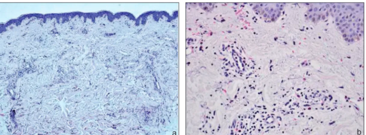
Informed consent has been obtained. Histopathological examination of the punch biopsy material taken from the border of the plaque lesion revealed a regular epidermis and perivascular and interstitial inflammation in the dermis (Figure 2-a). Perivascular and interstitial eosinophil, neu-trophil and lymphocytic infiltration accompanying with swelling and proliferation of the endothelial cells were seen in the dermis (Figure 2-b). Though, nuclear dusts were not noted. Direct immunofluo-rescence (DIF) was negative. Patch test examina-tion with Ginkgo bilobapills was negative.
FIGURE1: a-b: Firm, non-tender, erythemathous, polimorphic plaques in annular and semi-annular or polycyclic configurations on his chest, shoulders and back.
Based on these clinical and histological find-ings, the patient was diagnosed with NFE. He stopped taking Ginkgo biloba pills and he was treated with colchicine tablet 1.5 mg/day and lev-ocetirizine tablet 5 mg/day 3 months. The lesions almost completely improved. The patient was rechallenged with the same drug containing
Ginkgo bilobafor two weeks. This time, after 10th
day of the drug administration the lesions reap-peared on his back. In 1-year-follow up no flare up of the lesions was noted after the retreatment and discontinuation of the drug.
DISCUSSION
NFE is a rare inflammatory dermatosis, character-ized by indurated, erythematous, polycyclic or an-nular lesions, with rapid centrifugal enlargement and histological feature of neutrophilic infiltration with nuclear dusts in the dermis.1NFE is
charac-terized by a superficial and deep, perivascular and interstitial infiltrate of neutrophils associated with leukocytoclasis but without other signs of vasculi-tis.1,2It is characterized by papular erythematous
eruptions with rapid centrifugal enlargement to an-nular or polycyclic asymptomatic plaques with in-durated borders devoid of vesicles, crusts or desquamation.1,2Widespread or localized
distribu-tion, and transient or persistent behavior may all be seen in NFE.2 The patches tend to disappear
within 2-4 weeks, but the disease course is chronic. Relapses and recurrences are seen common.2
The differential diagnosis of NFE includes Sweet syndrome, plaque sarcoidosis, Jessner’s lym-phocytic infiltration, annular elastolytic giant cell granuloma, Sjögren’s syndrome, neutrophilic type of urticaria, annular variant of subacute cutaneous lupus erythematosus (SCLE), causes of figurate thema such as erythema chronicum migrans, ery-thema annulare centrifugum, eryery-thema gyratum repens and its variant familial annularerythema.1,3,4
Histopathological differential diagnosis of NFE con-sist some neutrophilic dermatosis such as rheuma-toid neutrophilic dermatosis, Behcet’s syndrome, urticarial vasculitis,bullous SLE, bullous variant of
pyoderma gangrenosum and subcorneal pustular dermatosis.2,5 Based on clinical, laboratory and
histopathological findings, all these diseases could be eliminated. The indurated character and neu-trophilic predominancy of the inflammatory infil-trate confirmed the diagnosis of NFE in our patient.
NFE belongs to those figurate erythemas of unknown etiology. As we know, only 4 adult cases of NFE have been reported in the literature.1,6,7 In
all of the previously reported cases of NFE, there was no history of insect bite or infection. The dis-ease may be idiopathic in otherwise healthy indi-viduals or present with some underlying diseases such as malignancies.6For instance, a 79-year-old
female presented with indurated figurate lesions in upper and lower limbs including palms and soles had been diagnosed with NFE accompanying with cryptogenic hepatic cirrhosis and Hodgkin’s
lym-FIGURE 2: a: Epidermis is regular. Note the perivascular and interstitial inflammation in the dermis (HEx40); b: Perivascular and interstitial eosinophil, neutrophil
and lymphocytic infiltration (HEx200).
phoma.6Moreover, another adult case of NFE
pre-sented with the morphology of erythema gyratum repens was also reported in a patient with systemic lupus erythematosus.7
Similar cold aggravation and presence of hot induced lesions were also remarkable in 1-year and 9-month-old white two boys with idiopathic NFE of infancy reported previously.8We believe that
the diagnosis of cryoglobulinemia has to be ex-cluded in the presence of this status. Complete res-olution of the lesions were previously reported during febrile episodes in these cases and in a few cases with erythema annulare centrifigum (EAC).8,9
After disregulation of some cytokine production in the skin triggered by cold, the skin lesions resolve with an increase in interferon if the body temper-ature rises to a degree higher than normal.9
More-over, NFE has been reported to show some clinical differences in paediatric cases such as benign, self-limiting course when compared with adult cases.10
It usually resolves within a few months in infants. First of all some other diseases such as neonatal lupus erythematosus and its variant erythema gy-ratum atrophicans transiens neonatale, erythema chronicum migrans, erythema marginatum rheumaticum, EAC and its variant familial annular erythema, tinea corporis, linear IgA dermatosis, granuloma annulare, erythema multiforme, neona-tal syphilis, annular erythematous lesions associ-ated with Still’s disease, and urticaria have to be excluded. Once a diagnosis of NFE of infancy is made, parents should be reassured, as this entity and its variants are unassociated with other condi-tions in most cases.8,10
Up to now drug induced NFE has not been re-ported before. In this report, we wanted to empha-size that our case is the first case of NFE which might have been induced by a herbal drug; Ginkgo
bilobathat has neuroprotective effects.11Ginkgo
biloba has been demonstrated to cause suppression
of nuclear factor kappa-B (NF-kB), and toll-like re-ceptor 4 (TLR4)/NF-kB, the up-regulation of heme oxygenase 1, erythropoietin secretion and anti-apoptotic protein expression, the inhibition of pro-apoptotic proteins expression, and the improve-ment of endothelial nitric oxide (NO) synthesis.11
Some of the latter molecular mechanisms such as the improvement of endothelial NO synthesis might have also act as a trigger for some of the histopathological findings such as swelling and pro-liferation of the endothelial cells and leucocytocla-sia, seen in our case. The presence of perivascular and interstitial eosinophil infiltration in this case also confirms the drug-induced pathogenesis. Each capsule contains; 90 mg standardizedGingko biloba
leaf extract (gingkoflavoglucoside) 21 mg 24%, ter-pene lactones 5 mg 6% and 45 mg elemental
Gingko bilobaleaf salt.9We experienced relapse of
the lesions after rechallenge with the same herbal drug containing Gingko biloba. Moreover, the patch test results were negative. However, some other preser- vative substances or dyestuffs in the ingredients of the herbal drugs might have also caused this neutrophilic dermatosis; NFE in this pa-tient. The good response of this case to colchicine was already expected as colchicine and dapson are well-known to be one the first choice treatments for neutrophilic dermatoses.12We think that some
cases with NFE might have been possibly misdiag-nosed or unreported. The reason of the relapsing course of NFE more or less at the same location is still not clear.
The exact pathological association of NFE with Ginkgo bilobaand/or cold is not clear and further more clinical cases are needed to clarify the exact pathology of this disease. NFE should also be considered in differential diagnosis of the erythematous annular plaques in adults and de-tailed investigation of a probable malignancy, an underlying disease or a drug association and cold exposure must be performed. In recent years the use of supplemental medications; containing merals, vitamins and anti-oxidant elements have in-creased worldwide without any prescription. We presented this case to focus on the importance of an accurate and detailed medication history and to remark a possible association of NFE and Ginkgo biloba.
C
Coonnfflliicctt ooff IInntteerreesstt
1. Ozdemir M, Engin B, Toy H, Demirkesen C. Neutrophilic figurate erythema. Int J Dermatol 2008;47(3):262-4.
2. Ríos-Martín JJ, Ferrándiz-Pulido L, Moreno-Ramírez D. [Approaches to the der-matopathologic diagnosis of figurate lesions]. Actas Dermosifiliogr 2011;102(5):316-24. 3. Naveen KN, Kalinga B, Pai VV, Athanikar SB,
Sori T, Raju K, et al. Erythema gyratum repens like figurate erythema responding to topical steroid in a healthy individual. Indian J Der-matol 2013;58(4):329.
4. Baglieri F, Scuderi G. Erythema annulare cen-trifugum: a “deep type” figurate eruption. G Ital Dermatol Venereol 2012;147(1):129-31. 5. Nischal KC, Khopkar U. An approach to the
diagnosis of neutrophilic dermatoses: a
histopathological perspective. Indian J Der-matol Venereol Leprol 2007;73(4):222-30. 6. Trébol I, González-Pérez R, García-Rio I,
Ar-regui MA, Saracibar N, Carnero L, et al. Para-neoplastic neutrophilic figurate erythema. Br J Dermatol 2007;156(2):396-8.
7. Khan Durani B, Andrassy K, Hartschuh W. Neu-trophilic dermatosis with an erythema gyratum repens-like pattern in systemic lupus erythe-matosus. Acta Derm Venereol 2005;85(5):455-6.
8. Patrizi A, Savoia F, Varotti E, Gaspari V, Pas-sarini B, Neri I. Neutrophilic figurate erythema of infancy. Pediatr Dermatol 2008;25(2):255-60. 9. Bottoni U, Innocenzi D, Bonaccorsi P, Carles-imo M, Faina P, Richetta A, et al. Erythema
annulare centrifugum: report of a case with neonatal onset. J Eur Acad Dermatol Venereol 2002;16(5):500-3.
10. Maurelli M, Colato C, Tessari G, Girolomoni G. Neutrophilic figurate erythema: report of a new case in an adult and comparison between adult and paediatric cases. Eur J Dermatol 2016;26(6):634-5.
11. Nabavi SM, Habtemariam S, Daglia M, Braidy N, Loizzo MR, Tundis R, et al. Neuroprotective effects of ginkgolide B against ischemic stroke: a review of current literature. Curr Top Med Chem 2015;15(21):2222-32.
12. Konda C, Rao AG. Colchicine in dermatology. Indian J Dermatol Venereol Leprol 2010; 76(2):201-5.
REFERENCES
A
Auutthhoorrsshhiipp CCoonnttrriibbuuttiioonnss
All of the authors contributed to the concept of the article. When Pelin Üstüner received clinical data, he designed the ar-ticle, analyzed it and contributed to his opinion. Pelin Üstüner
also prepared the article. Pelin Üstüner and Ali Balevi, revised the article for intellectual content. Aslı Ünlü collects histopathological data and regulates pathological patterns. Mustafa Özdemir approved the final version.