INVESTIGATION OF IMPACTED SUPERNUMERARY TEETH: A CONE BEAM COMPUTED TOMOGRAPH (CBCT) STUDY
Gömülü Süpernümerer Dişlerin İncelenmesi: Bir Konik Işınlı Bilgisayarlı Tomografi (KIBT) Çalışması
Gökhan GÜRLER 1, Çağrı DELİLBAŞI 1, Evren DELİLBAŞI 2 Received: 04/11/2016
Accepted:05/01/2017
ABSTRACT
Purpose: The purpose of this study was to investigate the
impacted supernumerary teeth which were initially detected on panoramic radiographs by using cone beam computed tomography (CBCT).
Materials and Methods: In this retrospective study,
supernumerary teeth diagnosed on panoramic radiographs taken from patients who had admitted for routine dental treatment were evaluated using CBCT. Patients’ age, gender, systemic conditions as well as number of supernumerary teeth, unilateral-bilateral presence, anatomical localization (maxilla, mandible, anterior-premolar-molar, mesiodens-lateral-canine, parapremolar-paramolar-distomolar) shape (rudimentary, supplemental, tuberculate, odontoma), position (palatal-lingual-buccal-labial-central), shortest distance between the tooth and adjacent cortical plate, complications and treatment were assessed.
Results: A total of 47 impacted supernumerary teeth in
34 patients were investigated in this study. Of these, 33 (70.2%) were unilateral and 14 (29.8%) were bilateral. Only 1 supernumerary tooth was found in 27 patients (79.4%) whereas 7 patients (20.6%) had 2 or more supernumerary teeth. Most of the teeth located in the anterior region (74.4%) of the jaws and maxilla (74.4%). Twenty teeth (42.5%) were mesiodens, 11 (23.4%) were lateral or canine, 14 (29.7%) were parapremolar and 2(4.4%) were distomolar. Twenty-seven teeth (57.4%) were rudimentary, 15 (31.9%) were supplemental and 5 (10.7%) were odontoma in shape. The shortest distance between the supernumerary tooth and adjacent cortical plate varied between 0 to 2.5 mm with a mean of 0.66 mm. The most common clinical complaint was the non-eruption of permanent teeth (42.5%). All supernumerary teeth were removed under local anesthesia. Orthodontic traction was performed for those impacted permanent teeth if necessary.
Conclusion: Impacted supernumerary teeth are usually in
close proximity to cortical bone. Although this may facilitate surgical access, there is a risk of damaging surrounding anatomical structures. Therefore, CBCT evaluation of impacted supernumerary teeth for accurate planning is recommended.
Keywords: Supernumerary tooth; hyperdontia; CBCT;
impacted teeth; cortical bone
ÖZ
Amaç: Bu çalışmanın amacı ilk olarak panoramik
radyografilerde fark edilen gömülü süpernümerer dişlerin konik ışınlı bilgisayarlı tomografiyle (CBCT) incelenmesidir.
Gereç ve Yöntem: Bu retrospektif çalışmada, rutin
dental muayene amacıyla panoramik radyografi çekilen hastalarda tespit edilen süpernümerer dişler CBCT ile incelendi. Hastaların yaş, cinsiyet, sistemik durumları ile süpernümerer diş sayısı, tek-çift taraflı oluşu, anatomik lokalizasyonu (maksilla, mandibula, anterior- premolar molar, meziodens- lateral, kanin-parapremolar- paramolar- distomolar), şekli (rudumenter, suplemetal, tüberküleyt, odontoma), pozisyonu (palatinal- lingual- bukkal- labial- orta hat), süpernümerer dişin komşu kortekse olan uzaklığı, yol açtığı sorunlar ve tedavi yaklaşımları değerlendirildi.
Bulgular: 34 hastada tespit edilen 47 adet süpernümerer
diş incelendi. Bu dişlerden 33’ü (%70.2) unilateral, 14’ü (29.8%) bilateraldi. Yirmi yedi (79.4%) hastada sadece 1 süpernümere diş mevcutken, 7 (%20.6) hastada 2 veya daha fazla diş mevcuttu. Dişlerin çoğu çenelerin anterior bölgesinde (%74.4) ve maksillada (%74.4) lokalizeydi. Yirmi bir (%44.6) diş meziyodens, 11 (%21.4) diş lateral ya da kanin, 14 diş (%29.7) parapremolar, ve 1 (%2.3) diş distomolar olarak tespit edildi. Yirmi yedi diş (%57.4) rudimenter, 15 diş (%31.9) suplemental, 5 diş (%10.7) odontoma şeklindeydi. Süpernümerer dişin komşu kortekse olan mesafesi 0 ile 2.5 mm arasında (ortalama 0.66 mm) ölçüldü. Bütün süpernümerer dişler lokal anesteziyle cerrahi olarak çıkartıldı. Gereken olgularda ortodontik tedavi uygulandı.
Sonuç: Gömülü süpernümerer dişler komşu kortikal
tabakaya yakın konumlanmaktadırlar. Bu durum cerrahi girişi kolaylaştırmasına rağmen, çevre anatomik yapıları ve dişlere zarar verme riski vardır. Bu nedenle doğru planlama yapabilmek için gömülü süpernümere dişlerin CBCT ile incelenmesi önerilmektedir.
Anahtar kelimeler: Süpernümerer diş; hiperdonti;
CBCT; gömük diş; kortikal kemik http://dx.doi.org/10.17096/jiufd.20098
1 Department of Oral and Maxillofacial Surgery Faculty of Dentistry Istanbul Medipol University 2 Department of Pediatric Dentistry Faculty of Dentistry Yeni Yüzyıl University
This work is licensed under a Creative Commons Attribution-NonCommercial-NoDerivatives 4.0 International License.
ORIGINAL RESEARCH
How to cite: Gurler G, Delilbasi C, Delilbasi E. Investigation of impacted supernumerary teeth: a cone beam computed tomography (CBCT) study. J Istanb Univ Fac Dent 2017;51(3):18-24.
Introduction
Supernumerary teeth term is defined as the presence of excessive number of tooth or tooth-like structures either in primary or permanent dentition. This condition may be due to the developmental disturbances during odontogenesis, which result in the formation of additional teeth (1-4). Although its reported incidence is less than 1% (5), presence of supernumerary teeth may also be overlooked by parents or children could have been taken to the dentist after permanent teeth are erupted (6). There is a slight male preponderance but male to female ratio is not known for certain. They are commonly found in maxillary anterior and mandibular premolar regions (2, 7). Frequency of supernumerary teeth, in decreasing order, has been reported as upper lateral incisors (50%), mesiodens (36%), upper central incisors (11%), and bicuspids (3%) (8). A study conducted in Turkish population has revealed that the most common supernumerary tooth was mesiodens, followed by premolar, lateral, distomolar, paramolar and canine (9). Shape of a supernumerary tooth may vary from an odontoma to a supplemental tooth. Multiple supernumerary teeth may be associated with some syndromes or conditions such as Gardner’s syndrome, cleidocranial dysostosis and cleft lip and palate (1, 2, 7). Physical examination as well as family and medical histories are essential to determine the genetic background of the syndrome. Supernumerary teeth may not have any effect on dentition and be discovered on routine radiographs incidentally. On the other hand, they may also cause crowding, failure of eruption, diastema, root resorption, dilaceration and displacement of adjacent teeth (1, 2, 6).
Panoramic and periapical radiographs are frequently used in routine dental examination. However, due to superimposition of anatomical structures, supernumerary teeth may be overlooked on conventional radiographs. Cone Beam Computed Tomography (CBCT) can provide precise and accurate information on normal and pathologic conditions such as odontomas, supernumerary teeth, developmental anomalies and traumatic injuries. The main advantages of CBCT are; multi-planar imaging of dental tissues, shorter acquisition time and less ionizing radiation dose compared to CT, and ease of data transfer. It is sometimes not possible to diagnose and make treatment planning by using routine radiographs (10-14). Detection of supernumerary teeth is best achieved by detailed clinical and radiographic
examination. Their anatomical shape and position should be recognized before the procedure in order to predict and overcome possible complications. Two dimensional techniques such as panoramic and periapical radiographies may be inadequate to determine exact localization of supernumerary teeth and their relations with neighboring structures. Therefore, multiplanar visualization may help practitioner to decide appropriate treatment plan. In recent years CBCT has been extensively used to detect impacted and supernumerary teeth. The purpose of this study was to investigate the impacted supernumerary teeth using CBCT which were detected on panoramic radiographs.
Materials and Methods Sample characteristics
This project was approved by the local ethical committee (Project number: 10840098-604.01.01-E.18858/465). Patients with impacted supernumerary who had been treated in our clinic were included in this study. All the patients had been admitted for routine dental treatment and supernumerary teeth were diagnosed on panoramic radiographs incidentally. Patients with previously diagnosed syndromes, cleft lift and palate or those with cleideocranial dyplasia were not included.
Imaging protocols
Radiological investigation was performed with
a CBCT scanner (i-CAT Next Generation, Imaging
Sciences International, Hatfield, PA, USA) by using the following settings: 6 cm x 16 cm field of view (FOV), 120 kV, 20.27 mA, 14.7 seconds exposure time and 0.25 mm slice thickness. Distances and presence of supernumerary teeth were evaluated on both sagittal and panoramic sections using i-CAT Vision software (Imaging Sciences International, Hatfield, PA, USA) with a 1920 x 1020 pixel resolution on a 24 inch monitor (Dell inc., Round Rock, TX, USA).
Study variables
Patients’ age, gender, systemic condition, number of supernumerary teeth, unilateral-bilateral presence, anatomical localization (maxilla, mandible, anterior-premolar-molar, mesiodens,
parapremolar-paramolar-distomolar), shape (rudimentary, supplemental, tuberculate, odontoma), position (palatal, lingual, buccal, labial, midline), and shortest distance between the most exterior part of the supernumerary tooth and the inner layer of neighboring cortical plate, as well as complications due to supernumerary tooth, were assessed. Mesiodens was defined as the supernumerary tooth located between the central incisors, parapremolar as the one located in the premolar region, paramolar tooth as the one located in the molar region and distomolar tooth as the one located distal to third molars. Rudimentary shape was defined as a peg shaped tooth, tuberculate shape as a tooth having multiple tubercules, supplemental shape as a tooth that resembles to a normal tooth, and odontoma as a mass of dental tissue which could not be described within the limits of standard tooth morphology (1, 15-17). Complications were classified as; impaction of permanent teeth, dislocation, dilacerations of teeth and root resorption.
Statistical analysis
Statistical Package for Social Sciences (SPSS)
version 21.0 (IBM SPSS Statistics for Windows, Armonk, NY, USA) was used for statistical analysis. Descriptive statistics such as mean, standard deviation, minimum, maximum and frequency were used to present the characteristics of the study sample.
Results
In this study, CBCTs of 34 (25 males and 9 females) patients were evaluated. The age range was 6 to 35 years with a mean of 14.7 years. None of the patients
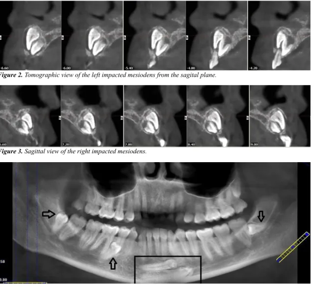
had uncontrolled systemic disease, cleft or syndrome. A total of 47 supernumerary teeth were detected in 34 patients. Of these, 33 (70.2%) were unilateral and 14 (29.8%) were bilateral. Single supernumerary tooth was found in 27 patients (79.4%) whereas 7 patients (20.6%) had 2 or more supernumerary teeth. Thirty-five teeth (74.4%) located in the maxilla whereas 12 teeth (25.6%) located in the mandible. Number of anterior supernumerary tooth was 35 (74.4%), premolar tooth was 11 (23.4%) and molar tooth was 1 (2.2%). Twenty teeth (42.5%) were mesiodens, 11 (23.4%) were lateral or canine, 14 (29.7%) were parapremolar and 2 (4.4%) were distomolar. There were 27 teeth (57.4%) having rudimentary shape, 15 (31.9%) supplemental and 5 (10.7%) odontoma. Eight teeth (17%) were positioned in buccal-labial direction, 34 (72.3%) in palatal-lingual direction and 5 (10.7%) in the midline. The shortest distance between the supernumerary tooth and adjacent cortical plate varied between 0 to 2.50 mm with a mean of 0.66 mm. When the complications due to supernumerary teeth were assessed; 20 teeth (42.5%) have been found to hinder the eruption of permanent teeth, 5 teeth (10.6%) caused dilacerations of neighboring teeth, 2 teeth (4.4%) caused tooth displacement, whereas 20 teeth (42.5%) caused no complication at all. All supernumerary teeth were removed under local anesthesia. Orthodontic traction was performed for those impacted permanent teeth if necessary. Samples of the supernumerary teeth were shown in Figure 1, 2, 3, 4 and 5.
Figure 2. Tomographic view of the left impacted mesiodens from the sagital plane.
Figure 3. Sagittal view of the right impacted mesiodens.
Figure 4. Panoramic view of the mandibular impacted parapremolar, distomolars (arrows) and impacted canines (rectangle).
Figure 5. Three dimensional volume rendering
reconstruction of the anterior supernumerary teeth.
Discussion
Supernumerary teeth (hyperdontia) are excess teeth found in primary or permanent dentition. Although its etiology still remains unclear; hereditary influence, disease processes, dichotomy of the tooth germ, excessive growth of the dental lamina have been suggested as possible causative factors (2, 4, 18, 19). Multiple supernumerary teeth in the same person is a rare occurrence. This condition may be related to a syndrome or a developmental disorder (6, 20, 21). Thus, patients having multiple supernumerary teeth should be carefully evaluated for the presence of a genetic disorder. The incidence of supernumerary teeth is low (0.06%). Most supernumerary teeth start developing later than other teeth of the same region; therefore, they usually have incomplete root formation
(22). Males are more affected than females. Most common location is the maxillary anterior (mesiodens being more common), followed by mandibular premolar region (23, 24). Supernumerary teeth can either erupt or remain impacted. As more than 75% of the anterior supernumeraries remain unerupted in mixed dentition, careful examination of the dentition is important. A delay in tooth eruption more than 6 months, particularly when the symmetrical tooth has erupted, should be radiographically examined (20, 25, 26). Supernumerary teeth are classified according to their shape and location and they are correlated with macrodontia. Patients with supernumerary teeth tend to have larger mesiodistal dimension (24). Most impacted supernumerary teeth are symptomless and can only be diagnosed on routine radiographs (6, 12, 17). Evidence for removal of supernumerary teeth suggests that it is prudent to remove these teeth where adequate space is available for adjacent teeth to erupt (8). Previously reported complications due to supernumerary teeth are as follows: eruption failure, displacement or rotation of the teeth in the dental arch, diastema, root-crown dilacerations, abnormal root formation, cyst formation and displacement of teeth (8, 20, 23, 24, 27-29).
Altuğ et al. (1) have investigated 78 supernumerary teeth in 54 patients. They found male predominance, majority of the teeth being single (77.8%), conical (51.3%) and impacted (52.5%). Most of the teeth were found to be in palatal/lingual position (53.9%). The most frequent complication was the eruption problems of adjacent teeth (44.4%). Treatment of these teeth consisted of surgical removal, orthodontic treatment or periodic follow-up. Tatlı et al. (20) have examined 156 supernumerary teeth in 112 patients. The percentage of impacted ones in this study was reported as 73.7%. Authors have found higher number of impacted teeth in females than males. Maxilla and mandible were equally affected. The other parameters of the study were related to erupted supernumerary teeth. Our findings were similar to that of Altuğ
et al. (1) but slightly different to that of Tatlı et al. (20). On the other hand, Vahid-Dastjerdi et al.
(30), who had investigated supernumerary teeth in orthodontic patients, have found that the prevalence of supernumerary teeth was 0.74% being highest in Class III malocclusions and lowest in Class II malocclusions. Maxilla (78.5%) was more affected than the mandible (21.5%) and conical (rudimentary) type supernumerary teeth had the highest frequency (43%). Spontaneous eruption of an impacted tooth
after extraction of supernumerary tooth depends on the distance of the apex of the impacted tooth relative to its estimated position, depth of impaction, stage of root development of the supernumerary tooth, angle of impaction, time of surgery, number of supernumerary teeth and loss of space for eruption. Patchett et al. (29) suggested that conical form supernumerary teeth have more chance to erupt spontaneously than tuberculate form
Panoramic, periapical and occlusal radiographs are conventional radiography techniques used in dentistry. Superimposition of the anatomical structures in two dimensional views sometimes makes the accurate diagnosis impossible. CBCT provides precise location of the impacted supernumerary teeth and allows accurate linear measurement of the distances from the anatomical structures and cortical plates. These advantages of CBCT enable correct and less invasive surgical planning with a less risk of damage (13, 14, 19, 31, 32). Initial diagnosis can be made using conventional techniques, but for further evaluation CBCT is recommended. Thanks to CBCT, clinicians can evaluate the impacted and supernumerary teeth from different planar orientations as well as in three dimensional reconstructions. In order to obtain best diagnostic efficiency and treatment outcome, CBCT is essential to clarify critical regions of interest and close anatomical structures where two dimensional view is not sufficient. Panoramic and periapical radiographies both magnify and distort the images often resulting in inadequate diagnostic certainty (14).
Liu et al. (12) investigated 626 supernumerary teeth in 487 patients using CBCT. Of these patients, 72% had 1 supernumerary tooth, 27.3% had 2, 0.6% had 3 teeth. These teeth mostly located in the anterior maxilla (92%) and had conical shape (83.5%)(12). They stated that the three dimensional reconstructions based on CBCT data could be used to examine dental and bony structures around the supernumerary teeth. Katheria et al. (33) compared the efficiency of conventional radiography and CBCT for the detection of impacted and supernumerary teeth in children. They concluded that CBCT provides more information on location, presence of root resorption and treatment planning. When supernumerary teeth are discovered, a decision should be made whether to remove or to follow-up (8, 22, 34). Surgical removal of supernumerary teeth may cause damage in adjacent anatomical structures or teeth. Therefore risks and benefits of surgery should be weighed (22). Some authors suggest follow-up unless the teeth cause any
complication or occlusal changes (18). The surgical-orthodontic treatment of supernumerary teeth needs accurate diagnosis and precise localization of the teeth and surrounding structures (7). In our patients, we preferred to remove all supernumerary teeth after consulting orthodontics.
Conclusion
Impacted supernumerary teeth are usually in close proximity to cortical bone. Although this may facilitate surgical access, there is a risk of damaging surrounding anatomical structures. Therefore, CBCT evaluation of impacted supernumerary teeth for accurate case planning is recommended.
Source of funding
None declared.
Conflict of interest
None declared. References
1. Altuğ HA, Altuğ H, Sarı E, Şençimen M, Altun C. Süt ve daimi dentisyonda süpernümere dişlerin teşhisi, cerrahi ve ortodontik olarak tedavileri. GÜ Diş Hek Fak Derg 2010;27(2):77-82. 2. Erdem MA ÇB, Güven G, Kasapoğlu Ç. Artı
dişler (süpernümerer dişler). İst Üni Dis Hek Fak Derg 2011;45(1):15-18.
3. Kaya GS, Yapici G, Omezli MM, Dayi E. Non-syndromic supernumerary premolars. Med Oral Patol Oral Cir Bucal 2011;16(4):e522-525. 4. Masih S, Sethi HS, Singh N, Thomas AM.
Differential expressions of bilaterally unerupted supernumerary teeth. J Indian Soc Pedod Prev Dent 2011;29(4):320-322.
5. Acikgoz A, Acikgoz G, Tunga U, Otan F. Characteristics and prevalence of non-syndrome multiple supernumerary teeth: A retrospective study. Dentomaxillofac Radiol 2006;35(3):185-190.
6. Bereket C, ÇakırÖzkan N, Şener İ, Tek M, Çelik S. Sürnümerer molar dişlerin retrospektif olarak incelenmesi: Klinik ve radyolojik bir çalışma. Atatürk Üniv Diş Hek Fak Derg 2010;20(3):176-180.
7. Brauer HU. Case report: Non-syndromic multiple supernumerary teeth localized by cone beam computed tomography. Eur Arch Paediatr Dent
2010;11(1):41-43.
8. Scheiner MA, Sampson WJ. Supernumerary teeth: A review of the literature and four case reports. Aust Dent J 1997;42(3):160-165. 9. Celikoglu M, Kamak H, Oktay H. Prevalence
and characteristics of supernumerary teeth in a non-syndrome turkish population: Associated pathologies and proposed treatment. Med Oral Patol Oral Cir Bucal 2010;15(4):e575-578. 10. Cevidanes LH, Styner MA, Proffit WR. Image
analysis and superimposition of 3-dimensional cone-beam computed tomography models. Am J Orthod Dentofacial Orthop 2006;129(5):611-618.
11. Jeremias F, Fragelli CM, Mastrantonio SD, Dos Santos-Pinto L, Dos Santos-Pinto A, Pansani CA. Cone-beam computed tomography as a surgical guide to impacted anterior teeth. Dent Res J (Isfahan) 2016;13(1):85-89.
12. Liu DG, Zhang WL, Zhang ZY, Wu YT, Ma XC. Three-dimensional evaluations of supernumerary teeth using cone-beam computed tomography for 487 cases. Oral Surg Oral Med Oral Pathol Oral Radiol Endod 2007;103(3):403-411.
13. Sawamura T, Minowa K, Nakamura M. Impacted teeth in the maxilla: Usefulness of 3d dental-ct for preoperative evaluation. Eur J Radiol 2003;47(3):221-226.
14. Ziegler CM, Klimowicz TR. A comparison between various radiological techniques in the localization and analysis of impacted and supernumerary teeth. Indian J Dent Res 2013;24(3):336-341.
15. Ashkenazi M, Greenberg BP, Chodik G, Rakocz M. Postoperative prognosis of unerupted teeth after removal of supernumerary teeth or odontomas. Am J Orthod Dentofacial Orthop 2007;131(5):614-619.
16. Biradar V, Angadi S. Supernumerary teeth: Review of case series. Journal of Interdisciplinary Dentistry 2012;2(2):113-115.
17. Ezirganlı Ş, Ün E, Kırtay M, Özer K, Köşger H. Sivas bölgesinde artı dişlerin yaygınlığının araştırılması. Atatürk Üniv Diş Hek Fak Derg 2011;21(3):189-195.
18. Cantekin K GH, Aydınbelge M. Üst çene keserler bölgesinde bulunan süpernümerer dişlerin neden olduğu komplikasyonlar ve tedavi yaklaşımları. Erciyes Üniversitesi Sağlık Bilimleri Dergisi 2014;23(1):54-58.
computer tomography technology: An update and case report of an impacted incisor in a mixed dentition patient. Pediatr Dent 2010;32(4):356-360.
20. Tatlı U, Evlice B, Damlar İ, Arslanoğlu Z, Altan A. Çukurova bölgesinin süpernümerer diş karakteristikleri: Çok merkezli retrospektif bir çalışma. Acta Odontol Turc. 2014;31(2):84-88. 21. Yague-Garcia J, Berini-Aytes L, Gay-Escoda C.
Multiple supernumerary teeth not associated with complex syndromes: A retrospective study. Med Oral Patol Oral Cir Bucal 2009;14(7):E331-336. 22. Cochrane SM, Clark JR, Hunt NP. Late
developing supernumerary teeth in the mandible. Br J Orthod 1997;24(4):293-296.
23. Peker I, Kaya E, Darendeliler-Yaman S. Clinic and radiographical evaluation of non-syndromic hypodontia and hyperdontia in permanent dentition. Med Oral Patol Oral Cir Bucal 2009;14(8):e393-397.
24. Santos AP, Ammari MM, Moliterno LF, Junior JC. First report of bilateral supernumerary teeth associated with both primary and permanent maxillary canines. J Oral Sci 2009;51(1):145-150.
25. Cho SY, So FH, Lee CK, Chan JC. Late forming supernumerary tooth in the premaxilla: A case report. Int J Paediatr Dent 2000;10(4):335-340. 26. Yassaei S, Goldani Moghadam M, Tabatabaei
SM. Late developing supernumerary premolars: Reports of two cases. Case Rep Dent 2013;2013:969238.
27. Hegde SV, Munshi AK. Late development of supernumerary teeth in the premolar region: A case report. Quintessence Int 1996;27(7):479-481.
28. Mittal M, Sultan A. Clinical management of supernumerary teeth: A report of two cases. J Indian Soc Pedod Prev Dent 2010;28(3):219-222. 29. Patchett CL, Crawford PJ, Cameron AC, Stephens CD. The management of supernumerary teeth in childhood--a retrospective study of practice in Bristol Dental Hospital, England and Westmead Dental Hospital, Sydney, Australia. Int J Paediatr Dent 2001;11(4):259-265.
30. Vahid-Dastjerdi E, Borzabadi-Farahani A, Mahdian M, Amini N. Supernumerary teeth amongst iranian orthodontic patients. A retrospective radiographic and clinical survey. Acta Odontol Scand 2011;69(2):125-128. 31. Nematolahi H, Abadi H, Mohammadzade Z,
Soofiani Ghadim M. The use of cone beam computed tomography (CBCT) to determine supernumerary and impacted teeth position in pediatric patients: A case report. J Dent Res Dent Clin Dent Prospects 2013;7(1):47-50.
32. Romano N, Souza-Flamini LE, Mendonca IL, Silva RG, Cruz-Filho AM. Geminated maxillary lateral incisor with two root canals. Case Rep Dent 2016;2016:3759021.
33. Katheria BC, Kau CH, Tate R, Chen JW, English J, Bouquot J. Effectiveness of impacted and supernumerary tooth diagnosis from traditional radiography versus cone beam computed tomography. Pediatr Dent 2010;32(4):304-309. 34. Nogami S, Miyamoto I, Yamauchi K, Kataoka
Y, Morimoto Y, Saeki K, Maki K, Takahashi T. Supernumerary decidious teeth with multiple maxillary impacted mesiodens: A case report. Pediatric Dental Journal 2012;22(2):193-197.
Corresponding Author: Gökhan GÜRLER
Department of Oral and Maxillofacial Surgery Faculty of Dentistry Istanbul Medipol University 34083, Unkapanı-Fatih-İstanbul/Turkey Phone: +90 212 453 49 40