IMAgENs EM REUMATOLOgIA
ÓRgÃO OFICIAL DA sOCIEDADE PORTUgUEsA DE REUMATOLOgIA 347
A pictorial view to tarsal coalition:
the presentation of two children with foot pain
ACTA REUMATOL PORT. 2014;39:347-348
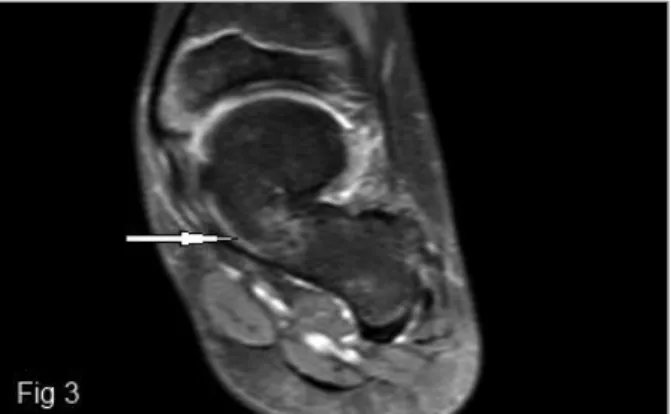
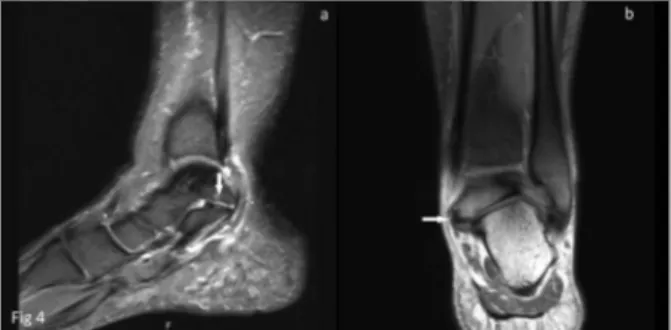
Two male patients aged 9 and 16 years were admitted with complaints of ankle pain and intoeing. There was no history of trauma and arthritis. In the first case, ra-diography examination revealed a “C sign” (a cortical continuity from the medial portion of the talus to the sustentaculum tali), a narrowing in the middle subta-lar joint space, and convexity in the lower margin of the sustentaculum tali (Figure 1). Magnetic resonance imaging (MRI) showed triple coalition in the anterior subtalar joint (talocalcaneonavicular joint) in the first case (Figures 2 and 3) and fibrocartilaginous coalition at the posterior subtalar joint in the second case (Figu-re 4). Narrowing in the joint spaces, findings of early degeneration, and medullary edema in the adjacent bone were observed at the sites of coalition (Figures 3 and 4). Nonoperative treatment, such as physical the-rapy, exercises, and analgesic medication were applied. Tarsal coalition is one of the most important causes of pes planus and may cause foot pain in children. Tar-sal coalition is the congenital or acquired fibrous, car-tilaginous or osseous fusion of one or more tarsal bo-nes. Although the described incidence of this condi-tion is approximately 1% of the populacondi-tion1,2, the
pro-portion of asymptomatic cases is much higher1. The
most common coalitions are talocalcaneal and calca-neonavicular coalitions, which account for 90% of the observed cases1-3. Talocalcaneal coalition is observed in
approximately half of the cases and often involves the middle subtalar joint, a finding similar to that observed in the first case2,3. Fusion at the level of the posterior
subtalar joint, as observed in the second case, is an ex-tremely uncommon occurrence3. Most cases are
asympto matic in the first years of life; however, the patho logy progresses gradually because of
biomecha-Ormeci T1, Kilicarslan R1, Durmus O2, Malkoc M3, Cakar E2
1. Department of Radiology/Istanbul Medipol University, Faculty of Medicine, Istanbul
2. Department of Physical Medicine and Rehabilitation/Istanbul Medipol University, Faculty of Medicine, Istanbul
3. Department of Orthopedics and Traumatology/Istanbul Medipol University, Faculty of Medicine, Istanbul
FIGURE 1.A) Lateral radiograph of the left ankle shows the talocalcaneal C sign, suggesting subtalar coalition across the middle subtalar joint. B) Deformed talus of the left foot is displaced in the antero-posterior view
FIGURE 2.Sagittal T1 weighted spin-echo (TSE) image shows talonavicular osseous coalition (arrow)
FIGURE 3.Coronal proton density (PD) fat saturated TSE image demonstrates the osseous connection with continuity of marrow fat from the talus to the calcaneus. There is mild bone marrow edema at the site of coalition (arrow)
nical stress during physical activity. The coalition be-comes osseous due to repetitive injury and remode-ling, resulting in rigidity or instability2,4. Ankle pain
and stiffness are the most common complaints in pa-tients with tarsal coalition. The limited movement of the affected ankle, in particular the impairment of sub-talar inversion or eversion, is the most common phy-sical examination finding. The coalition leads to pero-neal spasm or adaptive peropero-neal shortening, peropero-neal spastic flat foot or rigid flat foot deformity2. Direct
ra-diography is the first diagnostic method of choice for patients with suspected talocalcaneal coalition1. The
most useful radiological indicator for tarsal coalition is the “C sign” observed in lateral radiographs and the talar beak sign1,3. The absence of the subtalar facet and
dysmor hic sustentaculum tali in lateral radiographs are other useful findings for the diagnosis of talocal-caneal coalition1,2. The visualization of talocalcaneal
coalition on direct graphs is sometimes difficult due
to superposition of complex structures; thus, multi-planar imaging may be necessary3,4. Particularly, MRI
provides valuable information in diagnosing nonos-seous coalition, articular fusion, reactive changes such as arthritis, and periarticular bone marrow edema, as well as in detecting secondary degenerative changes that are important for surgery planning2,4. Computed
tomography is described as the gold standard and is a more cost-effective diagnostic method5however, MRI
can also be preferred for the initial diagnosis to avoid x-ray exposure, particularly in children.
CoRREspondEnCE to
O Durmus
Istanbul Medipol Universitesi Hastanesi, Fiziksel Tıp ve Rehabilitasyon Klinigi TEM Avrupa Otoyolu Goztepe Cikisi, No 1, Bagcilar Istanbul - TURKEY. 34214 E-mail: [email protected]
REFEREnCEs
1. Lawrence DA, Rolen MF, Haims AH, Zayour Z, Moukaddam HA. Tarsal Coalitions: Radiographic, CT, and MR Imaging Fin-dings. HSS J 2014;10:153-166.
2. Newman JS, Newberg AH. Congenital tarsal coalition: multi-modality evaluation with emphasis on CT and MR imaging. Radiographics 2000;20:321-332.
3. Staser J, Karmazyn B, Lubicky J. Radiographic diagnosis of pos-terior facet talocalcaneal coalition. Pediatr Radiol 2007;37:79--81.
4. Sijbrandij ES, van Gils AP, de Lange EE, Sijbrandij S. Bone mar-row ill-defined hyperintensities with tarsal coalition: MR ima-ging findings. Eur J Radiol 2002;43:61-65.
5. Emery KH, Bisset GS 3rd, Johnson ND, Nunan PJ. Tarsal coa-lition: a blinded comparison of MRI and CT. Pediatr Radiol 1998;28: 612-616.
ÓRgÃO OFICIAL DA sOCIEDADE PORTUgUEsA DE REUMATOLOgIA 348
A pictoriAl view to tArsAl coAlition: the presentAtion of two children with foot pAin
FIGURE 4.A) Coronal and B) Sagittal FS TSE PD images of the second patient show fibrocartilaginous talocalcaneal coalition (arrow) involving posterior facets of the subtalar joints. Minimal degenerative are changes associated with the coalition