IS THERE ANY RELATIONSHIP BETWEEN PHACOEMULSIFICATION
PARAMETERS AND INCREASED SECOND EYE PAIN COMPLAINT?
FAKOEMÜLSİFİKASYON PARAMETRELERİ İLE İKİNCİ GÖZDE ARTMIŞ AĞRI ŞİKAYETİ
ARASINDA HERHANGİ BİR İLİŞKİ VAR MI?
İletişim kurulacak yazar/Corresponding author: [email protected] Geliş tarihi/Received Date: 02.12.2017 • Kabul tarihi/Accepted Date: 18.04.2018 ©Copyright 2018 by J Ist Faculty Med - Available online at jmed.istanbul.edu.tr
©Telif Hakkı 2018 J Ist Faculty Med - Makale metnine jmed.istanbul.edu.tr web sayfasından ulaşılabilir.
ÖZET
Amaç: Katarakt ameliyatı sonrası ağrı şikayeti ile
fakoemülsifikas-yon parametreleri arasındaki ilişkiyi değerlendirmek.
Gereç ve Yöntem: Bu prospektif çalışmaya, 15 gün arayla
kata-rakt ameliyatı yapılan 47 hastanın 94 gözü dahil edildi. Ortalama nukleus sertliği, ameliyat süresi, total ultrason (U/S) zamanı, total enerji (U/S total equivalent power in foot position 3), kümülatif yayılan enerji, fakoemülsifikasyon zamanı, ortalama fakoemülsifi-kasyon enerjisi ve ayak pedalı 3. pozisyonda iken kullanılan orta-lama fakoemülsifikasyon enerjisi kaydedildi. Hastalardan ağrılarını postoperatif birinci gün, birinci hafta ve birinci ay kontrollerinde skorlamaları istendi.
Bulgular: Ortalama nukleus sertliği, kümülatif yayılan enerji, total
U/S zamanı ilk gözde ikinci göze göre anlamlı olarak artmış bulun-du. (sırasıyla p= 0.01, 0.016, ve 0.03). İlk göz ve ikinci göz arasında ortalama ameliyat süresi, fakoemülsifikasyon zamanı, total enerji, ortalama fakoemülsifikasyon enerjisi ve ayak pedalı 3. pozisyon-da iken kullanılan ortalama fakoemülsifikasyon enerjisi arasınpozisyon-da anlamlı fark yoktu. Postoperatif birinci gün ve birinci ayda ağrı skorunda birinci ve ikinci göz arasında istatistiksel olarak bir fark bulunmamasına rağmen, birinci hafta skorlarında anlamlı farklılık vardı. (sırasıyla p=0.080, 0.269 ve 0.004). Her iki ameliyattan sonra da fakoemülsifikasyon parametreleri ile ağrı skorları arasında her-hangi bir korelasyon yoktu.
Sonuç: Bu çalışma, hem birinci hem de ikinci ameliyatlardan
son-ra fakoemülsifikasyon pason-rametreleri ile postopeson-ratif ağrı skorları arasında korelasyon olmadığını gösterdi. Ek olarak, posoperatif birinci haftada ilk ameliyatın ağrı skorları ile ikinci ameliyatın ağrı skorları arasında anlamlı fark vardı.
Anahtar Kelimeler: Ağrı, postoperatif, katarakt ekstraksiyonu,
fa-koemülsifikasyon
Rukiye AYDIN* , Merve ÖZBEK** , Sevil KARAMAN ERDUR** , Mustafa ÖZSÜTÇÜ** , Eyyup KARAHAN***
*Columbia University, College of Physicians and Surgeons, Edward Harkness Eye Institute, Department of Ophthalmology, New York, USA **Medipol University Faculty of Medicine, Department of Ophthalmology, Istanbul, Turkey
***Health Sciences University, Van Training and Research Hospital, Clinic of Ophthalmology, Van, Turkey
Cite this article as: Aydın R, Özbek M, Karaman Erdur S, Özsütçü M, Karahan E. Is there any relationship between
phacoemulsifica-tion parameters and increased second eye pain complaint? J Ist Faculty Med 2018; 81(3): 71-76.
ABSTRACT
Objective: To evaluate the relation between pain complaints
af-ter cataract surgery and phacoemulsification parameaf-ters.
Materials and Methods: This prospective study was based on
studies of 94 eyes across 47 patients, who underwent cataract surgery at 15 day intervals. The mean values of nucleus grade, to-tal duration of surgery, U/S toto-tal time, U/S toto-tal equivalent power in foot position 3, cumulative dissipated energy, phacoemulsi-fication time, average phacoemulsiphacoemulsi-fication power and average phacoemulsification power in foot position 3 were recorded. Pa-tients were asked to score their pain on the first day, after the first week and after the first month following surgery on each eye.
Results: The mean nucleus grade, cumulative dissipated energy, and
U/S total time were found significantly higher in the first eye com-pared with the second eye (p= 0.01, 0.016, and 0.03, respectively). There was no statistically significant difference in mean operation time, phacoemulsification time, U/S total equivalent power position 3, average phacoemulsification power and average phacoemulsifica-tion power posiphacoemulsifica-tion 3 between the first and second eyes. Although there was no statistically significant difference in pain scores between first and second eyes on the first postoperative day and first month, there was a significant difference at one week (p=0.080, 0.269 and 0.004, respectively). There was no correlation between phacoemulsi-fication parameters and pain scores after both surgeries.
Conclusion: This study showed no correlation between the
phacoemulsification parameters and postoperative pain scores after both first and second surgeries. In addition, there was a sig-nificant difference between the pain scores of the first and that of the second surgery at the first postoperative week.
Keywords: Pain, postoperative, cataract extraction,
INTRODUCTION
Cataract surgery is one of the most common ophthalmo-logic surgical procedures in the world. Among the anes-thetic methods, topical anesthesia is the preferred meth-od due to its advantages (1-3). Clinical observations show that many patients who had cataract surgery with topical anesthesia felt more pain during the surgery on the sec-ond eye compared to their first eye (4-8). Several studies have examined the possible causes of the increased pain during second-eye surgery. Some researchers suggest that decreasing preoperative anxiety lies behind the increased pain in the second eye while other researchers suggest-ed that it is linksuggest-ed to patients’ perception (4-6). Recently, Zhu et al. (9) found that MCP-1 cytokine (a pain related inflammatory cytokine), exists in second eyes in increased amounts in aqueous humor compared to the first eye, and concluded that the increased pain might be related to a sympathetic ophthalmic type inflammation. Although the study carried out by Zhu et al. (9) is very interesting and valuable, we suggest that it is necessary to look into the effect of surgical time and other surgical parameters on the pain scores after first and second eye surgery before concluding that pain perceived after the fellow eye is more than the first eye. To our knowledge, there have been no studies into whether there is a relation between postoper-ative pain and surgical parameters.
In this study, we aimed to evaluate whether there is a relation between the pain complaints after cataract sur-gery and the parameters of phacoemulsification. We also compared the postoperative pain scores in the first oper-ation with those in the second operoper-ation on the first day, after the first week and after the first month.
MATERIALS AND METHODS
This prospective interventional study was based on stud-ies of 94 eyes across 47 patients, who visited Istanbul Medipol University, between September 2015 and Jan-uary 2016 and were found to have cataracts in both eyes after ophthalmologic examination. All patients signed their informed consent, and the study was performed in accordance with the Declaration of Helsinki. The proto-col was approved by the Research Ethics Committee of the Istanbul Medipol University.
Patients with corneal pathology, retinal pathology, glau-coma, uveitis, a high degree of hypermetropia, a high degree of myopia (axial length below 21 mm and above 26 mm), and systemic diseases were excluded from the study. Patients with intraoperative (eg iris prolapse, pos-terior capsular rupture, vitreous loss, iris hemorrhage, corneal abrasion, nucleus fragments and cortex drop in vitreous) and postoperative complications (eg serious corneal edema or anterior chamber inflammation) were excluded from the study. Phacoemulsification and
intra-ocular lens implantation with topical anesthesia were per-formed at 15-day intervals. Patients’ visual acuity, intra-ocular pressure values, biomicroscopic and fundoscopic examination findings were recorded preoperatively, at postoperative first day, first week and the first month.
Surgical Technique
All operations were carried out by the same right-handed surgeon (M.O.) in the same operating room. Tropicamide 5 mg/mL and cyclopentolate hydrochloride 10 mg/mL drops were administered one hour before surgery, and repeated 30 minutes later after first instillation to dilate the pupil. The conjunctival sac was rinsed with povidone iodine (0.02%) 5 minutes before surgery. The topical an-esthesia consisted of 3 applications of proparacaine hy-drochloride 0.5% ophthalmic solution. Oral and intrave-nous sedatives or analgesics were not used in any of the patients. After topical anesthesia, two side port corneal incisions were made, and 0.2 ml non-preserved lidocaine 0.5% anesthesia was injected into the anterior chamber. A 2.8 mm temporal clear corneal incision was created, followed by a viscoelastic (Easy Luron %1.6 Sodium Hy-aluronate) injection and 5-5.5 mm continuous curvilinear capsulorhexis.
All phaco procedures were done using the Infiniti phaco machine (Alcon Surgical). The Phacoemulsification pro-cedure was performed using a standard quick chop tech-nique. A foldable intraocular lens (SN60WF or SA60AT; Alcon Laboratories, Inc.) was implanted using a dedicat-ed injector. Following the aspiration of residual viscoelas-tic with bimanual irrigation/aspiration, the corneal wound was hydrated at the end of surgery and the tightness of the corneal wound was checked. At the end of surgery 0.1 mL of solution containing 500 mg moxifloxacin (Viga-mox®,Alcon Laboratories, Inc, Fort Worth, TX, USA) was
administered intracamerally.
Postoperatively, 0.5% moxifloxacin six times a day, pred-nisolone acetate 1% six times a day and nepafenac oph-thalmic suspension 0.1% three times a day were used. The steroid drop was gradually tapered over 1 month, the moxifloxacin and nepafenac ophthalmic suspensions were discontinued after one week.
The Lens Opacities Classification System II (LOCS II) was used to classify cataract severity, according to photo-graphic standards (10).A person with visually significant cataracts was defined by having, in either eye: any LOCS II grading of ≥2, best-corrected visual acuity of <20/40. The criteria for choosing the first eye was best-corrected visual acuity.
The total duration of surgery, ultrasound (U/S) total time during operation, U/S total equivalent power in position 3, cumulative dissipated energy, phacoemulsification
time, average phacoemulsification power and average phacoemulsification power in position 3 were recorded separately for both eye surgeries.
Postoperative pain questionnaires were completed on day 1, and at 1 week and 1 month. The questionnaires were administered to each patient before their ophthal-mic examinations. The physician explaining the question-naire and marking the responses did not change (M.Ö.). All patients were asked to grade the pain experienced during the surgery using a Visual Analogue Scale (VAS) – an 11-point numeric scale of pain. The questioning physician first explained to the patient that 0 represents no pain at all and 10 would be the worst pain imagin-able. The patients were told to report any change in their medication during first and second eye surgery and there weren’t any patients taking pain medication
Statistical Analysis
The normality of the distribution of each of the param-eters was checked using the Kolmogorov–Smirnov nor-mality test. The total duration of surgery, ultrasound (U/S) total time, U/S total equivalent power in foot po-sition 3, cumulative dissipated energy, phacoemulsifi-cation time, average phacoemulsifiphacoemulsifi-cation power and average phacoemulsification power in foot position 3 were compared using the independent t-test where-as the nucleus grade wwhere-as compared using the paired samples t- test between first and second eyes. The correlations between phacoemulsification parameters and pain scores were evaluated using Pearson’s cor-relation. A p-value less than 0.05 was considered sta-tistically significant.
RESULTS
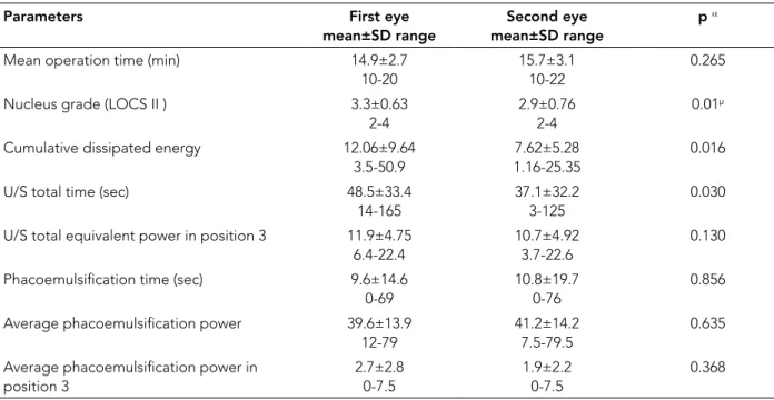
The study included 47 subjects,24 female and 23 male, aged between 43 and 82 years (mean 66.6±8.8). The pre-operative nucleus grades were 3.3±0.6 and 2.9±0.7 in the first operated eye and in the second eye, respectively (p=0.014). A comparison of intraoperative parameters between the first and second eyes is given in Table 1. There was a statistically significant difference in cumula-tive dissipated energy and U/S total time between first and second eyes (p=0.016, and p=0.030, respectively). There was no statistically significant difference in total duration of surgery, phacoemulsification time, U/S total equivalent power foot position 3, average phacoemulsi-fication power and average phacoemulsiphacoemulsi-fication power foot position 3 between the first and second eyes. Pain scores between the first eye and second eye after cataract surgery is given in Table 2 Although no statis-tically significant difference was found in pain score be-tween the first eye and second eye at the first day and
first month, there was a significant difference at one week (p=0.080, 0.269 and 0.004, respectively).
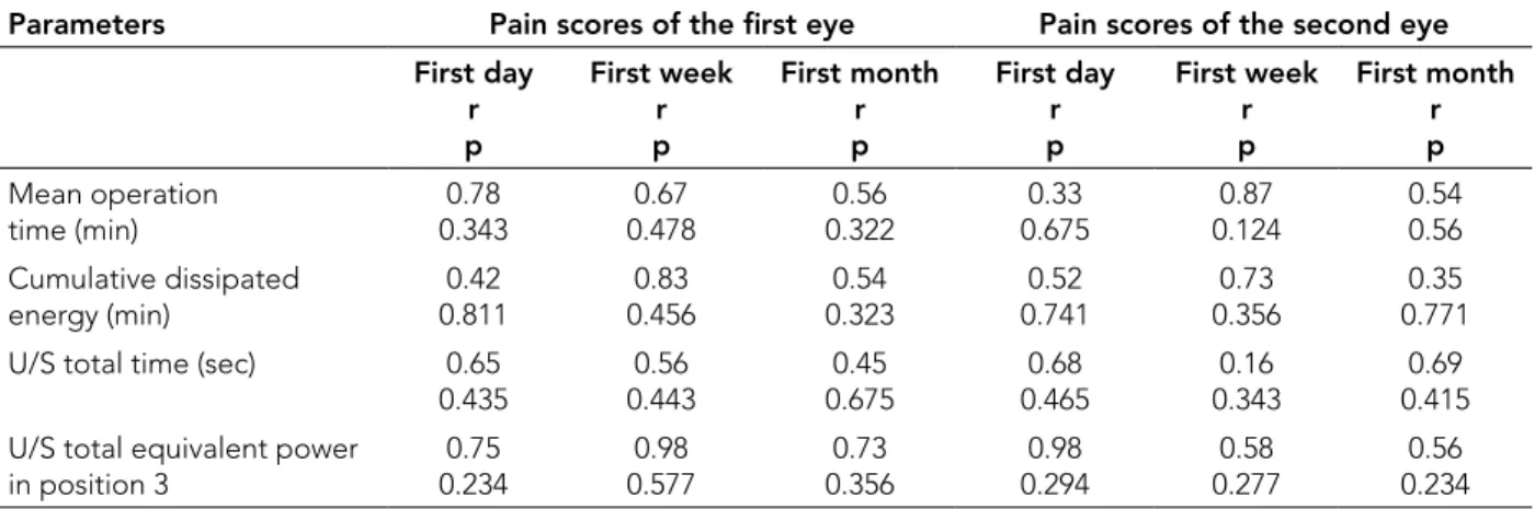
No significant correlations were found between phacoemulsification parameters of the first surgery and the pain scores of the first eye and second eye at postop-erative first day, first week and first month (Table 3). There was also no significant correlation between phacoemulsification parameters of the second surgery and the pain scores of the second eye at postoperative first day, first week and first month (Table 4). The correla-tion between the pain scores of the first and those of the second eye at postoperative first day, first week and first month was not statistically significant (r=0.256, p=0.126; r=0.754, p=0.663; r=0.155, p=0.361, respectively).
DISCUSSION
Our study evaluates the relation between the pain com-plaints of patients after first-eye and second-eye cataract surgeries and phacoemulsification parameters. When we evaluated the relation between pain and surgical condi-tions including U/S total time, U/S total equivalent power in position 3, cumulative dispersive energy, phacoemul-sification time, average phacoemulphacoemul-sification power and average phacoemulsification power in position 3 of phacoemulsification as well as duration of operation, we found no significant differences between phacoemul-sification parameters and pain scores. In our study, we observed that the majority of patients stated that they felt more pain in second eye during postoperative peri-od. Therefore we also compared the postoperative pain scores in the first operation with those in the second op-eration on the first day, at the first week and first month. We found that patients’ pain complaints in their second eye was significantly higher in the first postoperative week, whereas no significant difference was observed on the first day and at first month.
Ursea et al. (4) reported for the first time that there was a subtle increase in pain during second-eye surgery com-pared with first-eye surgery. They evaluated the pain scores immediately after the second eye operation and on the first postoperative day. They reported higher pain scores in the second eye on the day of the surgery; how-ever, there was no difference in pain scores between the two eyes on the first postoperative day. They concluded that more pain after second eye surgery was associated with decreased preoperative anxiety (4). Jiang et al. (5) also reported that cataract patients were likely to have more pain during second-eye surgery, which may be re-lated to lower preoperative anxiety.
Hari-Kovacs et al. (6) reported that consecutive phacoemulsification does not differ in perceived pain, but nevertheless, patients may believe the second eye
surgery more painful because they practically compare it with the lower remembered pain for the first eye pro-cedure.
However some other researchers have reported no sig-nificant differences between pain perceived in first-eye surgery and that in second-eye surgery (10, 11).
Previous studies usually focused on subjective psycho-logical mechanisms to explain why patients experienced pain during second eye cataract extraction. Zhu et al. (9) were the first ones to investigate this observation from a molecular perspective in 2015. In their study, they compared inflammatory cytokines by collecting aque-ous humor samples in the preoperative period from pa-tients who were having cataract surgery for the first time, and patients who had already had their first eye oper-ated on and had applied for the second-eye surgery. In
their study, they used the cytokine antibody array tech-nique and found that aqueous humor levels of MCP-1 (a pain-related inflammatory chemokine), were significantly increased in patients undergoing cataract surgery on the second eye compared with those undergoing surgery on the first eye. They thought that, this suggests there may be a sympathetic ophthalmic type uveitis in the contra-lateral eye after first-eye cataract surgery and that may help explain why second-eye phacoemulsification is of-ten more painful (9).
To our knowledge, there is no previous study which has investigated the relation of surgical parameters with postoperative pain scores. In our study there was no sig-nificant relation between the phacoemulsification param-eters of the first surgery and the pain scores of the first eye, and also there was no significant relation between the phacoemulsification parameters of the second sur-gery and the pain scores of the second eye at postoper-ative first day, first week and first month. However when we analyzed the relation between average phacoemulsi-fication power in foot position 3 of the first surgery and the pain scores of the second eye at postoperative first day and first week, we observed the differences (p=0.09 and 0.064, respectively) which might reach statistical sig-nificance in a larger sample. We may speculate that the increase in the amount of the energy used in phacoemul-sification surgery, especially in average phacoemulsifi-cation power in foot position 3, may be increasing the
Table 1. Comparison of phacoemulsification parameters between first and second eye surgeries Parameters First eye
mean±SD range
Second eye mean±SD range
p α
Mean operation time (min) 14.9±2.7
10-20
15.7±3.1 10-22
0.265
Nucleus grade (LOCS II ) 3.3±0.63
2-4
2.9±0.76 2-4
0.01µ
Cumulative dissipated energy 12.06±9.64
3.5-50.9
7.62±5.28 1.16-25.35
0.016
U/S total time (sec) 48.5±33.4
14-165
37.1±32.2 3-125
0.030 U/S total equivalent power in position 3 11.9±4.75
6.4-22.4
10.7±4.92 3.7-22.6
0.130
Phacoemulsification time (sec) 9.6±14.6
0-69
10.8±19.7 0-76
0.856
Average phacoemulsification power 39.6±13.9
12-79
41.2±14.2 7.5-79.5
0.635 Average phacoemulsification power in
position 3 2.7±2.8 0-7.5 1.9±2.2 0-7.5 0.368
SD: Standard deviation, U/S: Ultrasound, LOCS II: Lens Opacities Classification System α Independent t test
µ Paired samples t-test
Table 2. Comparison of pain scores between first eye and second eye after cataract surgery over time
First eye Second eye
p*
First day 2.24±2 3.05±2.4 0.080
First week 0.7±1.2 1.78±1.6 0.004
First month 0.38±0.8 0.65±1 0.269
amount of inflammatory chemokines. This situation may be giving rise to a kind of sympathetic ophthalmic type uveitis.
CONCLUSION
Pain scores after uneventful cataract surgery differ between first and second eye procedures. A possi-ble relation might be between the phacoemulsifica-tion parameters of the first surgery, especially average phacoemulsification power in foot position 3, and the second eye pain scores. More studies with larger sample sizes may strengthen the value of our study.
Ethics Committee Approval: Ethics committee approval was
received for this study from the Research Ethics Committee of the Istanbul Medipol University (Approval No: 290).
Informed Consent: Written informed consent was obtained
from patients who participated in this study.
Peer-review: Externally peer-reviewed.
Author Contributions: Concept – R.A., M.Ö., S.K.E., M.Ö., E.K.;
Design – R.A., M.Ö., S.K.E., M.Ö., E.K.; Supervision - R.A., M.Ö., S.K.E., M.Ö., E.K.; Resources – M.Ö.; Materials – M.Ö ; Data Collection and/or Processing – M.Ö., R.A., S.K.E., M.Ö.; Anal-ysis and/or Interpretation - R.A., M.Ö., S.K.E; Literature Search - M.Ö.; Writing Manuscript – R.A.; Critical Review - M.Ö., R.A.
Table 3. Correlation between phacoemulsification parameters of the first surgery versus the pain scores of the first eye and second eye
Parameters Pain scores of the first eye Pain scores of the second eye First day r p First week r p First month r p First day r p First week r p First month r p Mean operation time (min) 0.78 0.343 0.67 0.478 0.56 0.322 0.33 0.675 0.87 0.124 0.54 0.56 Cumulative dissipated energy (min) 0.8110.42 0.4560.83 0.3230.54 0.7410.52 0.3560.73 0.7710.35
U/S total time (sec) 0.65
0.435 0.4430.56 0.6750.45 0.4650.68 0.3430.16 0.4150.69 U/S total equivalent power
in position 3 0.75 0.234 0.98 0.577 0.73 0.356 0.98 0.294 0.58 0.277 0.56 0.234 U/S: Ultrasound
*Pearson correlation coefficient
Table 4. Correlation between phacoemulsification parameters of the second surgery and the pain scores of the second eye
Parameters
Pain scores of the second eye
First day r p First week r p First month r p
Mean operation time (min) 0.56
0.393
0.97 0.178
0.96 0.522
Cumulative dissipated energy 0.82
0.311
0.43 0.556
0.94 0.123
U/S total time (sec) 0.85
0.535 0.6430.46 0.3750.75
U/S total equivalent power in position 3 0.95
0.334 0.8770.18 0.3460.79
Phacoemulsification time (sec) 0.55
0.778
0.43 0.226
0.96 0.343
Average phacoemulsification power 0.23
0.697 0.821 0.54 0.6770.18
Average phacoemulsification power in
position 3 0.3210.66 0.7160.33 0.2770.78
U/S = Ultrasound *Pearson correlation
Conflict of Interest: No conflict of interest was declared by the
authors.
Financial Disclosure: The authors declared that this study has
received no financial support.
Etik Komite Onayı: Bu çalışma için etik komite onayı İstanbul
Medipol Üniversitesi’nden alınmıştır (Karar No: 290).
Hasta Onamı: Yazılı hasta onamı bu çalışmaya katılan
hastalar-dan alınmıştır.
Hakem Değerlendirmesi: Dış bağımsız.
Yazar Katkıları: Fikir – R.A., E.K., M.Ö., M.Ö.; Tasarım – R.A.,
E.K.; Denetleme – R.A., M.Ö., S.K.E., M.Ö., E.K.; Kaynaklar – M.Ö.; Malzemeler – M.Ö.; Veri Toplanması ve/veya İşlemesi – M.Ö., R.A., S.K.E., M.Ö.; Analiz ve/veya Yorum – R.A., M.Ö., S.K.E.; Literatür Taraması – M.Ö.; Yazıyı Yazan – R.A.; Eleştirel İnceleme – M.Ö., R.A.
Çıkar Çatışması: Yazarlar çıkar çatışması bildirmemişlerdir. Finansal Destek: Yazarlar bu çalışma için finansal destek
alma-dıklarını beyan etmişlerdir.
REFERENCES
1. Zhao LQ, Zhu H, Zhao PQ, Wu QR, Hu YQ. Topical anes-thesia versus regional anesanes-thesia for cataract surgery: a me-ta-analysis of randomized controlled trials. Ophthalmology 2012;119(4):659-67. [CrossRef]
2. Thevi T, Godinho MA. Trends and complications of local anesthesia in cataract surgery: an 8-year analysis of 12 992 patients. Br J Ophthalmol 2016;100(12):1708-13. [CrossRef]
3. Haddadi S, Marzban S, Fazeli B, Heidarzadeh A, Parvizi A, Naderinabi B, et al. Comparing the effect of topical anes-thesia and retrobulbar block with ıntravenous sedation on hemodynamic changes and satisfaction in patients under-going cataract surgery (phaco method). Anesth Pain Med 2015;28;5(2):e24780.
6. Hari-Kovacs A, Lovas P, Facsko A, Crate ID. Is second eye phacoemulsification really more painful? Wien Klin Wo-chenschr 2012;124(15-16):516-9. [CrossRef]
4. Ursea R, Feng MT, Zhou M, Lien V, Loeb R. Pain Per-ception in Sequential Cataract Surgery: Comparison of First and Second Procedures. J Cataract Refract Surg 2011;37(6):1009-14. [CrossRef]
5. Jiang L, Zhang K, He W, Zhu X, Zhou P, Lu Y. Perceived pain during cataract surgery with topical anesthesia: a compari-son between first-eye and second-eye surgery. Ophthalmol 2015;2015:383456.
7. Aslan L, Aslankurt M, Cekic O, Aksoy A, Yildiz H. The pain ex-perience and cooperation of patients in consecutive cataract surgery. Eur J Ophthalmol 2013;23(3):339-43. [CrossRef]
8. Bardocci A. Second-Eye Pain in Cataract Surgery. J Cata-ract RefCata-ract Surg 2012;38(9):1705. [CrossRef]
9. Zhu XJ, Wolff D, Zhang KK, He WW, Sun XH, Lu Y, et al. molecular ınflammation in the contralateral eye after cat-aract surgery in the first eye. Invest Ophthalmol Vis Sci 2015;56(9):5566-73. [CrossRef]
10. Bardocci A, Ciucci F, Lofoco G, Perdicaro S, Lischetti A. Pain during second eye cataract surgery under topical an-esthesia: an intraindividual study. Graefes Arch Clin Exp Ophthalmol 2011;249(10):1511-4. [CrossRef]
11. Sharma NS, Ooi JL, Figueira EC, Rosenberg ML, Massel-os K, Papalkar DP, et al. Patient perceptions of second eye clear corneal cataract surgery using assisted topical anaes-thesia. Eye (Lond) 2008;22(4):547-50. [CrossRef]