High resolution 3D magnetic resonance imaging of the visceral organs
in chicken (Gallus domesticus) by 3 Tesla MR unit and 15-channel
transmit coil
Okan EKİM
1, Çağdaş OTO
1, Oktay ALGIN
2, Caner BAKICI
11 Ankara University Faculty of Veterinary Medicine Department of Anatomy, Ankara; 2 Atatürk Training and Research Hospital
Department of Radiology, Ankara, Turkey.
Summary:
Imaging studies conducted on the modern imaging techniques for birds are limited and probably insufficient forthe clinicians. As in mammals, magnetic resonance (MR) imaging (MRI) can be used as a convenient method for the diagnosis and treatment of the avian diseases. In this study, the whole bodies of 2 male and 2 female chickens were imaged by a 3 Tesla superconductive magnet and 15-channel transmit-receive birdcage coil. After acquisition of three dimensional (3D) T1, T2 and proton density weighted (W) MR images; bodies were frozen in same position with the one in imaging process and sliced from matching sections with original and reformatted MR images. Anatomic structures were identified and labeled in both MR images and cadaver sections. After that, 3D multiplanar reconstruction was performed on the MR images. On T1W images, it was observed that the anatomical details were superior due to the high geometric resolution. On T2W images, the tissue contrast differences and fluid filled ducts were clearly detected. On three orthogonal and oblique planes reformatted and maximum intensity projection (MIP) colored images, the anatomic details were more clearly determined and the tissues were more easily distinguished from each other with high geometric and contrast resolution. The aim of this study was to define MRI features of the tissues, and to provide an overview of MRI anatomy of the avian body structures. Besides, the most convenient sequences for the avian MRI were also designated.
Keywords: 3 Tesla, 15 channel transmit-receive coil, anatomy, chicken, magnetic resonance imaging, three dimensional.
Tavukta (Gallus domesticus) visseral organların 3 Tesla manyetik rezonans ünitesi ve 15-kanallı coil ile
yüksek rezolüsyonlu 3B görüntülenmesi
Özet:
Kuşlarda modern görüntüleme teknikleri üzerine yürütülmüş çalışmalar kısıtlıdır ve özellikle klinisyenler için yetersizkalmaktadır. Manyetik rezonans (MR) görüntüleme (MRG), memelilerde olduğu gibi kanatlı hastalıklarının tanı ve tedavisi için de elverişli bir yöntemdir. Bu çalışmada; 2 horoz ve 2 tavuğun vücudu 3 Tesla MRG cihazı ile T1, T2 ve proton-dansite ağırlıklı (A) sekanslar ve 15-kanallı alıcı-verici koyil kullanılarak, 3 boyutlu (3B) olarak değerlendirildi. MRG işlemi sonrasında; görüntüleme işlemindeki pozisyonu ile aynı konumda dondurulmuş vücutlar, MR görüntüleri ile eşleşen bölümlerden dilimlendi. Anatomik yapılar, MR görüntüleri ve kadavra bölümlerinde tespit edildi ve işaretlendi. Daha sonra, MR görüntülerine 3B multiplanar rekonstrüksiyon uygulandı. T1A görüntülerde, anatomik detaylar yüksek geometrik çözünürlük nedeniyle daha iyi gözlendi. T2A görüntülerde, doku kontrastı farkları ve sıvı içerikli yapılar açıkça tespit edildi. 3B sekanslardan elde edilen oblik planlı ve maksimum yoğunluk gösteren renkli reformat görüntülerde, anatomik ayrıntılar daha net bir şekilde belirlendi ve dokular yüksek çözünürlük ile birbirinden daha kolay bir şekilde ayırt edildi. Bu çalışmadaki amaç; dokuların görüntüleme özelliklerini tanımlamak ve kuşların vücut yapılarının MRG’deki anatomisine genel bir bakış sağlamaktı. Bunların yanı sıra, kanatlılarda MRG için en uygun sekanslarda tayin edildi.
Anahtar sözcükler: 3 Tesla, 15 kanal alıcı-verici coil, anatomi, manyetik rezonans görüntüleme, tavuk, üç boyutlu.
Introduction
Similar to mammals’, magnetic resonance imaging
(MRI) has been a quite efficient method to understand
some physiological functions related with the anatomical
formation of birds and to evaluate the diagnosis and
treatment of the avian diseases. However, studies, either
clinical or experimental, focused on the modern imaging
techniques for birds seem to be insufficient. Not only
magnetic resonance (MR) studies but also computed
tomography (CT) or micro CT researches were also very
limited and most of these were based on osteology or
osteological modeling (6, 12, 23, 24, 25).
Although MRI distinguishes from CT as high
resolution and radiation free technique (1, 10, 18, 19, 20),
the MR studies on avian are mostly focused on the
clinical cases (8, 14, 21, 26). Some of the researches has
been carried out to calculate the different tissue densities
such as fat or water ratios in the body by using MR
techniques (9, 29). Especially tissues which have high fat
and water ratio can be readily visualized and quantified
in virtual slices with T1-weighted (W) MR sequences,
and then summed across slices to calculate body
composition (9). MRI gives us the opportunity to
investigate the avian body parts from a three-dimensional
(3D) capturing (22, 27, 28). MR can also provide
important data about the egg composition or the
embryonic development of the birds (4, 7, 13).
Dissimilarly to the domestic mammals (11), the
thorax and the abdomen appear to one single cavity in
most of the birds due to the rudimentary diaphragm.
Considering about the abdominal organs, for instance,
kidneys embedded into the lumbosacral bone, in males,
testicles take a large space inside the body cavity, and the
gaster consists of two different parts as is known (15,
17). Although current MR studies based on the anatomy
or physiology of domestic birds were available (2, 5, 20,
22, 27), an anatomic evaluation for the abdominal organs
comparing the MR images with the cadaver slices on
birds might be useful for the researchers working on
avian. With this study we tried to prepare an anatomical
reference which combines and compares the data on the
organic cross-sections and the MR images. We preferred
the species Gallus domesticus for its economic value and
it was thought that it could be an efficient animal model
for a comparative anatomic study.
Materials and Methods
In this study, the whole bodies of 2 male and 2
female chickens were investigated. For the induction, an
intramuscular application of 50 mg/kg ketamine
hydrochloride was used to the birds. The anaesthetized
birds were placed in “prone” position and were imaged
by 3T superconductive magnet (Siemens Magnetom
Trio, Erlangen, Germany) with using 15-channel receiver
and transmitter birdcage coil. T1W and T2W 3D data
were obtained with the optimized sequences (Table 1).
Multiplanar reformatted, maximum-minimum intensity
projection, and volume rendered images were achieved
using Leonardo Workstation software and 3D data. An
intravenous administration of 150 mg/kg thiopental
sodium was injected to the birds for euthanasia. After
MRI process, bodies were frozen to -25°C for 3 days.
Then bodies were sliced from same levels with the
matching MR image sections (Figure 1.A.1,2,3,4).
Macroscopic segments were acquired using 3 mm slice
thickness on frozen body. Afterwards, anatomic
structures of the chicken were identified and labeled in
both MR sections and cadaver slices. Nomina Anatomica
Avium (3) was used for nomenclature.
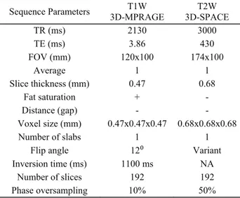
Table 1: MRI protocol of the authors.
3D: three dimensional; W: weighted; NA: not applicable; FOV: field of view; 3D-SPACE: 3D sampling perfection with application-optimized contrasts by using different flip angle evolutions; 3D-MPRAGE: 3D magnetization-prepared rapid gradient-echo.
Tablo 1: Yazarların MRG protokolü
3D: three dimensional; W: weighted; NA: not applicable; FOV: field of view; 3D-SPACE: 3D sampling perfection with application-optimized contrasts by using different flip angle evolutions; 3D-MPRAGE: 3D magnetization-prepared rapid gradient-echo.
Sequence Parameters 3D-MPRAGE T1W 3D-SPACE T2W
TR (ms) 2130 3000 TE (ms) 3.86 430 FOV (mm) 120x100 174x100 Average 1 1 Slice thickness (mm) 0.47 0.68 Fat saturation + - Distance (gap) - - Voxel size (mm) 0.47x0.47x0.47 0.68x0.68x0.68 Number of slabs 1 1
Flip angle 12⁰ Variant
Inversion time (ms) 1100 ms NA
Number of slices 192 192
Phase oversampling 10% 50%
Results
On T1W images, it was observed that the anatomical
data were superior due to the high geometric resolution
(Figure 1.B.1,2,3,4). Fat containing tissues were
appeared bright (hyperintense) on T1W images. Also
aqueous structures had lower intensity (hypointense) on
T1W images. The most hyperintense solid abdominal
organ was liver (Figure 1.B.1,2,3,4) but muscular tissues
such as muscular stomach and myocardium of the heart
(Figure 1.B.1,2,3,4) were also appeared hyperintense on
T1W images.
On T2W images, the tissue contrast differences and
fluid filled spaces were more clearly detected. Testicles
(Figure 1.C.1,3,4), cerebrospinal liquid, and also preen
gland (Figure 1.C.2,4) were observed as hyperintense on
these images (Figure 1.C.1,2,3,4). Fatty tissues were
appeared bright (hyperintense) on T2W images but these
tissues had the lower signal intensity compared to T1W
images.
The lumen of the hollow organs such as intestines,
stomach, and lungs (Figure 1.B.C) were prominent
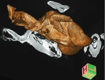
hypointense (very dark) on all sequences. On colored
reconstructed and volume-rendered T1W images, the
morphological details were more clearly determined
(Figure 1.D.1,2,3,4, Figure 2) and the tissues were easily
distinguished from each other due to high signal-to-noise
and contrast-to-noise ratios.
Figure 1: Comparing of the anatomic structures in cadaver body slices and the MR images. A/ 1,2,3,4; The sagittal slices of the body cavity obtained from specimens.
B/ 1,2,3,4; T1-weighted sagittal images obtained from the same level with cadaver slices. C/ 1,2,3,4; T2-weighted sagittal images obtained from the same level with cadaver slices.
D/ 1,2,3,4; Reformatted, colored, and volume rendered 3D T1-weighted images from the same level with cadaver slices.
a: lung (pulmo), b; liver (hepar), c; kidney (ren), d; testicle (testis), e; left caecum, f; right caecum, g; colon, h; spleen (lien), ı; jejenum, j; heart (cor), k; muscular stomach (ventriculus muscularis), l; trachea, m; glandular stomach (ventriculus glandularis). Figür 1: MR görüntüleri ile kadavra vücut kesitlerindeki anatomik yapıların karşılaştırılması.
A/ 1,2,3,4; Örneklerden elde edilen, vücut boşluğuna ait sagittal kesitler.
B/ 1,2,3,4; Kadavra kesitleri ile aynı düzeyden elden edilen T1A sagittal kesit görüntüleri. C/ 1,2,3,4; Kadavra kesitleri ile aynı düzeyden elden edilen T2A sagittal kesit görüntüleri.
D/ 1,2,3,4; Kadavra kesitleri ile aynı düzeyden elden edilen T1A, reformat, renklendirilmiş, volumetrik 3B sagittal kesit görüntüleri. a: akciğer (pulmo), b; karaciğer (hepar), c; böbrek (ren), d; testis, e; sol caecum, f; sağ caecum, g; colon, h; dalak (lien), ı; jejenum, j; kalp (cor), k; kaslı mide (ventriculus muscularis), l; trachea, m; bezli mide (ventriculus glandularis).
Figure 2. A 3D reconstructed, reformatted, colored, and volume rendered T1W image sample.
Figür 2. 3B rekonstrükte edilmiş, reformat, renklendirilmiş, volümetrik T1A görüntü örneği.
Discussion and Conclusion
As mentioned before, one of the purposes of this
study was to define imaging features of tissues and to
provide an overview of MRI anatomy of the avian
abdominal structures and their natural positions. It can be
said that MRI is quite efficient for the identification of
anatomical structures and also can be used for diagnosis
of pathological changes. The most convenient sequence
that enables to delineate the anatomic details was also
designated by this study. Wirestam et al. (29) had
indicated that MRI could be used for the measurement of
the variations in the spatial distributions of adipose tissue
in small migratory birds. Our research supports this
finding on domestic birds. Although MRI provides better
anatomical detail due to its high resolution when
compared to the CT, researches on the use of
cross-sectional imaging in avian species have been limited.
Ruffins et al. and Vellema et all. (22,27) have composed
atlases in detail using MR images, but they examined
prenatal and postnatal anatomic structures which are not
exactly corresponds to our research. Therefore, data
collected from our MR images can be used to create a
comparative avian body atlas for anatomy education or
avian researchers and clinicians in the future. However
the number of subjects should be increased to prepare a
detailed atlas.
On T1W images the clear displaying of organ
boundaries made us think that T1W sequences were more
efficient for anatomists. One to one correspondence
between the slices obtained from the cadavers and the
MR images couldn’t be provided in all cross-sections.
This mismatch might probably originate from the
volumetric changes in specimens after freezing process
and the positioning failures during the band saw process.
When compared with the previous MR researches,
the most important superiority and difference of our
study was to use of 3 Tesla MR device and 15-channel
transmit-receive birdcage coil. With the usage of this coil
in 3 Tesla MR unit, the 3D data which is composed from
isotropic voxels smaller than 1 mm could be obtained
with high signal-to-noise ratio. The quality and
resolution of the multiplanar images obtained from this
data was notably high. Because the all sequences that
we’ve used, were composed from submilimetric isotropic
voxels. The devices and the techniques we’ve mentioned
above are generally used in cases that require high
resolution such as human knee pathologies (16).
In conclusion, 3 Tesla MR unit with 15-channel coil
and optimized 3D sequences provides significantly
higher quality images compared to conventional 2D
techniques, particularly for fluid and fat containing
spaces, leading to improved diagnostic confidence in
birds.
References
1. Aagaard BD, Lazar DA, Lankerovich L, Andrus K, Hayes CE, Maravilla K, Kliot M (2003): High-resolution magnetic resonance imaging is a noninvasive method of observing injury and recovery in the peripheral nervous system. Neurosurgery, 53, 199-203.
2. Bartels T, Brinkmeier J, Portmann S, Baulain U, Zinke A, Junghanns MEK, Boos A, Wolf P, Kummerfeld N (2001): Magnetic resonance imaging of intracranial tissue
accumulations in domestic ducks (Anas platyrhynchos f.dom.) with feather crests. Vet Radiol Ultrasound, 42,
254-258.
3. Baumel JJ, King AS, Breazile JE, Evans HE, Vanden Berge JC (1993): Nomina Anatomica Avium, MA: Nuttall Ornithological Club, Cambridge.
4. Belton PS, Gordon RE, Jones JM, Shaw D. (1983): A
31P topical magnetic resonance study of embryonic development in hens' eggs. Br Poult Sci, 24, 429-33.
5. Boumans T, Theunissen FE, Poirier C, Van Der Linden A (2007): Neural representation of spectral and temporal features of song in the auditory forebrain of zebra finches as revealed by functional MRI. Eur J Neurosci, 26,
2613-2626.
6. Charuta A, Cooper RG (2012): Computed tomographic
and densitometric analysis of tibiotarsal bone mineral density and content in postnatal Peking ducks (Anas platyrhynchos var. domestica) as influenced by age and sex. Pol J Vet Sci, 15, 537-545.
7. Falen SW, Szeverenyi NM, Packard DS Jr, Ruocco MJ (1991): Magnetic resonance imaging study of the structure
of the yolk in the developing avian egg. J Morphol, 209,
331-342.
8. Graham JE, Werner JA, Lowestine LJ, Wallack, ST, Tell LA (2003): Periorbital liposarcoma in an African grey parrot (Psittacus erithacus). J Avian Med Surg, 17,
147-153.
9. Guglielmo CG, McGuire LP, Gerson AR, Seewagen CL (2011): Simple, rapid and non-invasive measurement of
fat, lean and total water masses of live birds using quantitative magnetic resonance. J Ornithol, 152, 75-85.
10. Herberholz J, Mishra SH, Uma D, Germann MW, Edwards DH, Potter K (2011): Non-invasive imaging of neuro anatomical structures and neural activation with high-resolution MRI. Front Behav Neurosci, 31, 16.
11. König HE, Liebich HG (2004): Veterinary Anatomy of
Domestic Mammals. Schattauer, Stuttgart- New York.
12. Leslie MA, Coleman RA, Moehn S, Ball RO, Korver DR (2006): Relationship between bicarbonate retention and bone characteristics in broiler chickens. Poult Sci, 85,
1917-1922.
13. Li X, Liu J, Davey M, Duce S, Jaberi N, Liu G, Davidson G, Tenent S, Mahood R, Brown P, Cunningham C, Bain A, Beattie K, McDonald L, Schmidt K, Towers M, Tickle C, Chudek S (2007): Micro-magnetic resonance imaging of avian embryos, 211,
798-809.
14. Misra LK, Entrikin RK (1988): Corticosteroid therapy in
avian muscular dystrophy: evaluation by magnetic resonance relaxation times. Exp Neurol, 102, 217-220.
15. Nickel R, Schummer A, Seiferle E (1977): Anatomy of
16. Notohamiprodjo M, Horng A, Kuschel B, Paul D, Li G, Raya JG, Reiser MF, Glaser C (2012): 3D-imaging of the knee with an optimized 3D-FSE-sequence and a 15-channel knee-coil. Eur J Radiol, 81, 3441-3449.
17. O'Malley B (2005): Clinical Anatomy and Physiology of
Exotic Species: Structure and function of mammals, birds, reptiles and amphibians. Elsevier Saunders, Edinburg.
18. Oto Ç, Ekim O, Algin O, Şenel OO, İnce N, Hazıroğlu RM (2011): 3 Tesla Magnetic resonance imaging and multiplanar reconstruction of the brain and its associated structures in pig. Vet J Ankara Univ, 58, 75-78.
19. Oto Ç, Hazıroğlu RM (2011): Magnetic resonance
imaging of the guttural pouch (diverticulum tubae auditivae) and its related structures in donkey (Equus asinus). Vet J Ankara Univ, 58, 1-4.
20. Pepperberg IM, Howell KS, Banta PA, Patterson DK, Meister M (1998): Measurement of grey parrot (Psittacus erithacus) trachea via magnetic resonance imaging, dissection, and electron beam computed tomography. J
Morphol, 238, 81-91.
21. Pye GW, Bennett A, Newell SM, Kindred J, Johns R (2000): Magnetic resonance imaging in psittacine birds
with chronic sinusitis. J Avian Med Surg, 14, 243-256.
22. Ruffins SW, Martin M, Keough L, Truong S, Fraser SE, Jacobs RE, Lansford R (2007): Digital Three-Dimensional Atlas of Quail Development Using High-Resolution MRI. ScientificWorldJournal, 7, 592-604.
23. Seki Y, Mackey M, Meyers MA (2012): Structure and
micro-computed tomography-based finite element modeling of Toucan beak. J Mech Behav Biomed Mater. 9,
1-8.
24. Shastak Y, Witzig M, Hartung K, Bessei W, Rodehutscord M (2012): Comparison and evaluation of bone measurements for the assessment of mineral phosphorus sources in broilers. Poult Sci, 91, 2210-2220.
25. Shipov A, Sharir A, Zelzer E, Milgram J, Monsonego-Ornan E, Shahar R (2010): The influence of severe prolonged exercise restriction on the mechanical and structural properties of bone in an avian model. Vet J, 183, 153-160.
26. Stauber E, Holmes S, DeGhetto DL, Finch N (2007):
Magnetic resonance imaging is superior to radiography in evaluating spinal cord trauma in three bald eagles (Haliaeetus leucocephalus). J Avian Med Surg, 21,
196-200.
27. Vellema M, Verschueren J, Meir VV, Linden AV (2011): A customizable 3-dimensional digital atlas of the
canary brain in multiple modalities. NeuroImage, 57,
352-361.
28. Verhoye M, Van der Linden A, Van Audekerke J, Sijbers J, Eens M, Balthazart J (1998): Imaging birds in a bird cage: in-vivo FSE 3D MRI of bird brain. MAGMA, 6, 22-27.
29. Wirestam R, Fagerlund T, Rosen M, Hedenström A (2008): Magnetic Resonance Imaging for Noninvasive
Analysis of Fat Storage in Migratory Birds. The Auk, 125,
965-971.
Geliş tarihi: 27.02.2013 / Kabul tarihi: 29.04.2013 Address for correspondence:
Dr. Okan Ekim Ankara University
Faculty of Veterinary Medicine Department of Anatomy, Ankara-TURKEY