Ankara Üniv Vet Fak Derg, 62, 161-163, 2015
Short Communication / Kısa Bilimsel Çalışma
Abomasal ulcer and jejunal ileus caused by trichobezoar in a
two-day-old calf
Serkan ÇATIK
1, Melike AKBALA
2, Hasan KURT
2, Hakan SALCI
2 Uludağ University, Faculty of Veterinary Medicine, 1Department of Internal Medicine; 2 Department of Surgery, Görükle Campus, 16059 – Bursa, Turkey.
Summary: A two-day-old, male Holstein calf was presented with a history of anorexia, abdominal distension and pain,
inability to urinate and defecate. In physical examination, depression, recumbency, dehydration, weak sucking reflex, abdominal distension accompanied by pain and fluctuation were found in the calf. Furthermore, mucous membranes were pale and capillary refill time was 5 seconds. Laboratory findings revealed sepsis and deficiency of the some minerals. However, the calf died right after clinical and laboratory examination. Necropsy showed a rough and solid formation in jejunum as well as abomasitis, local ulcer and erosions on the abomasal mucosa. As a result of these findings, abomasal ulcer and jejunal ileus caused by trichobezoar was diagnosed in the calf.
Key words: Abomasal ulcer, calf, jejunal ileus, trichobezoar.
İki günlük bir buzağıda trikobezoar nedenli jejunal ileus ve abomasum ülseri
Özet: İki günlük erkek Holstein ırkı bir buzağı iştahsızlık, abdominal gerginlik ve ağrı, idrar ve dışkı yapamama anamneziyle
getirildi. Fiziksel muayenede buzağıda depresyon, yan yatma, dehidrasyon, zayıf emme refleksi, ağrı ve fluktuasyonla birlikte abdominal gerginlik görüldü. Bundan başka, mukozal membran rengi solgundu ve kapillar dolum zamanı 5 saniyeydi. Laboratuar bulgularında sepsis ve bazı minerallerde eksiklik gözlendi. Ancak, klinik ve laboratuar muayenelerinin hemen sonrasında buzağı öldü. Nekropsi, abomasum mukozası üzerinde abomasitis, local ülser ve erozyon ile birlikte jejunumda sert ve kitlesel bir yapıyı gösterdi. Bu bulguların sonucu olarak, buzağıda trikobezoar nedenli abomasum ülseri ve jejuneum ileusu tanısı konuldu.
Anahtar sözcükler: Abomasum ülseri, buzağı, jejuneum ileusu, trikobezoar.
Trichobezoars are round masses that they form by animals licking themselves or each other (1). One of the other causes of the trichobezoar formation is nutrition with mineral deficiency (3,4). Pica and soil eating behavior are particularly resulted from phosphorus, iron, sodium and magnesium deficiencies in calves (3,5). The calf breast sucking, instead of drinking milk with bottle, is liable to hair ingestion. Particularly in cold climates, due to shaggy hair coat, licking each other in calves is the main cause of the hair ingestion resulting in the trichobezoar formation (4).
Trichobezoar can have round or oval shape effecting the gastrointestinal motility. Although they are usually encountered by chance in slaughterhouse in adult animals, it is reported in the esophagus, pyloric and reticulo-omasal passage (2). Acute intestinal obstruction is the main clinical finding of the trichobezoars and phytobezoars in calves (2,6).
Possibility of the trichobezoar presence is 3.81 times higher in young calves with abomasal ulcer (4).
Although primary cause of the abomasal ulcer is not only trichobezoar, several factors including nutritional deficiencies, microbiological agents, stress and poor quality forages should be considered (3,4). In this presented case, we aim to report with clinical, laboratory and necropsy findings of the abomasal ulcer resulted from jejunal ileus by trichobezoar in a two-day-old calf.
A two-day old, male Holstein calf was presented to Uludag University, Faculty of Veterinary Medicine, Large Animal Clinics of Animal Hospital with a history of anorexia, abdominal distension and pain, inability to urination and defecation. Only a small amount of mucus was coming through the rectum. Defecation of the calf was never seen by the owner; however the colostrum intake of the calf was approximately 4 liters.
The calf was depressed, recumbent, dehydrated (10%) and had weak sucking reflex. Abdominal distension accompanied by pain and fluctuation in the abdomen were distinctive. The “ping” sound was not felt during abdominal percussion. In general examination,
Serkan Çatık - Melike Akbala - Hasan Kurt - Hakan Salcı 162
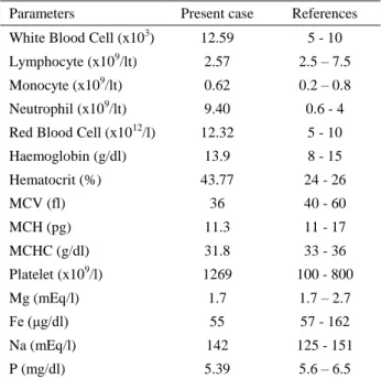
pulsation, respiration and body temperature were 84 beat/min, 80 breaths/minute, and 36.7°C, respectively. Abdominal palpation of the calf was painful, he had severe dyspnea, and also tongue was lolling out of his mouth. Peripheral pulse was weak. Mucous membranes were pale and capillary refill time was 5 seconds. After clinical examination, blood samples were taken and hematological parameters and serum phosphorus, iron, sodium and magnesium levels were analyzed (Table 1).
Table 1. Hematologic and serobiochemical findings of the calf. Parameters Present case References White Blood Cell (x103) 12.59 5 - 10 Lymphocyte (x109/lt) 2.57 2.5 – 7.5 Monocyte (x109/lt) 0.62 0.2 – 0.8 Neutrophil (x109/lt) 9.40 0.6 - 4 Red Blood Cell (x1012/l) 12.32 5 - 10
Haemoglobin (g/dl) 13.9 8 - 15 Hematocrit (%) 43.77 24 - 26 MCV (fl) 36 40 - 60 MCH (pg) 11.3 11 - 17 MCHC (g/dl) 31.8 33 - 36 Platelet (x109/l) 1269 100 - 800 Mg (mEq/l) 1.7 1.7 – 2.7 Fe (μg/dl) 55 57 - 162 Na (mEq/l) 142 125 - 151 P (mg/dl) 5.39 5.6 – 6.5
Based on the patients’ history, clinical and laboratorial findings, initial diagnose was atresia coli, bowel invagination or meconium constipation. However, the calf died right after clinical and laboratorial examinations. Necropsy was performed immediately.
Approximately 1 liter serosanguinous fluid was found in the peritoneal cavity. Abomasum was bigger than its normal size and had approximately 6 liters of fluidic content with cheesy particles. Abomasitis, local ulcer and erosions were distinctive on the mucosa of the major abomasal curvature (Figure 1). In the jejunum, a solid formation suspected as a foreign body was palpated. Following the incision of the jejunum, an oval-shaped hairball (2x4.5 cm in diameter) occluding to the bowel lumen was encountered (Figure 2). Furthermore, consistency of the other bowel segments was soft and there was no other pathological condition encountered in small and large intestines. The parenchyma organs (liver, kidney and spleen) of the abdomen had not any abnormality, as well.
When the intestinal obstruction is occurred, distinctive clinical signs such as anorexia, abdominal distension, fluctuation, inability to defecation and “ping “sound are remarkable (2). Although abdominal distension and fluctuation were observed in presented case, there was no
Figure 1. This view shows the local ulcerative areas and erosions (right arrows) and cheesy particles (left arrows) on the mucosa of the major curvature of the abomasum.
Şekil 1. Bu görüntü abomasumun curvature major mukozasında local ülseratif alanlar ve erezyonları ( sağ oklar) ve peynirimsi partikülleri (sol oklar) göstermektedir.
Figure 2. Appearance of the oval-shaped trichobezoar removed from the jejunal lumen (right arrow).
Şekil 2. Jejunum lümeninden çıkarılan oval şekilli trikobezoarın görünümü (sağ ok).
“ping” sound in abdominal percussion, and excessive fluid accumulation in abomasum and free peritoneal fluid were considered to be exact cause of the abdominal enlargement.
Incidence of the trichobezoar formation in calves is higher than the adult cattle. It has been implied that trichobezoar has not been shown in calves before 3 day old (4). Trichobezoar are often seen by chance after post mortem examination in calves died from tympani. After the other cause of the tympani has been eliminated, the possibility of the trichobezoar formation should always
Ankara Üniv Vet Fak Derg, 62, 2015 163
be considered (7). The present case was supported by literature data with the findings of compressive abdominal distension, severe respiratory distress, abomasitis, sepsis and abomasal ulcer.
As it has been shown in table 1, hematological findings were also clarified the severe inflammation and sepsis, which had slight increase in total number of leukocytes and extremely high neutrophil level (74.7%). This condition suggested the possibility of acute inflammation associated with abomasal ulcer, and the higher hematocrit value might be formed due to dehydration. Although serum magnesium and serum sodium was in normal values, serum iron and phosphorus levels were lower than reference ranges. These mineral changes could be related to animals’ age, colostum intake and mineral content of the colostrum. It was not clear whether these serum mineral values of the case led to the pica and the hair ingestion or not.
The pyloric obstruction due to trichobezoar is more common than the intestines. There is not any clinical importance up to obstruct the passage completely (5). Abomasal ulcers commonly form on the curvatura major of abomasum, because muscular structure of this area is weaker and abomasal peristaltic is slower (4). In our case, jejunal passage was completely blocked by trichobezoar; therefore, continuous hydrochloric acid secretion from abomasum and abomasal inactivity might cause abomasal ulcer on curvatura major.
References
1. Abutarbush SM, Naylor JM (2006): Obstruction of the
small intestine by a trichobezoar in cattle: 15 cases (1992– 2002). J Am Vet Med Assoc, 229, 1627-1630.
2. Abutarbush SM, Radostits OM (2004): Obstruction of
the small intestine caused by a hairball in 2 young beef calves. Can Vet J, 45, 324-325.
3. Brooks HV, Cook TG, Mansellf GP, Walker GA (1984): Phosphorus deficiency in a dairy herd. New Zeal Vet J, 32, 174-176.
4. Jelinski MD, Ribble CS, Campbell JR, Janzen ED (1996): Investigating the relationship between abomasal
hairballs and perforating abomasal ulcers in unweaned beef calves. Can Vet J, 37, 23-26.
5. Mesaric M, Modic T (2007): Dilation and torsion of the
caecum in a cow caused by a trichobezoar. Aust Vet J, 85,
156-157.
6. Patel JH, Brace DM (1995): Esophageal obstruction due
to a trichobezoar in a cow. Can Vet J, 36, 774-775.
7. Schweizer G, Ckiger MF, Metzger L, Braun U (2005):
Ruminal tympani due to a trichobezoar in a heifer. Vet
Radiol Ultrasound, 46, 500-501.
Geliş tarihi: 12.03.2014/ Kabul tarihi: 27.06.2014
Address for correspondence:
Hakan Salcı, DVM, PhD., Assoc. Prof. Dr. Uludağ University, Faculty of Veterinary Medicine Department of Surgery Görükle Campus
16059 - Bursa / TURKEY e-mail: [email protected]