Ank4nı Oni ••Vet Fak Derg
42: 357 -372, ı995
EFFECTS OF DIETARY DEPRIVATION ON SMALLAND
LARGE INTESTINAL ION TRANSPORT IN THE MOUSE*
Vedat SAGMANLIGİL ••
RJ.
LEVİN"'.Farede, açlığın ince ve kalın bağırsaktaki iyon transferi üzerine etkileri
Özet: Bu çalışmanın amacı, farede beslenme düzeylerinin ince ve kalın bağırsağın salgılatıcı fonksiyonları üzerine etkilerini araştırmaktır.
Araştırmada 20-40 gr. ağırlığında yetişkin, "swiss", erkekfareler (yaklaşık olarak 700 adet) kullanıldı. Fareler 48 saat aç bırakıldı ve 48 saat açlıktan son-ra tekson-rar yem verildi Farelere ayrıca 4 gün için günlük yem tüketimlerinin %50'si (3gr/her fare/her gün) verildi. Farelerin ince (medialjejunum ve proksi-mal ileum) ve kalın bağırsağından (proksimal, medial ve distal kolon) elde edi-len veriler değeredi-lendirildi.
Medialjejununıda ve proksimal ileunıda potansiyelfarklılık (PD.) doku di-renci (R) ve net iyon transferi (Isc) gibi biyoelektrik değerlere göre fonksiyonel olarak farklılıklar görüldü. Medial jejununıda bu değerler açlık durumunda değişti, fakat proksimal ileumda değişiklik yoktu. Kalın bağırsakta da fonksiyo-nel olarak farklılık gösteren 3 bölge tesbit edildi (proksimal, medial ve distal kolon) ve toklukta elde edilen değerler açlıkıa değişti.
48 saat aç ince bağırsakta, etkisini hücre için Ca2+'u arttırarak gösteren agonistler (bethanechol, 5-HT) ve DbcCAMP ile tok bağırsağa göre dahafarklı miktarda sekresyon oluşmadl.
Kolonda ise, 48 saat açlık sonucu "bethanecho/" toklukta görülenden daha fazla sekresyon doğurdu. Distal kolonda "bethanecho/" önce düşüş hemen ardından yükselme gösteren bir "Isc" oluşturdu. Proksimal ve medial kolonda açlık durumunda siklik AMP yoluyla etkisini gösteren uyarıcılar (DbcAMP, the-ophylline) tok kolonla kıyaslandığındafazla sekresyon oluşturmadılar. Aç prok-simal kolonda atropin. "5-HT' tarafindan oluşturulan Isc' de artışa neden oldu, bu da toklarda elde edilenden istatistikselolarak önemli bir şekildefarklıydı.
ince ve kalın bağırsakta in vitro olarak CI-' un yerine glukonaı koyarak yapılan deneyler, kolinerjik sekresyon doğuran maddeler kullanmadan önce ve kullandıktan sonra oluşan sekresyonun asıl kaynağının CI- iyon u olduğunu gösterdi.
ince bağırsakta in vivo koşullarda açlık durumunda, "basa/" ve "bethane-chol" tarafindan uyarılan sıvı transferinde sekresyonun olmaması in vitro sonuçları doğruladı. Fakat, kolonda açlıkta in vitro "bethanechol" ile elde edi-lenfazla sekresyon in vivo sonuçlar ile doğrulanmadı.
Summary: The aim of this work is to investigate the effects of nutritional levels on the secretory functions of the small and large intestine in the mouse.
The study was carried out on male swiss strain adult mice (of approximate 700) weighing 2040 gr. Mice starvedfor 48h refed af ter 48h starvation
allow-• Bu çalışma aynı adlı dokıora ıezinden özetlenmiştir.
•• Arş. Gvl. Dr., A.O. Veteriner Fakülıesİ Fizyoloji Anabilim Dalı, Ankara . ••• Dr. University of Sheffield, Department of Biomedical Science, Sheffield-England.
358 v.SAÖMANUGlL-R.J.LEVlN ing themfood ad lib. Mice were also undernourishedfor 4d with 50% (3grlper
mouselper day) of the fed control adult daily food intake. Experimental data was collectedfrom mice smaIl (midjejunum and proximal ileum) and large in-testine (proximal, mid and distal colon).
In mid jejunum and proximal Ueum, funetionaIly distinct differences were observed by their bioelectric properties [i.e potential difference (P D.), tissue resistance (R) and basal short-circiuit current (Isc)). These were altered by changes in nutritional status in the mid jejunum, while those of the proximal ile-um remained unchanged. Three functionaIly distinet eolonie segments (proxi-mal, mid and distal) were identified by their bioeleetrie properties (P.D., R and basal Ise) which varied along the eolon. These properties were altered by changes in nuıritional status.
When stimulated to secrete the 48h starved mid jejunum and proximal ile-um did not exhibit a hypersecretory response to the Ca2+ acting secretory
ago-nists (bethaneehol, 5-HT) and DbeAMP.
In the eolon, af ter 48h starvation bethaneehol aetivated a higher elee-trogneic seeretion that in the fed controls. The response of the distal colon to bethaneehol was biphasic. In the proximal and mid colon, in the case of food deprivation, cye/ic nue/eotide acting secretory agonists (DbcAMP, theophylline) did not aetivate a hypersecretory response compared to the fed colons. In the starved proximal colon, atropine potentiared 5-HT-indueed inerease in Ise and this was significantly larger than that in thefed.
In the sma// and large intestine, CI. replaeement by glueonate in vitro, in-dicated that the basa i and eholinergie secretagogue stimulated seeretion were mainly carried by the C]-ion.
In the ease of starvation, in the sma// intestine in vivo, the basal and be-thanechol-stimulated fluid transport eonflrmed the lack of secretion obtained in vitro. In the colon, however, the in vivo results did not confirm the hypersecre-tion induced by bethanechol in vitro.
Introduction
The intestine plays a major role in prevent-ing a variety of intraluminal substances such as food antigens, micro-organisms and toxins from penetrating the mucosal barrier and enter-ing the systemic circulation (35). Starvation and undemutrition result in intestinal mucosal thin-ning, damage and loss of its protective barrier functİon. For examle, fasting over a period of two to three days can result in significant de-creases in total villous volume and villous sur-face area as well as an apparent thinning of villi (1). Fastİng has also been reported to cause a reduction in the primary mucosal surface area and an apparent increase in the ce]] cycle time of enterocytes (1, 2, 31). The major effects of dietary restriction (fastİng) on selected aspects of small intestİne in relatian to fed control s in various species were reviewed by Levin (20).
In rat, starvation for 48h and nh creates a hypersensitive duodenum (27), jejunum (25, 29) and proximal ileum (28, 30, 49), but not ter-minal ileum (28), that react to secretagogues
and neurotransmitters with a greatly enhaneed secretory response in vitro and in vivo. More over, in the case of the ileum, even the fed ba-sal absorptive tane was convened to a secretory one after 24h of food withdrawal and its magni-tude increased on contİnued food deprivation (49). Young and Levin (48) also showed that 48 to
nh
starvation make the rat jejunum hyper-reactive to a variety of stimuli that elicit intesti-nal secretian despite the decreasing ce]] popula-tion of crypts and viII. Contirmapopula-tion of the hy-persecretory action of starvation on smail intestinal secretory function in vitro has been reported in the jejunum of starved piglets (9).Although, acute undemutritİon has no ef-fect on basal Ise along the smail intestine (51), acute and chronic undemutritİon hpyersensitize jejunum and ileum to secretagogues (40, 51).
Young et al (52) also reponed a prolonged actİ-vation of both Ise and area under the curve to E. Coli heat-stable enterotoxin, in vitro, of the
jejunal and ileal response in the chronically un-demourished rats.
EFFEcrs OF DIETARY DEPRIVATION ON SMAlL AND LARGE INTESTINAL IAN TRANSPORT IN TIlE MOUSE 359
Levin (21) discussed the possible reasons for hypersecretion in case of starved small in-testine. It has been shown by Debnam and Thompson (l2) that the P.D. across the luminal membranes of the staıved enterocytes in the both jejunum and the i1eum becomes hyperpo-larised after only 24h of fasting and is main-tained for at least 72h of staıvation. Levin (21) suggested that the membrane hyperpolarisation creates an increased driving force on the intra-cellular 0- ions when the CL- channels of the luminal membrane are opened by neurotrans-mitters or secretagogues. Thus the hyperpolari-sation of the starved entrocyte's luminal mem-brane can account for the hypersecretory activity shown by the small intestine in the food deprived state.
Further studies showed that the proximal colon (23), and even the rectum (24) from rats starved for three days responded to cholinergic agonists by electrogenic CI- hypersecretion, not the agents those acting through increases in intracellular cAMP or in mid calan through cyclic GMP (22, 36). Undemutrition (9d and 2td) causes hypersecretory response to the C2+acting agonists in the stripped proximal, mid and distal colon, but not in the unstripped mid colon (35, 38).
The rat model is used to mimic the starva-tion and malnutristarva-tion diannoea seen in humans exposed to these conditions. The present study uses the mouse to:-assess whether the effects observed previously are excIusive to the rat or have wider scope and create and animal model to allow bacteria1/viral diannoea to be studied (as the mouse employs much smaller amounts of toxins, chemicals etc.).
Materials and Methods
Male Swiss strain adult mice (of appraxi-mate 700 and body weight 20-40 gr) were used in all the experiments described. All mice were maintained until required on standard commer-cial diet with free access to tap drinking water. All werehoused in a room with a 12h light/dark cycle with controlled humidity (72%) and tem-perature 23:f:1°C.
Mice were starved for 48h and refed after 48h staıvation allowing them food ad lib. The foad intalce was checked for 24h, 48h and 72 h refeeding process. Mice were undemourished for 4d with 50% (3 gr/per mouse/per day) of the fed control adult daily food intake. All starved and undemourished mice were supplied throughout with tap drinking water ad lib.
In vitro studies: In vi/ro studies employed
the method of estimating the electrogenic ion
transfer (4, 43) using the short-circuit current technique. Experimental data was collected from mice small and large intestinal segments. The segments of small intestine included: -i) the mid jejunum, a 2 cm length of tissue 25-27 cm praximal to the ileo-caecal junction. The segments of large intestine included:-i)proximal calan located immediately distal to the ileo-caecal junction. This segments in readily identi-fied by its relatively thin and striated luminal surface, ii) mid colon located approximately 4-5 cm distal to the i1eo-caeaca1 junction. Identi-fied by its typica1ly thick extemal muscle layer and smooth luminal su rface , and iii) distal co-lan located approximately 6-8 distal to the ileo-caecal junction. It is characterised by its rela-tively thin extemal muscle layer, smooth lumi-nal surface and inclusive of the caudal lymph node.
In vivo studies: A modification of the
grav-imetric method of Strombeck (45) by Young and Levin (48, 49) was used to assess intestinal t1uid transport in vivo.
The luminal fluid collected after centrifu-gation was analysed for 0-concentration (with a digital chloridometer, Model 4-2500, Buchler Instruments).
Expression of results, on which parameter, and interpretation of experimental data: Levin
(I 9) and Nzegwu (35) reviewed the varies pa-rameters used to characterize intestinal func-tion. The choice can be crucial as different bas-es for calculaling the data can change the intcrpretation placed on the assessment
Statütical tesıs: In all cases results have
been expressed as mean :tS.E. Statistical tests incIude: -a) Student's unpaired t-test, b) Stu-dent's unpaired t-test with Bonferroni correc-tion (14, 15), and c) Kruskal- Wall is ANOV A followed by Conover's multiple comparison t-test (47).
Results
Changes in body weight: Starvation for 48h and acute undemutrilion for 4d caused a continuous fall in body weight At the end of the study period this was 22% (p<0.001) and 26% (P<O.OOl) respectivcly on the first day compared to the initial weight.
Changes in smaIl and large intestinal lengths: In the fed Swiss adult male mouse (of
approximate 35 gr), the entire length of small intestine (from pylorus to ileocaecal jnuction) is approximately 47 cm (46.8:f:{).6, n=12) and the large intestine (from caecum to caudal Iymph
360 V. SAÖMANUGtL-R.J.LEVlN
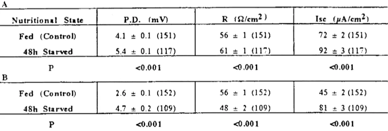
Table 1: Bioelecırie parameters ofmidjejınıum from fed and 48h starVed mice. Data show basal POO. (mY), R (Wern2) and Ise ijlNem2 serosal area) givcn as mean :tSEwith number of animals used in parentheses.
Nutrition.1 State
p.n.
(mV) R (Wem2) Ise (pA/em2)Fed (Control) 2.9 :t 0.1 (53 ) 38 :!: 2 (53) 81 :t 5 (53)
48h Starved 1.0 :t 0.1 (58) 28 4- 1 (58) 3i 4- 5 (58)
P <0.001 <0.001 <0.001
Table 2: Bioeleeırie parameıers of proximal (A) and mid eolon (B) from fed and 48h starved miee. Data show basal P.D. (m Y), R (n/em2) and Ise ijlNern2 serosal area) given as mean :t SE with number of anima1s used in parentheses.
A
NutritionaI State P.D. (mV) R (Q/em2 ) Ise (uA/em2)
Fed (Control) 4.1 :!: 0.1 (151) 56 :t 1 (151) 72 :t 2 (151) 4811 Starved 5.4 4- 0.1 (117) 61 4- 1 (11':') 92 4- 3 (11'7) B P Fed (Control) 48h Starved P <0.001 2.6 :!: 0.1 (152) 4.7 .•. 0.2 (109) <0.001
<o
.00 1 56:!: (152) 48 4- 2 (109) <0.001 <0.001 45 :!: 2 (152) 81 .•. 3 (109) <0.001Table 3: Bioelectrie parameters of distal eolon from fed, 48h and 4d acuıe undemourished (50% normal food intakc) mice. Data show basal P.D.(mY), R (n/ern2) and Ise (IıA/em2 scrosal area) given as mean :tSEwith number of animals used in
parenthcses.
NutritionaI State
p.n.
(mV) R (Wem2) Ise (IlA/em2)Fed (Control) 2.5 :!: 0.2 (90) 1 80 :!: 3 (90) a 33 :t 3 (90) A
48h Starved 6.1 :t 0.4 (92) 2 i9 :!: 2 (92) b 77 :!: 4 (92) B
4d .role U.N. 5.3 .•. 0.3 (80) 3 86 .•. 3 (SO)c 63 4-4 (80)C
p<O.O 1 p<O ,00
ı:
Bvs C
1\"5 2, 1vs3, AvsB, AvsC
node) is approximately 8 cm (8. B:O.2, n=12). In the small intestine, 48h starvation and 4d acute undemutıition caused similar, significant decreases (5%, p<O.02). In the large intestine, 48h starvation and 4d acute undemutrition caused significant. decreased of i 19 (p<O.Oi) and 12% (p<O.OO2)respectivcly.
In vitro bioelectric properties of the small
and large intestine in case of starvation:
Star-vation eaused deereases in the basal P.D. and Ise of the mid jejunum (Table I). The P.D. de-crease for 48h starvation was 66% (p<O.OOl). The deerease in basal Ise was 54% (p<O.OOl) compared to the fed control. in the proximal ile-um, starvation for 48h caused sm all decreases in all values compared fed group but thesc changes were not significant.
In the proximal colon, a significant in-crease 32% (p<O.OOl) in the P.D. occured in 48h starved group relative to the fed control (Table 2A). A significant increase in the tissue R (9%, p<O.OOI) and Isc (28%, p<O.OOI) were seen in the 48h starved group relative to the fed control.
In the mid colon, starvation for 48h gave highly significant increases (p<O.OOI) in both P.D. (81 %) and Isc (80%) while the R de-creased significantly (14%, p<O.OOI) compared to fed group (Tabı e 2b).
In distal colon beside the fed and 48h starved groups, the effccts of undemutıition was alsa investigated. Starvation for 48h and 4d acute undemuıriıİon (50% of food), when
com-(i)-A
EFFECfS OF DIETARY DEPRIVATION ON SMALL AND LARGE INTESTINAL ION TRANSPORT IN THE MOUSE 361
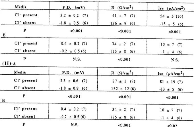
Table 4: Comparison of bioelectrie parameters in CI- present and absent media in mid jejunum (I) and proximaI ileum (ll) offed (A) and 48h starved (B) mice. Data show basal P.D. (mV), R (O/em2) and Ise(J.LAIem2 serosal area) given as mean
:tSEwith number of animals used in parentheses.
Medi.
p.n.
(mV) R m/em2) Ise (pA/cm2)cı-
present 3.2 :!:: 0.2 (7) 61 :!:: 7 (7) 54 :!: S (LO)cl-
absent -1.8 -+- 0.5 (6 ) 136 -+- 9 (6) -15 ~ S (6)P <{I.OOL <O.OOL <{I.OO 1
B
CI- present 0.4 :!:: 0.2 (i) 34 :!: 2 (7) 10 :!:: 7 (7)
ci-
.bsent -0.2 :!: 0.5 (6) 125 ot 8 (6) .1 o+- 4 (6)P N.S. <{I.OOl N.S.
(Li)-A
Medi.
p.n.
(mY) R (Q/emZ) he (pA/cm2)CI- present 2.3 :: 0.6 (7) 27 ::1 (7) 81 :!:: 19 (7 )
ci-
.bsent -1.8 :!: 0.8 (6 ) 152 :: 12 (6) -13 o+- 5 (6)P <{I.OOl <0.001 <{I.OOl
B
cı-
present 0.4 :!: 0.2 (7) 34 :!:"
"- (7) 10 :t 7 (7)cı-
.bsent -0.2 :!:: 0.5 (6) 125 o+- 8 (6) .1 -+- 4 (6)P N.S. <{I.OOL <{I.OZ
pared to fed and 91% compared to the fed con-trol group respectively. The Ise of undemour-ished group was less (18%, p<O.Oi) than of the starved group (Tabı e 3).
Effeet of the
cr
ion on smail and large in-testinal bioeleetrie parameters: Replacement studies of Cı- with glueonate in the mid jeju-num were undertaken in fed and 48h starved mice. Table 4- (I) shows eomparisons betweencı-
present and absent results both in fed (A) and 48h starved (B) animals. In the fed group without CI-, the P.D. and Ise decreased by 156% (p<O.OOl) and 128% (p<O.OOl) but the R value increased by 123% (P<O.OOl) compared to the values with Cı-. In the 48h starved group, in the absenee of Cı- the P.D. and Ise values were reduced but not significantly again R value increased by 268% (P<O.OOl) compared to the results obtained in the presence ofcı-o
In thecı-
free media, the P.D. and Ise polarity re-versed from +ve (serosa) to -ve in both fed and 48h starved groups. Compared to the fed con-trol, 48h starvation gaye an mcrease for the both P.D. (89%, p<O.05 and for the Isc (93%, p<O.05).Replaeement studies of 0- with gluconate were alsa made in proximal ileum from fed and
48h starved micc. Table 4- (II) shows com pari-sons between ci- present and absent both in fed (A) and 48h starved (B) animaIs. In the fed group without
cr,
the polarity of the P.D. and Ise reversed to -ve (scrosa) 178% and 116% (both p<O.OOl). The tissue R rose by 463% (p<O.OOI). After starvation for 48h, the Ise was redueed (91 %, p<O.02) and the tissue R increased withcı-
absence (729%, p<O.OOl) respcetively. In contrast to mid jejunum, in the absence of CI-, with 48h starvation the P.D. and Ise of the proximal ilcum shifted from -ve to +ve of 139% for the P.D. and 138% for the Ise (both p<O.05). A further comparison in fed and starved smaIl intestine was made between mid jejunum and proximal ileum of the values ob-tained in the 0- free media. No signifıcant changes were noted in any of the measure-ments.In the fed proximal eoion (Table5-1 A), re-moval of 0- eaused decreases in P.D. and Ise of 122% (P<O.OOl) and 111% (p<O.OOl) while the R increased by 116% (p<O.OOl). Similarly in the 48h starved group (B), removal of 0- de-creased the P.D. and Ise by 131% and 118% (both p<O.OOl) while the R increased by 77% (p<O.OOl). Removal of 0- in both fed and 48h starved micc produced a reversal of the polarity
362 V. SAÖMANUGtL-R.J.LEVİN
Table 5: Comparison of bioelectrie parameıers in CI- presenl and ahsent media in proximal colon (i) and mid colon (II) of fed (A) and 48h swved (B) mice. Data show basal P.D. (mY), R (Wem2) and IseuıNcm2 serosal area) given as mean:tSE
with number of animals used in parentheses.
Media P.D. (mV) R m/cm2) he (uA/em2)
cı-
present 4.1 :!: 0.3 (lO) 64 :!: 2 (lO) 63 :!: 4 (lO)CI- absent -0.9 .•. 0.4 (9) 138 .•. 7 (9) -7 .•. 3 (9)
(i)-A
P <0.001 <0.001 <0.001
B
CI- present 4.9 :!: 0.3 (lO) 70 :!: 3 (10) 72 :!: 5 (LO)
CI- absent -1.5 :i: 0.3 (9) 124 :i: 4 (9) -13 :i: 3 (9)
P <0.001 <0.001 <0.001
Medi. P.D. (mV) R (Q/cm2) Ise (uA/em2)
CI- present 2.3 :!: 0.3 (21) 63 :!: 3 (2i) 34 :!: 3 (21) CI- .bsent 0.1 .•. 0.1 (9) 198 .•. 9 (9) 0.4 .•. 0.7 (9) (I1)-A P <0.001 <0.001 <0.001 B CI- present 4.1 :!: 0.5 (9) 67 :!: 5 (9) 59 :!: 5 (9)
CI- .bsent 0.3 :!: 0.4 (9) 173 .•. 20 (9) 0.6 :i: 2 (9)
P <0.001 <0.001 <0.001
Table 6: Comparisan of bioelectrie parameıers in CL- presenı and absenı media in distal colon of fed (A), 48h starved CB)
and 4d acute undemourished mice. Data show basal P.D. (mV), R (Wem2) and Ise(IlNcm2 serosal area) given as mean :tSE with number of animals used in parentheses.
A
Merli. P.D. (mV) R (Q/cm2) Ise (uA/em2)
CI- present 2.5 :!: 0.5 (8) 63 :!: 5 (8) 48 :!: 6 (8)
CI- absent 2.8 :i: 0.4 (8 ) 145 .•. 10 (8) 20 .•. 3 (8)
P N.S. <0.001 <0.002
B
CI- present 6.4 :!: 0.7 (8) 68 :t 6 (8) 93 :!: 5 (8)
cı-
.bsent 5.1 :i: 0.5 (8) 129 :i: 10 (8) 39 :i: 3 (8)P N.S. <0.001 <0.001
C
cı-
present 8.4 :!: (7) 80 :!: 4 (7) 105 :!: 12 (7)cı-
.bsent 4.9 .•. (7) 118 .•. 9 (7) 40 .•. 9 ('7)EFFEcrs OF DIETARY DEPRIVATION ON SMAIL AND LARGE INTESTINAL ION TRANSPORT IN THE MOUSE 363
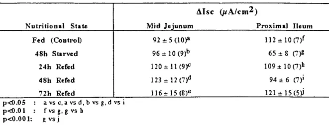
Table 7: Response to serosal beth~hoI (I mM) in mid jejunum and proximal ileum from fed, 48h starved followed by re-feeding for 24h, 48h and 72 h mice. Data show Msc (J.ı.Ncm2 serosal area) given as mean :tSE with number of animals
used in parentheses. Nutritional State Fed (Control) 48h Starved 24h Refed 48h Refed
nh
Rered p<ü.05 : a vs c. a vs d. b vs g. d vs i p<Q.Ol: fvsg.gvsb p<O.OO 1: £ vs.l Mid Jejunum 92 ~ 5 (l0)'l 96~10(9)b 120~11(9f 123:!: 12 (7)d 11 6 ..•.15 (8)e Proximal I1eum 112 ~ 10(7)f 65 ~ 8 (7)1: 109:t10(7)b 94 ~ 6 (7)1 121 ..•.15(5))Table 8: Response to serosal 5- hydroxytryptamine (5-fIT. 50jlM) in presence and absence of serosal atropine (10 jlM) by midjejunum (A) and proximal ileum (B) from fed, 48h starved mice. Data show tUsc (J.ı.Ncm2 serosal area) given as mean
:tSE with number of animals used in parentheses. A ~Isc (pA/cm2) Nutritional State Fed (Control) 48h Stan'ed B Fed (Control) 48h Stuved p<Ü.05 : b vdd, 3 vs 4 P<Q.OL : c vsd, 1 vs 2 5-HT 55:t4 (9)'l SO:!:: 9 (8)b 91:!:: 18 (S)l 40 +9 (9)2 5-HT + AtroDine 72 :!:: 8 (6)C 29:!:4 (6)d 64:!:10 (6)3 37+6 (8)4
of the P.D. and Ise from +ve to -ve. The differ-enees between fed and starved groups in basal parameters (p.D., R, Ise) were not signifieant.
In the mid calan, when CI- was removed the P.D. and Ise feIl signifieantly (p<O.OOl) in the fed group by 96% and 99%, in the 48h starved group by 93% and again 99%. The R on the other hand signifieantly inereased both in the fed group (214%. p<O.OOl) and in the 48h starved group (158%, p<O.OOl) (TabI e 5-II A, B). Comparison between fed and 48h starved mice in the
cr
free media showed no signifi-cant differences in the P.D., R and Ise values.in the fed distal colon, removal of 0- in-creased R by 179% (p<O.OOl) and dein-creased Ise by 58% (p<0.01) (Tabı e 6A). In the 48h starved group, R was inereased by 90% and the Ise was deereased by 58% (P<O.OOl) (Table 6B). With the undemourished group although
there was deerease in P.D. in CI- free media. this was not signifieant. The R inereased by 48% (p<0.OO2) while the Ise deereased by 62% (p<O.OOI) (Table 6C). Unlike the other parts of the colon, removal of CI- in starvation signifi-cantly increased the distal P.D. and the distal Ise by 82% (p<0.01) and 95% (P<O.OOl) com-pared to the fed control group.
Studies on smaIl intestinaljunction in vitro
and in vivo: In mid jejunum, bethanechol
cı
mM) indueed in 48h starved segment an insig-nificant increase in the ~Ise and a siginsig-nificant deerease in 48h starved proximal ile um (42%, p<O.OI) compared to the fed (Table 7). These responses in the mid jejunum and proximal ile-um after 48h starvation were increased by pro-gressive refeeding. In the mid jejunum the in-creases were 30% and 34% (both p<0.05) after 24h and 48h refeeding compared to fed control. In the proximal ileum the se were 68% (P<O.Ol)
364 V. SAÖMANUGll.-R.J.LEVlN
Table 9: Fluid andci-transport in vivo in mıstimulated (basa}) and bcthanechol stimulated (bch, 60 ~g/kg b.Wl.) state by mid jejunum (A) and proximal i1eum (B) from fed and 48h starVed miee. Data given as meantSE with number of animals used in parentheses. Positive values denote gain of luminal fluid and
cr;
negalive values denote loss of fluid andci-
fromlumen.
A
~utriıional Fluid (mglemllS min) Fluid (mg/lOOmg dr}' w) cı- (mmolfL/lS min)
Slate Basal +Beh Basal +Bdt Basal +Bch
+21:t2(Bf +15-+-2 (10)4 Fed +0.3:t1.6 (23)1 48h Stuved .9-3 (19)1 P<O.05 1vs 2,a vs b p<O.001 1vs 3, 2 vs 4, a vs c, b vs d B Fed .7:;:3 (20)1 +12:t2 (13f 4Sh Starved -4:;:3 (1S)1 .•.S:t1 (10)4 P<O.O i 2vs 4 p<O.001: 1vs 3, a vs c, b vs d +5:t15 (23)a +216:t22 (13)C .2.2:,:1.4 (23Y\ .•.3.2:t1.6 (13)C .136=19 (l9)b +251~6 (LO)d .2.8=:1.6 (19)13 .2.6:::1.4 (LO)D .76:t39 (20)a +148:,:22 (L3)C .3=2 (20,-\ .3:t1 (B)C .S9:t50 (8)b +179:t34 (LO)d +0.3 .•.1.6 (1s)B .3 .•.1 (10)0
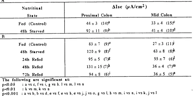
Table 10: Response to serosal bethanechol (I mM) by proximal and mid rolon from fed and 48h starVed (A) and a1so from fed and 48h starved followed by refceding for 24h, 48h andnh(B) mice. Data show tolse (IıNcm2 serosal area) givcn as
mean:tSE with number of animals used in parcntheses.
A
Nutritinal ~Isc (jJA/cm2 )
State Proximal Colon l\1id Colon
Fed (Control) 44 :!:3 (14jl 33:= 4 (15)'
.:I8h St:ırved 92 .•. ı ı (9'P 4 ı-+-4 (lofl
Fed (Conlrol) 83 :t '7 (9)e 27 :d (1 ı ~
48h Starved 120 :!: 9 (s,r 63 :!:8 (8;'
24h Refed 95 :!: 5 (7~ 55 :!: 7 (6~
48h Refed Bl:!: 15 (i~ 36:t 4 (7)m
nh Refed 94 .•. S (6)1 36 .•. 5 (5)11
The foııo~ing are significant at:
p<O.05 : a VS C, fvs i,g vs h.1 vs m, i vs n
p<O.Ol : k vs m, k vs n
p<O.OOI : a vs b, b vsd,evs f, evs h,e vsj,j vs 0, g vs I,h vs m, i vs n, ivs k,j vs i
and 86% (p<O.OOI) by 24h and nh refeeding compared to the 48h starved group respecıively.
in mid jejunum (Tabı e 8A), serasal 5-HT (50JlM) induced incrcases in Lllsc in the fed and 48h starved groups but they were not signifi-cantIy different. In the presence of serosa! atro-pine (ıoJllvf) , in the 48h starved group, the 5-HT-induced Ise was significant1y less (60%, p<O.OOI) than that in the fed group, In proximal ileum (Table 8B), the 5-HT-induced secretion was reduced in the 48h starved groups both in the absence (56%, p<0.01) and presence of aı-ropine (42%, p<0.05) when compared ıo ıhe fed group.
Sıarvaıion for 48h caused decreases in Lilsc induced by serosa! DbcAMP (I mM) in boıh mid jcjunum and proxima! ileum buı these were nal significant comparcd to the fed va!ues.
Role of
cr
in the secretory response of the smail intestine: In the mid jejunum the Lllsc inrespon~ to bethanechol was significant1y re-duced ın the CI- free bicarbonate saline com-parcd to the 0- present contrals by 91 % (p<O.OOI) in the 48h starved groups. SimiIarly in lhe proxima! ileum, removal of 0- from the bathing medium reduced the LlJsc in rcsponse to bethanechol by 95% (p<O.OOl) in the fed and
EFFECfS OF DIETARY DEPRIVATION ON SMALL AND LARGE IJI<'TESTINALION TRANSPORT IN THE MOUSE 365
by 91 % (p<O.OOl) in the 48h starved group. There was no significant differenee in the ~Isc between fed and 48h starved groups in 0- free medium either in the mid jejunum or the proxi-mal ileum.
Fluid and ion tranport in the smail intes-tine: Measurement of jejunaI fluid movement in vivo showed that (Table 9A), the basaI tone in the fed intestine was neutml (O.3J;L.6) and the 48h starvation indueed a significant increase in the basal absorption of fluid both per cm and per 100 mg dry weight. Bethanechol caused a net secretion in both fed and starved groups this was significant1y different (both same on a 1ength and dry weight basis, p<O.OOl) from fed and starved basaI states, unstimulated, but not significant1y different from each other whether placed on a length basis or on a dry weight ba-sis. Basal and bethanechol-induced final 0-concentration values were not significant1y dif-ferent from each other and also from zero.
Fluid and CI- transport in the ileum are also shown in Table 9B. Basal absorptİve tone in the fed and starved basal states of the ileum was significant1y reversed by bethanechol to the secretory tone in the fed (both length and dry weight bases, p<O.OOl) and 48h starved groups (on a length basis, p<O.OI; on a dry weight basis, P<O.OOl) but there was no signifi-cant differenee between them either on the length or weight basis of expressing the results. Basal final 0- conccntrations in the fed and in the 48h starved groups were not significant1y different from each other and also from zero. There was the same amount of bethanechol-indueed CI- absorptİon in fed and 48h starved groups.
Studies on large intestinaljunction in vitro and in vivo: The response to secretory
stimula-tion was examined in porximal and mid colon taken from mice in the two main nutntional groups; fed control and 48h starved.
The results (Tablc 10A). show that in the proximal colon, progressiye starvation resulted in a hypersecrctory response to bethanechol (1 MM, serosaI). There was a significant elevation in ~Isc above that of the fed control after 48h starvation (109%, P<O.OOl). in the mid colon, com pa red to the fed control 48h starvation did not cause a significant increase. Comparisan be-tween these two segments of the large intestine showcd that the proximal colon had larger in-creascs in bethanechol induced ~Isc than the mid colon (fed, p<O.05; 48h group, p<O.ool). The response to bethanechol was also examined in proximal and mid colon from mice starved for 48h and in a group refed up to nh. The re-sults (Table IOB) show that starvation for 48h resulted in a hypersecretory response to be-thanechol (1 mM, serosal), significant inereases in ~Isc by 45% (p<O.OOI) in the proximal colon and by 133% (p<O.OOl) in the mid colon com-pared to the fed controls. This ~Isc indueed by bcthanechol in the 48h starved proximal colon was not shown after 24h refeeding, however, in the mid colon, after 24h refeeding the ~Isc in-duced by bethanechol was stiıı significant1y larger (104%, p<O.OOI) than that of the fed group. In the proximal colon after 48h refeed-ing, increased ~Isc was restored to 58% (p<O.OOI) but nh of refeeding retumed the ~Isc to the fed leveL. Proximal colon had sig-nificanlly larger ~Isc induced by bethanechol than mid colon in all nutntional conditions (same for aıı, p+O.OOl). Typical time course re-sponse of the proximal colon to bethanechol (similar observed in the mid colon) shows that adding serosal bethanechol resulted in an in-stantaneous increase in Ise, which reachcd a peak within 2-3 foııowed by a gradual fall back to the basal level after 10-15 min.
The response of distal colon to 1 mM (se-rosal) bethanechol was different from proximal and mid colon. Initially, there was a transient rapid decrease lasting about 15 see which was
Table 11: Biphasie response to 1 mM serosal bethanechol, -AIse (maximal fall below basallevel over 15 see) and +ölsc by distal colon from fed, 48h sta!Ved and 4d aeute undemourished miee. Data show ölse ÜlNem2 serosal area) given as
mean:!:SE with number of animals used in parenthescs.
:!'-iutritional
Sıa te
Fed (Control)
48h Starved
4d Acute V.N.
The foııomng are significant at:
p<O.05 : 1 vs 2, 2 vs 3 p<O.O
ı
:
a vsb, b vs c -ölse -14 ot 4 (19)1 -30 ot 6 (23~ .13:t4 (14~ Betltanechol +Msc 31=3 (26)'l 49=
5 (29)b 31=5 (15fV. SAÖMANUGll.-R.1.LEVlN 366
Table 12: Response to serosal bethanechol (I mM) fırstIy maximal fall below basa! level over 15 (A) and and increase (B) by distal colon from fed, 48h starved and 4d acute undernourished mice in the absence (control) and prence of serosa.! tetro-dotoxin (ITX. 1j.1M) and diphenylamine-2-carboxylic acid (DPC. 25 mM). Data show Msc (JLNcm2 serosal area) gıven as
meaıdSE with number of animals used in parentheses.
A
Nutritional .1is e (pAlem2)
St. te Control TTX DPC
Fed (Control) -17 == 6 (9)a .0.6 == OJ (i)d .3 :!: 2 (6)g
48h St.n'ed -37 == 9 (12)b .0.6 == 0.4 (7)e -8 :!: 3 (6)h 4d Acute U.N. -15 .•. 5 (7)c .3
...
2 (6)f -4 :!: 2 ('7)1 B Fed (Control) 38 :!: 7 (9)1 48h St.noed 59 :!: 7 (12)2 4d Acute U.N. 37 .•. 8 (7)3The foııol'ing are signific.nt at:
p-dl.05 :a vs g,e vs h, 1vs2, 2 vs3,4 vs 8 p-dl.O
ı
:
bvs h,c vsr.
c vsi,3 vs9, 7vs8 p<O.ooı :
a vsd, b vs e,ıvs
4, 1vs 7, 2vs 5, 2vs8 13 == 2 (7)4 20 == 2 (6,? 24 .•. 4 (6)6 7 == 3 (6)7 21 == 5 (6)8ı
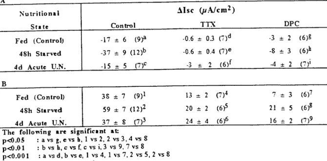
6 .•. 2 (7)9followed by an immediate rise in the Ise and plateaued at least 10 min. The maximum de-creases and inde-creases are shown in Table 1I. The responses to bethanechol indicated that 48h starved distal calan had significantly greater de-crease by 114% and 131% (both, p<0.05) and increase by same 58% (both, P<O.OOL)when compared to the fed and 4d acute undemour-ished groups respectively,
in order to investigate if there was any neural mediatian of the basal Ise and of the de-crease caused by serosal bethanechol, TTX (1 ~ was added serosally after recording the basal Ise. It caused a rapid decrease in Ise espe-cially in the foo d deprived intestine. Mareaver, in the presence of TTX, the bethanechol-induced deereases (-D.Isc) were practically abol-ished in the fed (97%, p<O.OOl), in the 48h starved (98%, p<0.001) and in the 4d acute un-demourished groups (80%, P<O.Ol) compared to those in the control groups (Table l2A). The actian of the CL- channel bloeker DPC (2.5 mM, mucosal) was alsa monitared. DPC caused a rapid fall especially in the foad de-prived intestine and a decrease in the bethane-chol-induced Msc as it was expected but alsa it reduced the -D.lsc caused by bethanechoL. The reductions caused by DPC in the -D.Isc induced by bethancchol were 82% (p<0.5) in the fcd, 78% (p<0.0I) in the 48h starved and 73% (p<0.01) in the 4d acute undemourished groups compared to the control groups. Aıı
thCSCper-centage reductions caused by DPC were less than that caused by TTX but there was only sig-niticant difference between TTX and DPC treated starved groups (P<0.05). TTX and DPC reduced the D.Isc caused by bethanechol (Table 12B). In the presence ofTTX, the bethanechol-induced D.Isc was redueed in the fed group by 66% (p<O.OOI) and in the 48h starved group by 66% (p<O.OOl) compared to the control groups respectively. There was no significant reduction in D.lsc of the 4d acute undemourished group compared to that of the control undemourished group but it was significantly greater than the reduction in D.Isc of TTX treated fed group (85%, p<0.05). In the presence of TTX, the D.Isc induced by bethanechol was the direct ef-fect of bethanechol on ceııs not the neural com-ponent. In the 4d acute undernourished intes-tine, it seemed the neural mechanism did not play big role in the bethanechol-induced secre-tian as it did in fcd and starved groups. in the presencc of DPC, the bethanechol-induced D.Isc was reduced in the fed (83%, p<0.00l) 48h starved (64%, p<0.01) and 4d acute undemour-ished groups (57%, p<O.Ol). In the presence of DPC, there was still a significant increase (200%, p<0.01) in D.Isc caused by bethanechol with 48h starvation compared to the fed group.
Neural involvement in proximal and mid
colonic secretion: Food deprivation (starvation
for 48h) did not change the D.Isc induced by 5-HT in the praximal calan (Table 13). In the
EFFEcrs OF DIETARY DEPRIVATION ON SMALL AND LARGE INTESTINAL IAN TRANSPORT IN TIIE MOUSE 367
Table 13: Response to serosal 5-lIT (5 ~) by proxirnal colon in the absence (conırol and presence of serosal atropine (lO ~). pirenzepine 50~) and hexamethonium (LOO~) from fed and 48h starved mice. Data show ölse u.ı.Ncm2serosal
area) given as mean:l:SE with number of animals used in parentheses.
CoLonie response to cyeLie nucieotide secretory agonists: In the proximal and mid
co-lon from fed and 48h starved mice. the effects of serosal DbcAMP (1 mM) and theophylIine (10 mM), a phosphodiesterase inhibitor, added to both mucosal and serosal solutions to raise the intracellular level of cAMP, were assessed on the Ise. In the case of proximal calan, DbcAMP did not cause a significant increase in the shon circuit current in the starved calan compared to the fed calan. Theophylline treat-ment showed an increase but this was not sig-nificant. In the mid colon, DbcAMP gaye a small increase in the starved (25%), but this was not significant. Theophylline did not change filsc significantly in the starved calan. In distal calan in the presence of serosal hexamethonium (100 JlM) and serosal and mucosal indometha-cin (1 JlM), DbcAMP (1 mM) did not cause any signifıcant increases with food deprivation.
and presence of antagonists, 5-HT induced more secretian (filsc) in the mid calan than in the proximal calan. These significant differenc-es were: in the presence of atropine 153% (p<0.002), pirenzepine 200% (p<O.05) and hex-amethonium 229% (p<0.001) respectively. The same comparison in 48h starved groups indicat-ed that the mid colon again showindicat-ed larger 5-HT-induced secretian in all groups but the only significant filsc was see n in the presence of hexamethonium (184%. p<0.01).
~Isc (pA/cm!)
Aıro ine Pirenze ine He" amethoni um
32::!: 3 (l2 )C 21 :!: 5 (6)e 2 i :!:3 (S)g 64 .•. 6 (14)d 39 .•. S (8)f 32 .•. S C)h Control Siale Nutriıionıl Fed (Control) 39 :!:6 (gr 48h Starved 37 .•.'7 (8)b
The following are significant at:
p<Ü.05 : avse,cvs g,d vsf p<Ü.O
ı
:
a vs g,e vs fp<O.OO 1 : b vsd,c vs d, dvs b
presence of serosal atropine (10 JlM). 5-HT-induced secretian was elevated in 48h starva-tion by 100% (p<O.OOI) compared to the fed group. In the presence of serosal atropine. star-vatian for 48h showed significantly larger re-sponse to 5-HT (73%, p<0.(01) than that in the absence of atropine. although fed response was similar. In the presence of pirenzepine (50 JLM. serosal), 5-HT filsc was significantly reduced (46%. p<0.05) in the fed group but in the 48h starved group it was unaffected, Starvation for 48h caused a significand increased response to serosal 5-HT in the presence of pirenzepine (86%, p<0.01) when compared to the fed group. Similar to pirenzepine. hexamethonium (100 JlM, serosal) signifıcantly reduced the 5-HT-induced secretion in the fed group 46%, p<0.01). but hexamethonium did not give a sig-nificant increase in the 5-HT filsc in the 48h starved group compared to the [ed group. Com-parison of 5-HT-induced filsc with the various antagonists indicated that 5-HT caused more se-cretian in the presence of atropine than pirenze-pine and hexamethonium in both fed and 48h starved groups. In the fed group. atropine insig-nificantly increased the effect of 5-HT by 52% compared to pirenzepine and same by 52% (p<0.05) compared to the hexamethonium re-spectively. In the 48h starved group, these dif-ferences were 64% (p<0.05) compared to piren-zepine and 100% (p<0.00l) compared to hexamethoni um respecti vel y.
in the mid calan, similar studies showed that in the presence of atropine the fed tissue gaye the highest response to 5-HT, but in the 48h starved group, the biggest filsc was ob-tained in the presence of hexamethonium. Al-though in the presence of antagonists 5-HT filsc was greater than in their absence both in fed and 48h starved groups these responses were not significantly different from each other and the control groups.
Comparison between proximal and mid ca-lan in the fed groups showed that in the absence
RoLe of CL- in seeretory response of the co-Lon: In the proximal calan, removal of 0- from bathing medium reduced the bethanechol-induced fiIsc by 80% (p<0.001) in the fed and by 90% (p<0.002) in the 48h starved groups. The significant increase in fiIsc induced by be-thanechol with starvation of 173% (p<0.001) with 0- present was not seen in absence of
cı-.
In the mid calan the absence of 0- caused de-creases in bethanechol induced fiIsc by 91 % (p<O.OOI) in the fed and by 93% (p<0.001) in the 48h starved groups compared to the CI-present controls. In the presence ofcı-,
star-368 V. SAÖMANUGll.-RJ.LEVlN
Table 14: Fluid andCI-transport in vivo in unstimulaıed (basal) and bethanechol stimulaıed (bch, ~g/k:g b.WL) colons in fed and 48h starVed mice. Data given as mean:l:SE with number of animals used in parentheses. Positive values denote gain
of luminal fluid and
cr;
negative values denoıe loss of fluid andcr
from lumen.Nutritional Fluid (mg/cm/ISmin) Fluid (mgllOOmg dry w) C 1- (mmollLllS min)
State Basal +Bch Basal +Bch Basal +Bch
Fed 0:1:2(17)1 +12:1: 2 (10)3 -11:1:14 (17)8 +137:1:20 (10)C -6:1:U(17)A -7:1: 2(lO)C
48h Starved -2 +2 (189 +4 :t 1 (l 0)4 -1"''''2208ıb .•46"'14 (lo)d -3 :i: 2 (l8)B 0.4 + 2 OOP
P<O.OS p<O.Ol p<O.OOI 2 '1154, 3 vs4, C V5cl b V5 cl aV5c
vation for 48h gaye a significant increase of 67% (p<0.05) in the bethanechol-induced illsc compared to the fed group but this was not observed in the absence of 0-. Comparisons between proximal and mid colonic illsc in-duced by bethanechol in the absenee and pres-ence of showed that in the fed groups in CI--free medium, the proximal colon had signifi-cantly larger illsc (+200%, p<0.(01) than the mid colon. In the 48h starved groups the proxi-mal colonic illsc in response to bethanechol was larger than in the mid colonic ilIs c in both
cr
-containing (+119%, p<0.0l) andcr
free medium (+200%, p<0.(01). In the distal colon, the increases in Isc (illsc) caused by behane-chol were also reduced in the 0- -free medium by 85% (p<0.001) in the fed group, by 76% (p<0.00l) in the 48h starved group and by 60% (p<0.05) in the 4 d acute undemourished group.Fluid and ian (Cn transport in the calan:
Fluid and ion (0-) transport were measured in the who le large imestine. No division with three segments could be accomplished because of short lengths involved. The net fluid trans-port was calculated in both per cm imestine and per 100 mg dry weight of intestine in both un-stimulated (basal) and seeretagogue stimulated states. Secretion was indueed by i.p. belhane-chol (601ıg/kg b. wl. in 501ı1 KBS) (TabI e 14). The results indicated that there was no basal fluid transport in either the fed or starved colon but bethanechol-induced secretian (mg/cm) was significantly reduced by 48h starvation (67%, p<O.05) compared to the seeretion in the fed group. The second comparison of fluid trans-port (mg/lOOmg dry. Wl.) confirmed the previ-ous decrease and this time deerease in the be-thanechol-induced secretion was reduced by starvation again significantly 66% (p<0.05) compared to the fed group. In the case of the bethaneehol-stimulation in the fed colon, the fi-nal concentration of 0- was negatiye although an increase of f1uid transport was observed. In
the starved colon also final 0- concemration was not consistem with the f1uid transport in-duced by bethaneehol. One explanation, there are other ion (s) (like HC03-) and in the case of belhanechol-stimulation they come to the lumi-nal fluid, not
cı-o
Discussion
In was shown in this study that dietary dep-rivation (48h starvatian and 4d acute undemu-trition) effeets the basal and stimulated bioelec-tric properties of the mouse sm all and large intestine in vitro and in vivo. Starvation for 48h eaused 22% fall in body weight, the deerease in body weight caused by 4d aeute undemutrition was 26%. The largest deereases (11-13%) dur-ing the 4d acute undemutrition and starvation for 48h happened on the first day of their peri-od. There were also changes in the lengths of mouse imestine after foo d deprivation. These findings are consistem with the starvation in-duced mucosal thinning and reduction in eeil proliferatian alsa lass of body weiht previously reported in several studies (5, 8, 17, 35, 41, 46). lt was alsa reported by Goodlad et al. (16) that after 4d starvation the percentage of the lass in body weight was 23% in the rats which was lost by miee only after Id starvation. That is prob-abIy due to different surface/volume ratio. Mouse is smailer and metaboIism has to fune-tion, especially in the case of starvafune-tion, at high-er level beeause of inhigh-ereased he at loss.
In the ease of starvation, basal electrogenie seeretion of the mid jejunum was significantly reduced but the bethaneehol-stimulated secre-tion was not signifieantly different from the fed control. In the proximal ileum, however, starva-tion did not reduee the basal secrestarva-tion, but be-thaneehol-stimulated secretion was signifieantly redueed. In the fed eondition, basal seeretion of mid jejunum was significant1y larger than that of the proximal ilcum, but not in the starvation.
EFFEcrS OF DIETARY DEPRIVATION ON SMALL AND LARGE INTESTINAL ION TRANSPORT IN TIIE MOUSE 369
Experiments with mouse intestine showed that after starvatian the mid jejunum and proxi-mal ileum did not exhibit a hypersecretory re-sponse to bethanechol and 5-HT. In contrast to the mouse, in rat jejunum and ileum, starvatian for 48h and nh increased the electrogenic secretion induced by bethanechol and 5-HT in
vitro (25, 26, 48, 49). There might be several
reasons for the deereased response to bethane-ehol and 5-HT in ease of starvation by mouse small intestine compared to the rat, apart from just saying "species difference".
in the fed and 48h starved mid jejunum and proximal ileum, atropine produeed no sig-nitkant effeet on the basal Ise. Sheldon et al. (42) alsa reparted that in fed mouse jejunum. atropine did not eause any signifieant change. The 5-HT-induced ~Isc was qualitatively simi-lar to bethanechol-induced ~Ise. Atropine did not black the effeet of 5-HT in the starved prox-imal ileum but interestingly blocked it in the starved mid jejunum. Atropine inhibils the re-sponse to 5-HT in the guinea-pig ileum in vitro in the fed condition (LO). One passibility for this is that in starved mouse mid jejunum and in the guinea-pig ileum, the stimulatin of 5-HT might be via the release of acetylcholine wruch then stimulates secretian by muscarinic recep-tors. In contrast to mouse and guinea-pig ileum, in the rat ileum 5-HT -induced electrogenic se-cretian potentiated by atropine (6). The expla-natian by Beesley and Levin, (6) was prcsence of alacal enteric neural pathway in rat smaIl in-testine, like rat colon (37), and that inhibits electrogenic ian secretion induced by 5-HT and various secretagogues.
in respanse to DbcAMP, starved mid jeju-num and proximal ileum again failed to show any enhaneed secretian compared to fed con-trol. This indicates that hypersecrctory phenom-ena of the mouse small intestine can not be elic-ited by raising the eylie AMP concentrations of the starved enteroeyte. Whereas in rat jejunum and ileum, beside various secretagogues whose action are mediated through cyclic AMP, DbcAMP per se caused significant increases af-ter nh starvation compared to fed condition (29,48,49).
The ion replacement studies suggest that in the fed and starved mid jejunum and proximal ileum, the bethanechol-stimulated Ise eould be aecounted for alma st entirely by the CI- ion. The significant reduction in bethanechol-induced ~Isc seen in the 48h starved proximal ileum in CI- containing saline, was not ob-served in the CI- free media. The major role CL-in rat small CL-intestCL-inal secretian was also shown
by Young and Levin (48, 49). Marcover, in vivo, the final Cı- eoncentration values did not contirın that the bethanechol-induced secretion in both fed and starved jejunum and ileum was carried by Cl-o What are the possible species of ions that eould be the basis of the seeretory mavement observed in vivo? Lack of fluid did not make possible to measure other ions. Possi-bly, in vivo bethaneehol like same secreta-gogues (7, ll, 18) induces secretion by inhibit-ing electroneutral NaO absarption without changing electcogenie Cl-secretion.
The increases in the mid and distal colonic basal Isc after starvation were consistent those in the rat (35), but in rat the proximal colonic basal Ise was redueed after nh starvation al-though it was increased in mouse after 48h star-vation.
The biphasie response was also reported in rat stripped distal colon, but not in unstripped one, by Nzegwu (35). Serosal TTX and mucosal DPC redueed the basal Ise and both the deereas-es and increasdeereas-es indueed by bethanechol. In the presenee of TTX, the increases in Ise indueed by bethaneehol decayed after the maximum had been reaehed unlike the plateaus observed in the absence of TTX. These findings suggested that bethanechol activates electrogenic ion transport in fed and dietary-deprived distal eolon by neu-ral and non-neuneu-ral mechanisms. The initial de-crease in the basal Ise appears to be neurally mediated while the increase in Isc has both non-neural (direct action on colonoeytes?) and neu-ral components. The latter influences not only the maximum response but alsa its duration. The tindings observed in ease of refeeding were similar to those found in rat proximal calan af-ter starvation and following refeeding, only dif-ference increased Ise was not reduced by 24h refeeding (39).
The increased secretian in the starved mouse calan is interesting as this condition causes adecrease in colonic erypt eell produc-tion rate (l8. 40). Crypts are generaııy aceepted as the secretory cells, although there is same ev-idence against the view of compartmentaliza-tion of all absorptive cells to the villus and all secretory eeııs to the crypts (13, 44).
In the ease of foad deprivation, the basal Ise of the distal calan was decreased by various antagonists, this was especially so by TTX, a nerve toxin blocking neural transmission and by DPC, a
cı-
channel blocker. This indicates that there is aneural control mechanism of the basal distal colonic secretion which appears com-po sed mainly of eleetrogenic CI- secretion.370
TIX- induced decreases in the basal Ise of fed rat distal oolon was also reported by Andres et al. (3) and Nobles et al. (33). in their work the magnitude of the decrease was larger but simi-larly. a plateau was reached in less than 5 min.
in the starved proximal colon. atropine potentiated 5-HT-induced increase in Ise and this was significantly larger than that in the ab-sence of atropine. in the starved distal colon. hexamethonium potentiated bethanechol-induced increase in Ise. These potentiated in-creases in the starved eolons were significantly larger than those in the fed tissues. This result suggests that the re may be an ENCAP (enterie neural cholinergie adrenergie pathway) in mouse colon which becomes effective especial-ly in the starved condition. The results appear very similar to the mechanism said previously in the rat colon (35, 36).
The mechanism(s) of the secretory re-sponses induced by the cyclic nucleotide acting agonists during starvation and undemutrition are not known. in the proximal colon the secre-tory responses to the secretagogue action of DbcAMP and of theophylline were generally lower than those produced by the Ca2+ acting agonist bethanechoL. This was also found in rat colon (35, 36).
The reasons for the differences between in
vitro and in vivo results are not known, but
pos-sible explanations are: -) differences in neural and/or hormonal influences on fluid transfer and electrogenic secretion. For example the in
vitro colonie preparation is removed from its normal hormonal and neural interactions, 2) al-though. in the case of starvation, proximal, mid and distal oolon show ed hypersecretion induced by bethanechol above the fed control, using whole oolon in vivo any cause different re-sponse to bethanechol because of different transfer mechanisms in different parts of colon. The final concentration of 0- in the luminal fluid were not signifieantly different in the fed and 48h starved colon in both the basal and bethanechol-stimulated states. Bethaneehol did not inerease the final
cı-
concentration in both fed and starved oolon unlike the luminal fluid volume. It is likely that these discrepancies between fluid volume and 0- eoncentrations are due either to the net movement of other ion-ic species (e.g. Na+, K+, HC3- and H+) or to large amounts of water passing into the lumen.Basal pararneters of electrogenic ion trans-port and the responses to the different secreta-gogues und er fed and food deprived conditions vary from the proximal to the distal end of the
V. SACMANUGll..-R.J.LEvtN
mouse colon (summarized previously). Similar fındings were also reported for rat colon under the fed eondition (32, 33, 34, 50) and under the fed and food deprived conditions (35).
Kaynaklar
i. Aldewaehl, H.S., Wrlght, N.A., appleton. D.R. and Walson. A.J. (1975).The ~ff~cls ofstarwuiorı and r~.
fuding on c~1l populaıion kiMıies in ıhe ral smail bow~1frW.
cos~.J Anal, 119, ıo5.121.
2. Alıman, G.G. and Eneseo, M. (1967).C~llltwmbu as a
m£aswre of dislribWlion and renewal of ~pillı£lial ulis inılı£
smail inıesıine of growing and adulı rals. Am JAnat, 121,
319.336.
3. Andres, H., Boek, R., Brldges. R.J. Rummel, W. and Sehrcincr, J. (1985).SwbfrWCosal pluw and ~Itclro.
Iyıe /ransporı across ral colonic mweosa. J Physiol, 364,
301-312.
4. Armstrong. W.M. (1987).Cellular Mechanisms of ion
ıransporl in ılı£smail inlesıine. In: Physiology of ıhe Gas.
troinıesıinal Tracl, 2nd edn, cd. Johnson, L.R. Ravcn Press, New York.
5. Bcek, LT. and Dinda, P.K. (1973).Sodiıun and waler
Iransporl across ılı£ jejwnwm of fasıed ral. can J Physiol and
Pharmacol, 51, 405-509.
6. Bcesley, A. and Levin, R.J. (1991). 5.
Hydroxırypıamine indwees elecırogenie secr~/ion and sifrWl.
ıaneowsly acıivales a modulaıing inhibilory n~wral circuil in
ral small inlesıine in viıro. Ex Physiol, 76, 607-610.
7. Bcublcr, E., Caupar, LM., HardeasUe, J. and HardeasUe, P.T. (1990). Sıimulaıory efftcls of 5.
hydroxyırypıamine on flwid secreıian and /rallSfrWral polen.
ıial differenc~ in raı smail inlesıine ar~ m£dialed by differenı
recepıor swbıypes J Pharm Pharmaeol, 42, 35-39.
8. Brown, H.O., Levine, M.L. and Llpkln, M. (1963).
Inhibition of inıesıinal epiıhelial cell reMwal and migraıion
induced by starvaıion. Am J Ptıysiol, 205, 868-872.
9. Carey, H.V. and Tueker, K.E. (I99I).lnıesıinal secre.
ıion is allered by ıhe abunce of lıuninal conıenls inpigleıs.
Gasırocnıerol, 100, A68i.
LO. Cooke, H.J. and Carey, H.V. (1985).Pharmacologieal
analysis of 5-Hydroxyırypıamine acliollS on Gwinea.Pig iJeal
mwcosa Eur J Pharmacol, lll, 329-337.
IL. Coupar, I.M .• HardeasUe. J. and HardeasUe, P.T.
(1988). The respolISe of ıhe inlesıinal frWCosa ıo
prostaglan-din E2 during wilhdrawal from morphiM. J Phann Pharma.
eol, 40, 262-266.
12. Debnam, E.S, and Thompson, C.S. (1982).TM ~ffuı
of fasıing on po/~nıial difference across tM brwsh.border
m£mbrane of ~nıerocyıes in rat smail intestw. J Physiol,
355, 449-456.
13. Donowilz. M, and Madara. J. (1983).Eff~cı
ofexıracel-lıdar calciıun depl~ıiorı on epillı£lial slrwellU~ andfıuıclion in
rabiı i/~um: A rnıXUI for s~lecıiv~ crypı or vil/w ~pitlı£lial
ce/I damage and swggesıion of secreıion by vil/w ~pillı£lial
eells. Gastroenterol, 83,1231-1243.
14. Elashoff,
ı.D.
(1981).Dowlt with muJtjp/~ Hesls. Gas-i
' __ rol, '<>.6"-620. ~EFFEcrs OF DIETARY DEPRIVATION ON SMAIl.. AND LARGE INTESTINAL ION TRANSPORT IN THE MOUSE 37i
LS. Glantz, S.A. (1980).Biosıaıis/ia: How to deıeeı, co"ecl
and prevCfIJ e"ors iiııhe~dit:al literatwc. Ciraılation, 61,
ı-s.
16. Goodlad, R.A., Plub, J.A. and Wrlght, N.A. (1988).
EpiıhelilJl ecll proliferaıion and Wuıilıal absorpıiııe /wncıwn
dluilıg sıarvaıwn and ufeeding iiıiM raı.Oin Sci, 74,
301-306.
17. Goodlad, R.A. and Wrlght, N.A. (1984).TM effecu of
sıarvaıwn and refeedilıg on inlulüıal ecll praliferaıilı iiııhe
mouse. Wirchows Ardı [Cell Pathoı),45, 63-73.
18. Kaurman, M.E., Dınno, M.A. and Huang, K.C.
(1980). Effeeı of gllU."agon on wn Iransporl iiımouse iniu.
ıine. Am J Physiol. 238. G491-G494.
19. Levln, R.J. (1967).Teclıniques, ıerminology and parame.
ıers iiıilıleslinal absorpıion. Briı Med Bulleıin. 23. 209.212.
20. Levln, R.J. (1984)./nıesıinal adaptaıion ıo dieıary change
as eumpllfıed by dietary resıricıion sıwi~s. In: Funeıion and
Dysfunetion of the Smail Inıestine, eds. Bau, R.M. and law-rence,T.J.Liverpool Universıy Press.
21. Levln, R.J. (1992).The dia"hoea of famine and seııere
malnwrition- is gllU."agonıhemajor cwpriı? Guı, 33,
432-434.
22 Levln, R.J., Nzegwu, H.C., Perelra, M. and Young, A. (1988).MalnolUis~nl enhances ıhe invilro
electrogenic secrelory response ıo Escherichia coli TSa en.
ıerolozln inral smail and large Wesıine. JPhysiol, 406,S6P.
23. Levin, R.J., lüegwu, H.C. and Young, A. (1987).
Pror.imal colon secreıion infed and fasICd ralS. J Physiol,
396. 33p.
24. Levln, R.J. and Parker, A.J. (1990).Rectal elecırogen.
ic secreıwn-is iı a pUlalive indicaıor of inıcslinal secrelory
stalUS iNiJu:ed by nwriıional deprivaıion in ıheraı.Ex
Physi-01,75, 609-{il
ı.
25. Levln, R.J. and Young, A. (1986).The effeclS of fasıing
upan ral jejwıal secreıwn in ııiıro inııiııo. J Physiol. 378,
23P.
26. Levln, R.J. and Young, A. (1987).Differenıial changes
iiıelecırogenic wn ıransporı associaıcd ..,iıh progessive sıar.
valwn and refeeding inıhe raı smail inluıine. JPhysiol. 396
i49P.
27. Levln, R.J. and Young, A. (1988).NwriıwnaJ level and
ral pror.imal duodı!nal secreıwn inııi/ro and invivo. J
Physi-ol, 403,47P.
28. Levln, R.J. and Young, A.(1990).Pror.imalbUlnoıdis-tal ilell11l shows adapıive responsu lo sıarvaıwn. J Physiol, 420,14SP.
29. Levin, R.J. and You ng, A. (1990).Cyc/ic AMP levels
slinUlJated by secretagogues are greaICr in ileal enlerocyles
isolaıed from starved compared lo fed ralS. J Physiol, 424
8P.
30. Levln, R.J., Wragg, M.S. and Young, A. (1987).
Sıarvaıiorı and iieal secrelwn iniM ral.J Physiol, 386, 63P.
31. Mayhew, T.M. (1988).A geo~ıric model for ulimaling
ııiilOllS swfaec area in rat smail bowel is jusllfıed by
WI-biased esıimaıes obtained using verıical secıions. JAnaı,
161,187-193.
32. Mezorr, A.G., GI.nnella, R.A., Eade, M.N. and Cohen, M.B. (1992).Eschericlıia cali enlerolor.iIı (STa)
binds lo receplors, sıimwates gumıyl eyewc, and impairs
ab-sorpıwn iiıral calan. Gaslroenterol, 102,816-822.
33. Nobles, M., Diener, M., Mestres, P. and Rummel, W. (1991).Segmental heıerogeneily of iM ral calon in ıhe
respense lo acıivalors of secreıion on ıJU cAMP-IM cGMP.
and ıheCa2+ paıhway. Aela Physiol Scaııd, 142,375-386.
34. Nobles, M., Dlener, M. and rummel, W. (1990).
Mechanisms of acıian of ıhe heat.sıable enlerOlar.iIı of E. coli
inrat calan. Gasırocnıerol, 28, 431.
35. I"zcgwu, H.C. (1990).The effeclS ofnwriıional leııel on
inlCSlüıal fwncıion. PhD Thesis. University of Sheffield.
36. I"zegwu, H.A. and Leving, R.J. (1990).Dielary re.
sıricıion increases secreıion in ral distal colon induced l7y se.
creıagoglU!S acıing via calcium bUlnOl by cAMP. Prog NUlr
Sac, 49, 178A.
37. Nzegwu, H.C. and Levln, R.J. (1990). Anenleric
neu-ral 17WScarinic-adrenergic paıhway (ENCAP) ıhaı inhibiıs se.
rOlonin elecırogenic ian secreıion in raı proDmaI, mid and
disıal Co/Oflinviıro. J Physiol, 430, 18P.
38. I"zegwu, H.C., Perelra, M.C., Warren, M.A., Le-vin, R.J. and Young, A. (1988).Acwe and chronic Wl.
derfWlrilwn and ıheir effeclS on ral colomc secreıion. proc
NuırSoc,47,I64A.
39. Nzegwu, H.C., Young, A. and Levin, R.J. (1987).
EffeclS of sıarvaıion and refeeding on elecırogemc ion
Irans-port inıhe ral colofl; a model for famine diarrhoea. Guı, 28,
A1395-AI396.
40. Pereira, M.M.C., Young, A., Warren, M.A. and Levin, R.J. (1988).Chronic malnwriıion inraı induces a
hypersecreıory response lo cholinergic sıinudaıwn in Ihe
smail inıesıine. Guı, 29, A714-A7IS.
41. Rothman, D., UdaJl, J.I"., Pang, K.Y., Kırkham, S.E. and Walker, W.A. (1985).The effeeı ofshorı.ıerm
starvaıwn on mucosal barrier funcıwn in ıhenewborn rabbi/o
Pediaırk Res, 19,727.73i.
42. Sheldon, R.J., Malarchik, M.E., Fox, D.A. Burks, T.F. and Porreca, F. (1989).PJıarmacological
characler-iıaıion of f1elUal mechanisms regulating f1Wi:osal wn
ırans-parI inmouse jejWlum J Pharmaeol Ex Therap,249,572.582.
43. Sleward, M.C. and Case, M. (1989).Principles of iofl
and waıer ıransport across epiıhelias. In: Gasırointesıinal
Se-erction, ed. Davison, J.S. Wrighı, London.
44. Slewart, C.P. and Turnberg, L.A. (1989).A mlcroelec.
Irade sıudy of responses lo secreıogoguu by epiıhelial cells
on wiilus and crypı of raı sma// inlesıine. Am J Physiol. 257,
G334-G343.
45. Strombeck, D.R. (1972).The producıwn ofinlcsıüıalfluid
by cholera 10Dninıhe ral. Prov soc Exp Biol Med,
140,297-303.
46. Sun, T.P. (1927).HisıophysiologU:al sıudy of ıhe epiıhelial
changes inıhe sma// inlesıine ofıheAlbino Mouse af/er slar.
vaıwn and refeeding. The Analomical Record, 34, 341.349.
47. Theodorsson-Nordhelm, E. (1986).Kruslrııl-Wa//is lest:
Basic compwer program lo perform nonparameırit: one-way
analysis of variance and mulıiple comparisons on rlJllks of in.
372
48. Young, A. and Levln, R.J. (1990).Di4rrhoea offatTUM
and mallıutriliOll: 11IvesligaıiollS usiııg a ral model: I.
Jeju-Mi hy~rsecıioıı induced uySllJTYlJıiolı.Gut, 31. 43.53.
49. Young, A. and Levln, R.J. (1990).Diarrhoea offamiııe
and malllUlriliolı: 11IvesıigaliollS usiııg a ral model: I. Ilea/
hy~rsecreıioıı induced uyslarwııioıı. Gut, 3 I. 162.169.
50. Young, A. and Levln, R.J. (1991).Segmelital heıeroge.
lUiiy of ral colollU: elecırogellic secrelioıı iııresponse lo ıhe
bacıerial eıııeroıoıciıı E. co/i STa iıı vilro. Ex Physiol. 76.
979.982.
V. SAÖMANUGll..R.J.LEVlN
SI. Young, A., Nzegwu, H.C. and Levln, R.J. (1988).
The effects of resıricıed food ilııaU 011iıııesıiııal secrelioıı iıı
ıhe ral small illiesıiM. Proc Nutr Soc. 47.127.
52. Young, A., Warren, M.A., Perelra, M.M.C. and Levin, R.J. (1988).Hypersecreıioıı associaled wilh ıhe ac.
ıioıı of Escherichio coli STa elııerOlOıciıı on jejU1lum and ile.
um from sıaryed and chronica/ly wıtUrllOurished rals. Med