Comparison of minimally invasive plate versus intramedullary nailing
in the treatment of distal tibial metaphyseal fractures
Ferhat Say, Erkan İnkaya, Deniz Gürler, Murat BülBül, Emre Gültaç
From Department of Orthopaedics and Traumatology, Faculty of Medicine, Muğla Sıtkı Koçman University, Training and Research Hospital, Muğla, Turkey
n Ferhat Say1, MD. n Erkan İnkaya2, MD. n Deniz Gürler2, MD n Murat Bülbül3, MD. n Emre Gültaç4.
1Department of Orthopaedics and Traumatology, Faculty of Medicine, Ondokuz Mayıs University, Samsun, Turkey. 2Department of Orthopaedics and Traumatology, Samsun Training and Research Hospital, Samsun, Turkey.
3Department of Orthopaedics and Traumatology, Faculty of Medicine, Medipol University, Istanbul, Turkey.
4Department of Orthopaedics and Traumatology, Faculty of Medicine, Muğla Sıtkı Koçman University, Training and Research Hospital, Muğla, Turkey.
Correspondence : Muğla Sıtkı Koçman Üniversitesi Tıp Fakültesi Hastanesi, Ortopedi ve Travmatoloji Anabilim Dalı, Menteşe, Muğla, Turkey. Tel.: +90 5316016787
E-mail : [email protected] © 2020, Acta Orthopaedica Belgica.
With conventional open reduction and plate-screw internal fixation in the surgical treatment of distal tibia metaphyseal fractures, as the soft tissue coverage on the tibia anteromedial surface is slight, there is an increased risk of wound complications and infection (4). With minimally invasive plate osteosynthesis (MIPO), union is achieved with stable fracture fixation, and protecting the periosteal circulation, irrespective of soft tissue coverage (6).
Intramedullary nailing in the treatment of distal tibia fractures provides the advantages of being able to be applied in a minimally invasive method and can provide biological, stable fixation (14). Treatment was made by locked anatomic tibial plate
with MIPO technique on 17 patients (Group1), closed intramedullary nailing on 18 patients (Group2). The cases were evaluated with the AFAS score.
The median operation duration was 90(59-139)mins in Group1, 77.5(59-145)mins in Group2. The median union time was 15(11-20)weeks in Group1, 18(12-22)weeks in Group2. The median AFAS score was 84(47-90) in Group1, 82(65-90) in Group2. The most common complication was infection in Group1 and malunion in Group2. Only the difference in operation time was found to be statistically significant between the two groups
The clinical and functional results of both treatment methods were similar and effective. Malalignment should be kept in mind when treating with intramedullary nail; infection when treating with MIPO.
Keywords : Fracture ; distal tibia ; intramedullary nailing ; minimally invasive plate osteosynthesis.
INTRODUCTION
Fractures of the tibia are frequently seen fractures. Anatomically, as there is scant soft tissue coverage surrounding the bone, and because the metaphyseal region bone structure is more circular and wide than the triangular bone structure of the diaphyseal region, the treatment of distal tibia metaphyseal fractures is more complicated than that of diaphyseal fractures (12).
However, reduction is achieved and maintained in the most difficult direction with treatment with intramedullary nailing (12). It is possible to narrow
the canal diameter with polar screws or obtain a more stable fixation in different axes with multiple distal locking (7).
There are few studies in literature which have compared intramedullary nailing and MIPO in distal tibia metaphyseal fractures (12-17). Both
methods have advantages and disadvantages and there is no consensus on the subject of which method should be chosen for the treatment of distal tibia fractures.
The aim of this study was to compare the clinical results and complications of cases treated with intramedullary nailing or MIPO for a diagnosis of distal tibia fracture.
METHODS
A retrospective evaluation was made of patients operated on with a diagnosis of distal tibia metaphyseal fracture (Orthopaedic Trauma Association classification 43-A9) between January 2012 and August 2013. A total of 35 patients were included who were operated on for a diagnosis of distal tibia fracture between 4 and 10 cm from the tibial joint surface. Patients were excluded with open fractures, pathological fractures, multiple injuries, vascular or nerve injuries and those determined on tomography examination to have fractures extending to the ankle joint. The demographic data of the patients are given in Table 1.
The MIPO group (Group 1) comprised 17 patients. In the surgical technique, after closed reduction of the fracture line under fluoroscopy, temporary fixation was obtained with Kirschner wires (K-wire) or percutaneous reduction clamps. With small incisions, a locking anatomic distal tibia plate was placed over the periosteum and fixation was achieved with locking screws under fluoroscopy.
The intramedullary nailing group (Group 2) comprised 18 patients. In the surgical technique, after closed reduction under fluoroscopy, reamed tibial nails were applied. In 3 patients, block screw was applied and in all patients, locking was applied
with 2 proximal static screws. In 14 patients, 3 distal screws were used and in 4 patients, 4 screws.
In 10 patients (28%) a fibula fracture in the distal third was determined. Plate-screw was used for fixation in 4 patients and intramedullary K-wire in 6 patients.
In both groups, ankle and knee movements were started as soon as tolerated. The patients were followed up clinically and radiologically at 4-week intervals. The decision for partial and full weight-bearing was made according to the union status clinically and radiologically. Union was accepted as the visualisation of callus tissue on three of four cortices on routine anterior posterior and lateral radiographs. Patients with no signs of union at 6 months were accepted as non-union. Malunion was accepted as more than 5° angulation in any plane or more than 1cm shortness (2). Complications were
evaluated in both groups.
The duration of operations was determined from the hospital computer records and time to union and complications from the patient records.
At the final follow-up examination, clinical evaluation was made with the American Foot and Ankle Score (AFAS)5. To obtain the AFAS score, patients were questioned and examined
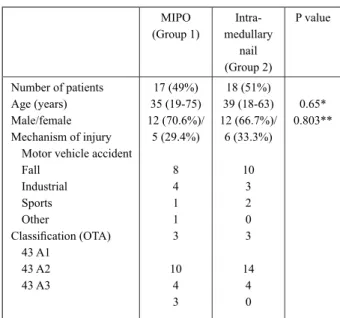
Table 1. – Patient’s demographics.
MIPO (Group 1) Intra-medullary nail (Group 2) P value Number of patients Age (years) Male/female Mechanism of injury
Motor vehicle accident Fall Industrial Sports Other Classification (OTA) 43 A1 43 A2 43 A3 17 (49%) 35 (19-75) 12 (70.6%)/ 5 (29.4%) 8 4 1 1 3 10 4 3 18 (51%) 39 (18-63) 12 (66.7%)/ 6 (33.3%) 10 3 2 0 3 14 4 0 0.65* 0.803**
*Independent Samples test ; **Chi-Square test ; OTA : Orthopaedic Trauma Association.
in respect of pain, function, maximum walking distance, walking surface, gait impairment, sagittal movement, hindfoot movement, alignment and ankle-hindfoot stability.
Statistical Evaluation
In the data evaluation, the SPSS (Windows version 16.0) software program was used. Results were stated as median (minimum-maximum). In the statistical comparison of values between groups, the Independent Sample t-test, the Mann Whitney U-test and the Chi-square test were used. A value of P < 0.05 was accepted as statistically significant.
RESULTS
The median follow-up period was determined as 11 months (range, 6-17 months) for the whole group and as 11 months (range, 8-16 months) in Group 1 and 10 months (range, 6-17 months) in Group 2 (Table 2).
Duration of surgery was determined as 90 mins (range 59-139 mins) in Group 1 and 77.5 mins (range, 59-145 mins) in Group 2.
Time to union was determined as 15 weeks (range, 11-20 weeks) in Group 1 and 18 weeks (range, 12-22) in Group 2.
Complications were determined in 9 patients (52.9%) in Group 1 and 9 patients (50%) in Group 2.
Superficial infection was determined in a total of 6 patients, 4 (23.5%) in Group 1 and 2 (11.1%) in
Group 2. All fully recovered with antibiotic therapy and wound care.
Implant irritation was determined in 2 patients (11.8%) in Group 1 and anterior knee pain in 3 patients (16.7%) in Group 2.
In Group 1, 2 patients (11.8%) and in Group 2, 4 patients (22.2%) were evaluated with malunion. At 13 months postoperatively, iliac wing autograft was applied to 1 patient in Group 1 for a diagnosis of non-union. Union was achieved in the third month after grafting.
In the whole study sample, the most common complication was infection. When the groups were evaluated separately, the most common complication in Group 1 was infection and in Group 2 it was malunion.
The mean AFAS scores were determined as 84 (range, 47-90) in Group 1 and 82 (range, 65-90) in Group 2.
In the statistical evaluation of the data of the two groups, the difference between the groups was determined as statistically significant only in respect of the duration of the operation (P = 0.04).
DISCUSSION
In the choice of surgical treatment for distal tibia metaphyseal fractures, the indicating factors are the fracture configuration, fracture displacement, bone quality, extent of soft tissue damage, the surgeon’s experience and adequate equipment. Whichever
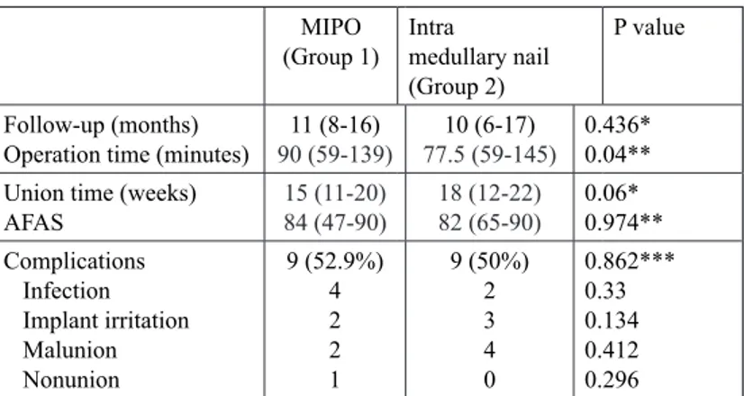
Table 2. – Patient’s outcomes and complications.
MIPO
(Group 1) Intramedullary nail
(Group 2)
P value Follow-up (months)
Operation time (minutes) 90 (59-139)11 (8-16) 77.5 (59-145)10 (6-17) 0.436*0.04** Union time (weeks)
AFAS 84 (47-90)15 (11-20) 18 (12-22)82 (65-90) 0.06*0.974** Complications Infection Implant irritation Malunion Nonunion 9 (52.9%) 4 2 2 1 9 (50%) 2 3 4 0 0.862*** 0.33 0.134 0.412 0.296
*Independent Samples test ; **Mann-Whitney test ; ***Chi-Square test ; AFAS : *American Foot &Ankle Score
method is preferred by the surgeon, by adhering to the basic principles of orthopaedic trauma surgery, fracture healing will be achieved which protects the soft tissue, does not impair the blood supply of the fracture and provides stable fixation. Nowadays intramedullary nailing and MIPO are two frequently used treatment choices for these fractures. This study was designed to compare the treatment results of patients to whom intramedullary nailing or MIPO had been applied for distal tibia metaphyseal fractures. The clinical and functional results of both methods were found to be similar in this study.
Using indirect reduction techniques in the MIPO method may lengthen operating time (4). In the
current study, the operating time of the MIPO group was determined to be longer. In literature, the time to union with MIPO and intramedullary nailing has been determined as similar (3). When the groups of
the current study were compared in respect of time to union, they were similar, with a slightly longer time determined in the intramedullary nailing group. The AFAS scores for the functional evaluation of the patients were consistent with literature and similar for the two groups (3).
The most frequently seen complications in the current study were infection, malunion and implant irritation, which are all related to the anatomic properties of the distal tibia region. In different studies which have compared MIPO and intramedullary nailing, the MIPO infection rate has been reported as 1-16.7% 10-16,17 and the infection rate for intramedullary nailing has been reported as varying from 0-25%10-16,17. In the current study, the infection rate in the MIPO group was determined as 23% and in the intramedullary nailing group as 11%. All the infections in the MIPO group were superficial infection and no deep infection was encountered in any patient. That the infection rate was high compared to literature can be explained by the inclusion of superficial infections and that this technique was chosen for high energy injuries.
Previous studies have reported malunion rates as 2.8-12.9%15,16,17 for MIPO and 16-29% for intramedullary nailing. In the current study, malunion was determined as 11.8% in the MIPO group and as 22.2% in the intramedullary nailing group. Despite all the advantages of intramedullary nailing, sufficient
stability could not be obtained for fractures in this area. In the results of the current study, although the malunion rate of the intramedullary nailing group was not statistically significant, it was high. The use of polar screws and locking distal screws in different axes increases the stability of tibial fractures (7). The
use of these techniques in the current study could have reduced the malunion rate.
In previous studies, non-union rates have been reported as 2.7%-5.6%15,16,17 for MIPO and as 4-9.8% for intramedullar nailing (15,16,17). In the
current study, non-union was determined at the rate of 5% in the MIPO group and no cases of non-union were determined in the intramedullary nailing group. It has been reported that non-union may be seen more when fibula fixation is applied in tibia fractures (16-17). In the 1 patient with non-union in
the current study, fibula fixation had been applied. However, further studies on this subject are required with greater numbers of patients. The use of a single distal locking screw in treatment with intramedullary nailing for distal tibia fractures has been reported to increase the possibility of non-union (11). In the
current study, distal locking in the intramedullary nailing group was achieved with at least 3 screws. That no cases of non-union were observed in the intramedullary nailing group can be explained by the locking of distal screws in different axes and the use of at least 3 screws.
Fibula fixation in distal tibia fractures is controversial (1). In displaced and fragmented tibia
fractures, fibula fixation provides length and can correct angulated and rotational deformities (1-12).
The likelihood of malunion may be reduced. In the current study, fibula fixation was applied to 28% of the patients.
Implant irritation may be observed often in the MIPO technique due to the scant soft tissue coverage in the distal tibial region (3,8). One of
the most common complications in patients with intramedullary nailing is anterior knee pain (2). In
the current study, implant irritation was determined in 12% of the MIPO group patients and anterior knee pain in 17% of the intramedullary nailing group patients.
Apart from intramedullary nailing and MIPO, other treatment choices for non-displaced stable
6. Krackhardt T, Dilger J, Flesch I et al. Fractures of the
distal tibia treated with closed reduction and minimally invasive plating. Arch Orthop Trauma Surg 2005 ; 125 : 87-94.
7. Krettek C, Stephan C, Schandelmaier P et al. The use
of Poller screws as blocking screws in stabilising tibial fractures treated with small diameter intramedullary nails.
J Bone Joint Surg Br 1999 ; 81 : 963-8.
8. Lau TW, Leung F, Chan CF et al. Wound complication
of minimally invasive plate osteosynthesis in distal tibia fractures. Int Orthop 2008 ; 32 : 697-703.
9. Marsh JL, Slongo TF, Agel J et al. Fracture and
dislocation classification compendium - 2007: Orthopaedic Trauma Association classification, database and outcomes committee. J Orthop Trauma 2007 ; 21 : 1-133.
10. Mauffrey C, McGuinness K, Parsons N et al. A
randomised pilot trial of “locking plate” fixation versus intramedullary nailing for extra-articular fractures of the distal tibia. J Bone Joint Surg Br 2012 ; 94 : 704-8.
11. Mohammed A, Saravanan R, Zammit J et al.
Intramedullary tibial nailing in distal third tibial fractures: distal locking screws and fracture non-union. Int Orthop 2008 ; 32 : 547-9.
12. Newman SD, Mauffrey CP, Krikler S. Distal
metadiaphyseal tibial fractures. Injury 2011 ; 42 : 975-84.
13. Nork SE, Schwartz AK, Agel J et al. Intramedullary
nailing of distal metaphyseal tibial fractures. J Bone Joint
Surg Am 2005 ; 87 : 1213-21.
14. Ruecker AH, Hoffmann M, Rupprecht ME et al. Distal
tibial fractures: intramedullary nailing. Eur J Trauma
Emerg Surg 2009 ; 35 : 520-6.
15. Seyhan M, Unay K, Sener N. Intramedullary nailing
versus percutaneous locked plating of distal extra-articular tibial fractures: a retrospective study. Eur J Orthop Surg
Traumatol 2013 ; 23 : 595-601.
16. Vallier HA, Cureton BA, Patterson BM. Randomized,
prospective comparison of plate versus intramedullary nail fixation for distal tibia shaft fractures. J Orthop Trauma 2011 ; 25 : 736-41.
17. Vallier HA, Le TT, Bedi A. Radiographic and clinical
comparisons of distal tibia shaft fractures (4 to 11 cm proximal to the plafond): plating versus intramedullary nailing. J Orthop Trauma 2008 ; 22 : 307-311.
fractures are plaster cast, open reduction with plate-screw fixation and monolateral or circular external fixator.
Limitations of the current study are that it was retrospective and non-randomised with a relatively low number of patients and short follow-up. Although the operations were performed by three different surgeons, the same techniques were used.
In conclusion, both intramedullary nailing and MIPO can be recommended in the surgical treatment of distal tibia metaphyseal fractures. Both are effective methods, clinically and functionally. In terms of complications, attention must be paid to impaired alignment in treatment with intramedullary nailing and infection in MIPO. Further prospective randomised, multi-centre studies are required to be able to obtain clearer results.
REFERENCES
1. Bedi A, Le TT, Karunakar MA. Surgical treatment of
nonarticular distal tibia fractures. J Am Acad Orthop Surg 2006 ; 14 : 406-16.
2. Court-Brown CM. Fractures of the tibia and fibula.
Rockwood and Green’s Fractures in Adults, 6th edn. Lippincott Williams & Wilkins, Philadelphia 2006 ; 2129. 3. Guo JJ, Tang N, Yang HL et al. A prospective, randomised
trial comparing closed intramedullary nailing with percutaneous plating in the treatment of distal metaphyseal fractures of the tibia. J Bone Joint Surg Br 2010 ; 92 : 984-8.
4. Im GI, Tae SK. Distal metaphyseal fractures of tibia:
a prospective randomized trial of closed reduction and intramedullary nail versus open reduction and plate and screws fixation. J Trauma 2005 ; 59 : 1219-23.
5. Kitaoka HB, Alexander IJ, Adelaar RS et al. Clinical
rating systems for the ankle-hindfoot, midfoot, hallux, and lesser toes. Foot Ankle Int 1994 ; 15 : 349-53.