Beneficial effects of agomelatine in experimental
model of sepsis-related acute kidney injury
Nurşah Başol, M.D.,1 Oytun Erbaş, M.D.,2 Türker Çavuşoğlu, M.D.,3 Ayfer Meral, M.D.,4 Utku Ateş, M.D.5 1Department of Emergency Medicine, Gaziosmanpaşa University Faculty of Medicine, Tokat-Turkey
2Department of Physiology, İstanbul Bilim University Faculty of Medicine, İstanbul-Turkey 3Department of Histology and Embryology, Ege University Faculty of Medicine, İzmir-Turkey
4Department of Biochemistry, Dumlupınar University Evliya Çelebi Training and Research Hospital, Kütahya-Turkey 5Department of Histology and Embryology, İstanbul Bilim University Faculty of Medicine, İstanbul-Turkey
ABSTRACT
BACKGROUND: Sepsis-related acute kidney injury (AKI) is a serious complication of sepsis. Problems persist regarding early diag-nosis and treatment of AKI. The aim of the present study was to evaluate the efficacy of agomelatine, which is primarily known for its positive effects on depressive and anxiety disorders in sepsis-related AKI.
METHODS: Sepsis model was created with cecal ligation puncture (CLP). Rats were separated into 4 groups of 8 each: the control group, the sham-operated group, the CLP+saline group, and the CLP+agomelatine group. Agomelatine was administered intraperitone-ally in doses of 20 mg/kg.
RESULTS: In the agomelatine group, reductions were observed in levels of tumor necrosis factor α (TNF-α), malondialdehyde (MDA), blood urea nitrogen (BUN), and creatinine, as well as in histological kidney scores, compared to the non-treated group. In addition, it was demonstrated that agomelatine treatment had positive effect on sepsis-induced morphological damage to renal and tubular tissues.
CONCLUSION: Agomelatine showed strong efficacy in sepsis-related AKI, demonstrated with histological and biochemical results in an experimental model. It is believed that antioxidant and pro-inflammatory effects of agomelatine are responsible for the improve-ment in kidneys.
Keywords: Acute kidney injury; agomelatine; cecal ligation puncture; sepsis.
complication that increases mortality and is characterized by rapid kidney failure due to micro- and macro-hemodynamic impairment and immune toxicity in kidney tissue cells.[5,6]
Pathophysiology of AKI is not clear, though multifactorial mechanisms containing ischemia–reperfusion injury, direct inflammatory injury, coagulation, endothelial cell dysfunction, and apoptosis are generally considered to be causes.[7]
Agomelatine, N-[2-(7-Methoxy-1-naphthyl)ethyl]acetamide, is specified for the treatment of major depression, with dual effects on sleep problems and depressive disorders.[8] It is a
synthetic drug, an agonist of melatonin receptors (MT1 and MT2) and an antagonist of serotonin receptor (5-HT2C).[9–11]
Oxidative stress, which plays a key role in pathogenesis of sepsis, is a leading cause of organ dysfunction in particular.
[12] Melatonin at high doses is known for its strong
antioxi-dant and anti-inflammatory activity.[13] In several experimental
studies, beneficial effects of melatonin have been demonstrat-ed with sepsis models.[13–16] Although the exact mechanism is
not clear, it is thought that the antioxidant, anti-apoptotic, anti-inflammatory, and immunomodulating effects of melato-Address for correspondence: Nurşah Başol, M.D.
Gaziosmanpaşa Üniversitesi Tıp Fakültesi Hastanesi, Acil Tıp Anabilim Dalı, 60000 Tokat, Turkey
Tel: +90 356 - 212 95 00 / 3418 E-mail: [email protected]
Qucik Response Code Ulus Travma Acil Cerrahi Derg 2016;22(2):121–126
doi: 10.5505/tjtes.2015.29499 Copyright 2016
TJTES
INTRODUCTION
Sepsis is a complex clinical syndrome with high rates of mor-bidity and mortality.[1] According to criteria of the
Ameri-can College of Chest Physicians (ACCP) and the Society of Critical Care Medicine (SCCM), the elementary definition of sepsis is “a complex immune reaction against a microorgan-ism.”[2] Sepsis is associated with multiple organ dysfunctions.[3]
Renal functions are particularly affected in the early stages of sepsis.[4] Acute kidney injury (AKI) is a common and serious
nin are responsible for good effects on sepsis.[14] In addition,
reports have indicated protective effects of melatonin on re-nal functions.[17–19] However, exact pathophysiology of
sepsis-related AKI remains unclear and, in spite of new treatment options, AKI continues to occur with high mortality.[20]
The aim of the present study was to research effects of agomelatine, a melatonin analogue, on sepsis-induced AKI. Cecal ligation puncture (CLP) was performed on rats to cre-ate an animal model of sepsis, and agomelatine effects on renal functions were evaluated with biochemical and histo-pathological testing.
MATERIALS AND METHODS
Animals
Used in the present study were 42 male Sprague Dawley ma-ture albino rats, each weighing 200–220 g. They were fed ad libitum and housed in pairs in steel cages in a temperature-controlled environment (22±2°C) with light/dark cycle of 12 hours each. Protocol was approved by the committee for ani-mal research and the study strictly conformed to the aniani-mal experiment guidelines of the Committee for Human Care.
Drugs
All drugs were freshly prepared. Agomelatine (Valdoxan®,
Servier Laboratories Ltd., Slough, UK) was dissolved in saline. Saline (0.9% NaCl) was used as control solution. All solutions were administered intraperitoneally (IP) in a volume of 1 mL/ kg body weight.
Experimental Design
Rats were separated at random into 2 initial groups, and CLP was performed on 26 rats to induce a sepsis model. Ten rats (7 rats in the CLP+saline group and 3 in the CLP+agomelatine group) died in the first 24 hours after surgical procedure and were excluded from the study. No mortality occurred in the sham-operated group. Study groups were designed as follows: Group 1: Normal (non-operated and orally fed control, n=8) Group 2: Sham-operated (n=8)
Group 3: CLP and 1 ml/kg 0.9 NaCl (saline) IP (n=8) Group 4: CLP and 20 mg/kg agomelatine IP (n=8)
Rats were anesthetized IP with injection of combination 80 mg/kg ketamine hydrochloride (Alfamine®; Alfasan
Interna-tional BV., Woerden, Holland) and 7 mg/kg xylazine hydrochlo-ride (Alfazyne®; Alfasan International BV, Woerden, Holland).
Under aseptic conditions, 3-cm midline laparotomy was per-formed to expose the cecum with the adjoining intestine. The cecum was ligated tightly with a 3.0 silk suture at its base under the ileocecal valve and punctured once with a 22-gauge needle. The cecum was then gently squeezed to extrude a small amount of feces from the perforation site. The cecum
was returned to the peritoneal cavity, and the laparotomy incision was closed with 4-0 polyglactin 910 sutures. Follow-ing surgery, the animals were permitted a period to recover before being placed in their cages. In the sham group, under aseptic conditions, only laparotomy was performed; the ce-cum was neither ligated nor punctured. Rats were consid-ered septic 5 hours after CLP.[21] Treatments were performed
within the first hour of surgical procedure. The study was concluded after 24 hours. The rats were euthanized with an overdose of pentobarbital sodium, and blood samples were collected by cardiac puncture for biochemical analysis.
Determination of BUN and Creatinine Levels
Blood urea nitrogen (BUN) and creatinine concentrations were determined spectrophotometrically, using an auto-mated system of analysis. BUN and creatinine concentrations were expressed as mg/dL.Determination of Plasma TNF-α Levels
Plasma TNF-α levels were measured using commercially avail-able ELISA enzyme-linked immunosorbent assay kit (Quansys Biosciences, Logan, UT, USA). Plasma samples were diluted 1:2, and TNF-α was determined in duplicate, according to manufacturer’s guide. Detection limit for TNF-α assay was <2 pg/mL.
Determination of Lipid Peroxidation
Lipid peroxidation was determined in plasma samples by mea-suring malondialdehyde (MDA) levels as thiobarbituric acid-reactive substances (TBARS).[22] Briefly, trichloroacetic acid
and TBARS reagent were added to the plasma samples, then mixed and incubated at 100°C for 60 minutes. After cool-ing on ice, the samples were centrifuged at 3000 rpm for 20 minutes, and the absorbance of the supernatant was read at 535 nM. MDA levels were expressed as nM, and tetraethoxy-propane was used for calibration.
Histopathological Studies of Kidney
For histological and immunohistochemical studies, all animals were IP anesthetized with 40 mg/kg ketamine and 4 mg/kg xylazine, and were perfused with 200 mL of 4% formaldehyde in 0.1 M phosphate-buffered saline. Formalin-fixed kidney sections (4 μm) were stained with hematoxylin and eosin. All sections were photographed with Olympus C-5050 digital camera mounted on Olympus BX51 microscope.
Morphological evaluation was performed by computerized image analysis system (Image-Pro Express 1.4.5; Media Cy-bernetics, Inc., Rockville, MD, USA) on 10 microscopic fields per section, examined with ×20 magnification by an observer blinded to the study group.
Kidney sections from all groups were evaluated semi-quan-titatively to determine extent of tubular epithelial necrosis,
luminal necrotic debris, tubular dilatation, hemorrhage, and interstitial inflammation, rated as follows: 0-5% = score 0; 6-20% = score 1; 21-40% = score 2; 41-60% = score 3; 61-80% = score 4; and 81-100% = score 5.[23,24]
Statistical Analysis
Data are presented as mean±standard error of the mean (SEM). Data analyses were performed using SPSS software for Windows (version 15.0; SPSS Inc., Chicago, IL, USA). Data were analyzed with non-parametric Mann-Whitney U test, and p values of 0.05 or less were considered statistically significant.
RESULTS
Serum TNF-α, MDA, BUN, and creatinine levels of all groups
are shown in Table 1. MDA is a predictor of lipid peroxi-dation, and in cases of sepsis, high levels indicate oxidative stress. Plasma MDA levels were markedly elevated in the CLP+saline group, compared to the normal and sham-oper-ated groups (p<0.000). A significant decrease was observed in the CLP+agomelatine group, compared to the CLP+saline group (p<0.000). No differences were observed between the normal and sham-operated groups.
TNF-α, which causes harmful effects of inflammation, is a pro-inflammatory cytokine that can be used to determine severity of sepsis as organ dysfunction. In the present study, plasma TNF-α was also found to be significantly higher in the CLP+saline group, compared to the normal and sham-oper-ated groups (p<0.000). A significant decrease in TNF-α was
Table 1. Malondialdehyde, TNF-α, BUN and creatinine levels
Normal group Sham-operated group CLP + saline group CLP + 20 mg/kg agomelatine group
Malondialdehyde (nM) 65.3±3.2 71.2±3.5 218.9±7.5** 212.5±6.5##
TNF-α (pg/mL) 21.07±1.9 22.8±2.14 274.5±7.9** 119.9±5.8##
Plasma BUN content (mg/dL) 24.7±1.2 21.6±1.5 55.01±3.2** 37.8±3.6#
Plasma creatinine content (mg/dL) 0.35±0.02 0.37±0.03 0.79±0.03* 0.56±0.05#
Results are presented as mean ± SEM. *p<0.01, **p<0.000 (different from normal and sham-operated groups), ##p<0.000, #p<0.05 (different from CLP + saline Group).
TNF-α: Tumor necrosis factor alpha; BUN: Blood urea nitrogen; CLP: Cecal ligation and puncture.
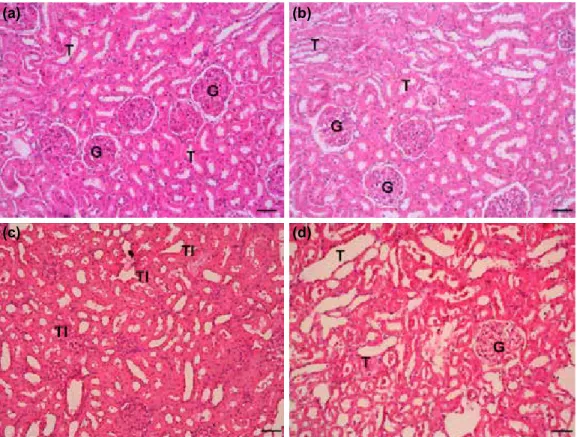
Figure 1. Kidney histolopathology. (a) Kidney from normal group, H&E x10 magnification, renal tubuls (T), (b) sham group showed minimal histopathological alteration, (c) CLP+saline group showed severe
histopatho-logical alteration related to tubular injury (TI). (d) CLP+agomelatine group showed decrease in tubular injury. (a)
(c)
(b)
also observed in the CLP+agomelatine group (p<0.000). No differences were observed between the normal and sham-operated groups.
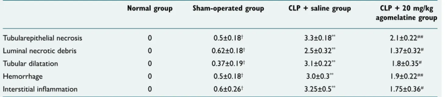
AKI was assessed by measurement of BUN and creatinine lev-els. BUN levels were compared among the 4 groups, with the highest in the CLP+saline group. BUN was significantly lower in the CLP+agomelatine group, compared to the CLP+saline group (p<0.05). Creatinine levels were markedly increased in the CLP+saline group, compared to the normal and sham groups (p<0.01). In the CLP+agomelatine group, a significant decrease was observed in creatinine levels, compared to the CLP+saline group (p<0.05). No difference in BUN or creati-nine levels was observed in the normal or sham groups. Histopathology of kidney tissue in all groups is shown in Fig. 1. Alteration of kidney tissue in sham group was minimal. His-topathological indicators of AKI (tubular epithelial necrosis, luminal necrotic debris, tubular dilatation, hemorrhage, and interstitial inflammation) were observed in the CLP+saline group. Tubular injury was observed in the CLP+agomelatine group, though to a lesser extent than in the CLP+saline group. Mean±SD of kidney tissue tubular epithelial necrosis scores were (0.5±0.18), (3.3±0.18), and (2.1±0.22); scores for luminal necrotic debris were (0.62±0.18), (2.5±0.32), and (1.37±0.32); scores for tubular dilatation were (0.37±0.19), (3.1±0.22), and (1.8±0.35); scores for hemorrhage were (0.5±0.18), (3.0±0.3), and (1.9±0.22), and scores for interstitial inflam-mation were (0.6±0.26), (3.25±0.5), and (1.75±0.36) in the sham, CIP+saline, and CIP+agomelatine groups, respectively (Table 2). A marked increase in tubular epithelial necrosis, lu-minal necrotic debris, tubular dilatation, hemorrhage, and in-terstitial inflammation was observed in the sham group, com-pared to the normal group (p<0.05). Scores of all histological parameters in the CLP+saline group were higher than those in the sham and normal groups (p<0.000). There was a sig-nificant decrease in scores of all histological parameters in the CLP+agomelatine group, compared to the CLP+saline group (p=<0.05 for luminal necrotic debris scores, tubular dilata-tion scores, and interstitial inflammadilata-tion scores; p=<0.01 for tubular epithelial necrosis scores and hemorrhage scores).
DISCUSSION
The curative effect of agomelatine on the kidney was demon-strated in the present study with biochemical and histological parameters in an experimental model of sepsis-induced AKI. AKI occurs in nearly half of all septic patients and is associated with increased mortality.[25] It has been suggested that AKI in
sepsis is correlated with important destructive effects. While detection of acute lung injury is not easy in early stages, the authors suggested that it can indicate severity of sepsis.[26]
Increased levels of BUN and creatinine were used to define AKI. Craciun et al. reported that creatinine is a predictor of glomerular filtration rate, which, though influenced by fac-tors such as gender and age, can be used to detect AKI. It was also reported that BUN can indicate kidney function, but is less specific than creatinine. The authors suggested that high levels of BUN can reflect mortality of sepsis.[27] In the
present study, in addition to BUN and creatinine levels, histo-pathological changes in renal and tubular tissues were used to determine renal injury. Olguner et al. reported that an asso-ciation between high kidney injury scores and sepsis has been histologically demonstrated.[28] For example, the histological
kidney score was found to be higher in the sepsis group than in the sham group of a study by Koca et al. These parameters were also used to evaluate an agent in AKI.[29] In the present
study, histological findings were similarly used in the diagnosis stage, as well as to evaluate effects of agomelatine on AKI. Certain mechanisms are believed to contribute to the patho-genesis of AKI, which has yet to be clearly understood. In the present study, TNF-α levels were higher in the sepsis group than in the normal and sham groups. Luo et al. demonstrat-ed that pro-inflammatory cytokines such as TNF-α increase with sepsis-related AKI. The authors suggested that the in-flammation largely contributed to the pathogenesis of AKI.
[30] Similarly, Chancharoenthana and et al. demonstrated that
hypercytokinemia plays a more distinct role in sepsis-related AKI than in non-sepsis related AKI.[31]
It has been determined that oxidative stress plays an impor-tant role in sepsis-related AKI.[32] The results of the present
Table 2. Changes in histopathological kidney injury scores
Normal group Sham-operated group CLP + saline group CLP + 20 mg/kg agomelatine group
Tubularepithelial necrosis 0 0.5±0.18† 3.3±0.18** 2.1±0.22##
Luminal necrotic debris 0 0.62±0.18† 2.5±0.32** 1.37±0.32#
Tubular dilatation 0 0.37±0.19† 3.1±0.22** 1.8±0.35#
Hemorrhage 0 0.5±0.18† 3.0±0.3** 1.9±0.22##
Interstitial inflammation 0 0.6±0.26† 3.25±0.5** 1.75±0.36#
Results are presented as mean ± SEM. †p<0.05 (different from normal group), **p<0.000 (different from normal and sham-operated groups), #p<0.05, ##p<0.01 (different
study support this conclusion. MDA, the end-product of lipid peroxidation, was higher in the sepsis group than in the nor-mal and sham groups.
TNF-α, MDA, BUN, and creatinine levels were used to evalu-ate the effect of agomelatine on sepsis-induced AKI. Histo-logical investigations and scores to determine kidney injury were used for further assessment. Levels of TNF-α, MDA, BUN, and creatinine were reduced, as were histological kid-ney injury scores, in the AKI group treated with agomelatine, compared to the AKI group that was not. Put simply, de-crease in BUN and creatinine can indicate the positive effect of agomelatine on renal function. This suggestion is support-ed with histopathological kidney investigations.
Agomelatine is a melatonin analogue, known to be more po-tent than melatonin in antidepressant models.[33] The effect of
agomelatine on inflammation was studied by Molteni et al.[34]
Pro-inflammatory cytokines were found to be reduced in rats treated with agomelatine, a finding similar to that of the pres-ent study. The authors reported that agomelatine modified the expression of enzymes involved in the kynurenine path-way. It has been suggested that the kynurenine pathway is aggravated in cases of septic shock.[35] Agomelatine may have
effects beneficial to this pathway, in addition to its antioxidant and pro-inflammatory effects on sepsis-related AKI.
The efficacy of melatonin in sepsis models has been docu-mented.[13,14,16] Shang et al. reported that use of melatonin as
a therapeutic agent in endotoxemic rats decreased incidence of acute lung injury by reducing lipid peroxidation, neutro-phil infiltration, TNF-α release, and IL-10 production. It has been demonstrated that melatonin affects sepsis by reducing inflammation and inhibiting nuclear factor κB (NF-κB) activa-tion.[16] Srinivasan noted that Gitto et al. demonstrated
posi-tive effects of melatonin on oxidaposi-tive stress in newborns with sepsis, utilizing MDA, 4-HDA concentration, and nitrate level. Melatonin was recommended because of its immunomodula-tory, antioxidant, and anti-apoptotic effects on sepsis caused by multiple organ failure.[14] Similarly, in a study by Lowes et
al., melatonin was administered to rats with sepsis induced by lipopolysaccharide/peptidoglycan G (LPS/Pep G) in an acute model, and it was reported that melatonin protected mito-chondria from oxidative stress and inflammation.[13] In
addi-tion to those using sepsis models, other studies have dem-onstrated beneficial effects of melatonin on renal damage as ischemia-reperfusion injury or acute renal failure.[11,18,36]
Agomelatine has generally been studied in relation to depres-sive and anxiety disorders, and, to the best of our knowl-edge, the present study is the first to research its effects on sepsis-related AKI. Agomelatine has effects similar to those of melatonin, due to its melatonergic receptors. Hence, the mechanism responsible for these beneficial effects is generally attributed to the antioxidant and anti-inflammatory effects of melatonin. Though it is outside the scope of the present
study, the authors suspect that agomelatine is more effective than melatonin, due to its impact on the kynurenine pathway in cases of sepsis-related AKI.
Conclusion
Results of the present study indicate that pro-inflammato-ry and antioxidant mechanisms play important roles in the pathogenesis of sepsis-induced AKI. While the mechanism is not entirely clear, the present data suggest that agomelatine may have beneficial effects on kidneys. Further studies are needed before agomelatine can be suggested as treatment of sepsis-related AKI.
Conflict of interest: None declared.
REFERENCES
1. Wang HE, Shapiro NI, Safford MM, Griffin R, Judd S, Rodgers JB, et al. High-sensitivity C-reactive protein and risk of sepsis. PLoS One 2013;8:e69232. CrossRef
2. Hecker A, Uhle F, Schwandner T, Padberg W, Weigand MA. Diagnos-tics, therapy and outcome prediction in abdominal sepsis: current stan-dards and future perspectives. Langenbecks Arch Surg 2014;399:11–22. 3. Bozza FA, D’Avila JC, Ritter C, Sonneville R, Sharshar T, Dal-Pizzol
F. Bioenergetics, mitochondrial dysfunction, and oxidative stress in the pathophysiology of septic encephalopathy. Shock 2013;39 Suppl 1:10–6. 4. Seija M, Baccino C, Nin N, Sánchez-Rodríguez C, Granados R, Ferruelo A, et al. Role of peroxynitrite in sepsis-induced acute kidney injury in an experimental model of sepsis in rats. Shock 2012;38:403–10. CrossRef 5. Mayeux PR, MacMillan-Crow LA. Pharmacological targets in the
re-nal peritubular microenvironment: implications for therapy for sepsis-induced acute kidney injury. Pharmacol Ther 2012;134:139–55. CrossRef 6. Legrand M, Dupuis C, Simon C, Gayat E, Mateo J, Lukaszewicz AC, et
al. Association between systemic hemodynamics and septic acute kidney injury in critically ill patients: a retrospective observational study. Crit Care 2013;17:R278. CrossRef
7. Adembri C, Selmi V, Vitali L, Nosi D, Tani A, Thyrion GD, et al. Expres-sion and characterization of anionic components in the tubulointerstitial compartment of rat kidney during polymicrobial sepsis. Acta Histochem 2014;116:94–105. CrossRef
8. Jakovljević M. Agomelatine as chronopsychopharmaceutics restoring cir-cadian rhythms and enhancing resilience to stress: a wishfull thinking or an innovative strategy for superior management of depression? Psychiatr Danub 2011;23:2–9.
9. Boyce P, Barriball E. Circadian rhythms and depression. Aust Fam Physi-cian 2010;39:307–10.
10. Fornaro M, Prestia D, Colicchio S, Perugi G. A systematic, updated review on the antidepressant agomelatine focusing on its melatonergic modulation. Curr Neuropharmacol 2010;8:287–304. CrossRef
11. Eser D, Baghai TC, Möller HJ. Agomelatine: The evidence for its place in the treatment of depression. Core Evid 2010;4:171–9.
12. Wheeler DS. Oxidative Stress in Critically Ill Children with Sepsis. Open Inflamm J 2011;4:74–81. CrossRef
13. Lowes DA, Webster NR, Murphy MP, Galley HF. Antioxidants that protect mitochondria reduce interleukin-6 and oxidative stress, improve mitochondrial function, and reduce biochemical markers of organ dys-function in a rat model of acute sepsis. Br J Anaesth 2013;110:472–80. 14. Srinivasan V, Pandi-Perumal SR, Spence DW, Kato H, Cardinali DP.
Melatonin in septic shock: some recent concepts. J Crit Care 2010;25:656. e1–6. CrossRef
15. Srinivasan V, Mohamed M, Kato H. Melatonin in bacterial and viral in-fections with focus on sepsis: a review. Recent Pat Endocr Metab Immune Drug Discov 2012;6:30–9. CrossRef
16. Shang Y, Xu SP, Wu Y, Jiang YX, Wu ZY, Yuan SY, et al. Melato-nin reduces acute lung injury in endotoxemic rats. Chin Med J (Engl) 2009;122:1388–93.
17. Farías JG, Zepeda AB, Calaf GM. Melatonin protects the heart, lungs and kidneys from oxidative stress under intermittent hypobaric hypoxia in rats. Biol Res 2012;45:81–5. CrossRef
18. Nava M, Romero F, Quiroz Y, Parra G, Bonet L, Rodríguez-Iturbe B. Melatonin attenuates acute renal failure and oxidative stress induced by mercuric chloride in rats. Am J Physiol Renal Physiol 2000;279:F910–8. 19. Aslaner A, Gunal O, Turgut HT, Celik E, Yildirim U, Demirci RK, et al. Effect of melatonin on kidney cold ischemic preservation injury. Int J Clin Exp Med 2013;6:794–8.
20. Liu B, Chen YX, Yin Q, Zhao YZ, Li CS. Diagnostic value and prognos-tic evaluation of Presepsin for sepsis in an emergency department. Crit Care 2013;17:R244. CrossRef
21. Işeri SO, Sener G, Saglam B, Gedik N, Ercan F, Yegen BC. Oxytocin pro-tects against sepsis-induced multiple organ damage: role of neutrophils. J Surg Res 2005;126:73–81. CrossRef
22. Demougeot C, Marie C, Beley A. Importance of iron location in iron-in-duced hydroxyl radical production by brain slices. Life Sci 2000;67:399– 410. CrossRef
23. Ashrafi F, Nematbakhsh M, Safari T, Talebi A, Nasri H, Khazaei M, et al. A combination of vitamin C and losartan for cisplatin-induced neph-rotoxicity in rats. Iran J Kidney Dis 2012;6:361–5.
24. Eren Z, Coban J, Ekinci ID, Kaspar C, Kantarci G. Evaluation of the ef-fects of a high dose of erythropoietin-beta on early endotoxemia using a rat model. Adv Clin Exp Med 2012;21:321–9.
25. Holthoff JH, Wang Z, Patil NK, Gokden N, Mayeux PR. Rolipram im-proves renal perfusion and function during sepsis in the mouse. J Phar-macol Exp Ther 2013;347:357–64. CrossRef
26. Bhargava R, Altmann CJ, Andres-Hernando A, Webb RG, Okamura K, Yang Y, et al. Acute lung injury and acute kidney injury are established by four hours in experimental sepsis and are improved with pre, but not post, sepsis administration of TNF-α antibodies. PLoS One 2013;8:e79037.
27. Craciun FL, Iskander KN, Chiswick EL, Stepien DM, Henderson JM, Remick DG. Early murine polymicrobial sepsis predominantly causes renal injury. Shock 2014;41:97–103. CrossRef
28. Olguner CG, Koca U, Altekin E, Ergür BU, Duru S, Girgin P, et al. Isch-emic preconditioning attenuates lipid peroxidation and apoptosis in the ce-cal ligation and puncture model of sepsis. Exp Ther Med 2013;5:1581–8. 29. Koca U, Olguner ÇG, Ergür BU, Altekin E, Taşdöğen A, Duru S, et al.
The effects of dexmedetomidine on secondary acute lung and kidney in-juries in the rat model of intra-abdominal sepsis. ScientificWorldJournal 2013;2013:292687. CrossRef
30. Luo CJ, Zhang FJ, Zhang L, Geng YQ, Li QG, Hong Q, et al. Mesenchy-mal stem cells ameliorate sepsis-associated acute kidney injury in mice. Shock 2014;41:123–9. CrossRef
31. Chancharoenthana W, Tiranathanagul K, Srisawat N, Susantitaphong P, Leelahavanichkul A, Praditpornsilpa K, et al. Enhanced vascular endo-thelial growth factor and inflammatory cytokine removal with online he-modiafiltration over high-flux hemodialysis in sepsis-related acute kidney injury patients. Ther Apher Dial 2013;17:557–63. CrossRef
32. Wang Z, Holthoff JH, Seely KA, Pathak E, Spencer HJ 3rd, Gokden N, et al. Development of oxidative stress in the peritubular capillary micro-environment mediates sepsis-induced renal microcirculatory failure and acute kidney injury. Am J Pathol 2012;180:505–16. CrossRef
33. Srinivasan V, Zakaria R, Othman Z, Lauterbach EC, Acuña-Castroviejo D. Agomelatine in depressive disorders: its novel mechanisms of action. J Neuropsychiatry Clin Neurosci 2012;24:290–308. CrossRef
34. Molteni R, Macchi F, Zecchillo C, Dell’agli M, Colombo E, Calabrese F, et al. Modulation of the inflammatory response in rats chronically treated with the antidepressant agomelatine. Eur Neuropsychopharmacol 2013;23:1645–55. CrossRef
35. Zeden JP, Fusch G, Holtfreter B, Schefold JC, Reinke P, Domanska G, et al. Excessive tryptophan catabolism along the kynurenine pathway precedes ongoing sepsis in critically ill patients. Anaesth Intensive Care 2010;38:307–16.
36. Shih YC, Lee PY, Cheng H, Tsai CH, Ma H, Tarng DC. Adipose-derived stem cells exhibit antioxidative and antiapoptotic properties to rescue ischemic acute kidney injury in rats. Plast Reconstr Surg 2013;132:940e–51e. CrossRef
OLGU SUNUMU
Sepsis nedenli akut böbrek hasarında agomelatinin etkilerinin değerlendirilmesi
Dr. Nurşah Başol,1 Dr. Oytun Erbaş,2 Dr. Türker Çavuşoğlu,3 Dr. Ayfer Meral,4 Dr. Utku Ateş51Gaziosmanpaşa Üniversitesi Tıp Fakültesi, Acil Tıp Anabilim Dalı, Tokat 2İstanbul Bilim Üniversitesi Tıp Fakültesi, Fizyoloji Anabilim Dalı, İstanbul 3Ege Üniversitesi Tıp Fakültesi, Histoloji ve Embriyoloji Anabilim Dalı, İzmir
4Dumlupınar Üniversitesi Evliya Çelebi Eğitim ve Araştırma Hastanesi, Biyokimya Anabilim Dalı, Kütahya 5İstanbul Bilim Üniversitesi Tıp Fakültesi, Histoloji ve Embriyoloji Anabilim Dalı, İstanbul
AMAÇ: Sepsis nedenli akut böbrek hasarı sepsisin önemli komplikasyonlarından biridir. Sepsisin erken dönemlerinde gelişir ve mortaliteyi artırır. Hem erken tanı sürecinde hem de tedavi aşamasında hala problemler vardır. Bu çalışmanın amacı, genellikle anksiyete ve depresyonda kullanılan agomelatinin sepsis nedenli akut böbrek hasarındaki olası etkilerinin değerlendirilmesidir.
GEREÇ VE YÖNTEM: Sepsis modeli çekal ligasyon ve delme (CLP) tekniği ile oluşturuldu. Sıçanlar her biri sekizerli dört gruba ayrıldı. İlk grup normal, ikinci grup sham grubu olarak belirlendi. Üçüncü grup sepsis modeli oluşturulup sadece salin verilen grup, dördüncü grup ise sepsis mo-delini takiben agomelatin uygulanan grup olarak belirlendi. Yirmi dört saatin sonunda böbrek doku örnekleri ve kanlar alınarak histopatolojik ve biyokimyasal yöntemlerle analizleri yapıldı.
BULGULAR: Çalışmada agomelatine uygulanan grupta sadece salin uygulanan gruba göre TNF-α, MDA, BUN, kreatin ve histolojik böbrek skor-laması daha düşük olarak bulundu. Aynı zamanda, histopatolojik değerlendirmede agomelatin tedavisinin böbrek dokularında sepsisin oluşturduğu hasarlanmayı düzelttiği görüldü.
TARTIŞMA: Bu çalışmada, agomelatinin sepsis nedenli akut böbrek hasarında faydalı olduğu savunulmaktadır. Bu hem biyokimyasal hem de histo-lojik değerlendirmeler ile gösterilmiştir. Agomelatinin anti-oksidan ve proenflamatuvar etkilerinin böbreklerdeki bu düzelmeden sorumlu olduğu düşünülmektedir.
Anahtar sözcükler: Agomelatin; akut böbrek hasarı; CLP; sepsis.
Ulus Travma Acil Cerrahi Derg 2016;22(2):121–126 doi: 10.5505/tjtes.2015.29499