51
Neutrophil to Lymphocyte and Platelet to Lymphocyte Ratio and Their Relationship with Canadian Cardiovascular Society Angina Classification in
Patients with Chest Pain
Nötrofil Lenfosit ve Platelet Lenfosit Oranlarının Göğüs Ağrısı Olan Hastalarda Kanada Kardiyovasküler Cemiyeti Angina Sınıflaması ile Olan İlişkisi Emrah İpek1, Erhan Yıldırım1, Mahir Cengiz2, Selami Demirelli1, Emrah Ermiş1, Bingül Dilekçi Şahin1,
Mustafa Yolcu3
1Erzurum Regional Training and Research Hospital, Departmentof Cardiology 2Istanbul University Cerrahpasa Medical School, Department of Internal Medicine
3Istanbul Arel University, Medicana Camlica Hospital, Department of Cardiology
Abstract
Objectives: We examined the association between neutrophil to lymphocyte ratio (NLR) and platelet to
lymphocyte ratio (PLR) with the Canadian Cardiovascular Society (CCS) angina grade of patients in outpatient setting.
Materials and Methods: The study population included patients with chest pain who had admitted to
our outpatient clinic. The study group consisted of noncardiac chest pain and atypical angina (group 1), CCS class 1 and class 2 angina (group 2) and CCS class 3 and class 4 (group 3) patients.
Results: The neutrophil lymphocyte ratio was positively correlated with extent of CAD and angina
grade (p<0.001). To test the power of neutrophil lymphocyte ratio for distinguishing group 1 and group 2+3, and group 1+2 and group 3, ROC analysis was performed. The NLR had AUC values of 0.714 and 0.698 (p<0.001), which demonstrate its sufficiency to distinguish patients in group 1 from individuals in group 2+3 and patients in group 1+2 from individuals in group 3, respectively. The platelet lymphocyte ratio did not differ among groups (p>0.05).
Conclusion: The NLR is a cheap and practical inflammatory marker and is associated with CCS angina
grade of patients in outpatient setting.
Key words: Neutrophil/lymphocyte ratio, platelet/lymphocyte ratio, coronary artery disease, CCS
angina classification
Öz
Amaç: Bu çalışmada polikliniğe başvuran ve göğüs ağrısı olan hastalarda nötrofil lenfosit (NLO) ve
platelet lenfosit oranlarının (PLO) Kanada Kardiyovasküler Cemiyeti (KKC) anjina sınıflaması ile ilişkisi incelenmiştir.
Materyal ve Metot: Çalışma hastaları polikliniğimize göğüs ağrısı ile başvuran hastaları içermektedir.
Çalışma grupları, kardiyak olmayan göğüs ağrısı ve atipik anjina (grup 1), KKC sınıf 1 ve sınıf 2 (grup 2), KKC sınıf 3 ve sınıf 4 (grup 3) hastalardan oluşmuştur.
Bulgular: NLO koroner arter hastalığının yaygınlığı ve anjina derecesi ile pozitif korelasyon
göstermiştir. (p<0,001). NLO’nun grup 1’deki hastaları grup 2+3’tekilerden ve grup 1+2’deki hastaları grup 3’deki hastalardan ayırt edebilme gücü ROC analizi ile test edilmiştir. NLO’nun grup 1’i grup 2+3’ten ve grup 1+2’yi grup 3’ten ayırt etmedeki yeterliliğini gösteren eğri altında kalan (EAA) değerleri sırası ile 0,714 ve 0,698 (p<0,001) bulunmuştur. PLO gruplar arasında anlamlı farklılık göstermemiştir (p> 0,05)
Sonuç: NLO, ucuz ve pratik bir enflamatuar belirteç olup poliklinik koşullarında hastaların KKC anjina
dereceleri ile ilişkili bulunuştur.
Anahtar kelimeler: Nötrofil/lenfosit oranı, platelet/lenfosit oranı, koroner arter hastalığı, KKC anjina
sınıflaması
Correspondence/Yazışma Adresi:
Emrah İpek, MD
Erzurum Region Training and Research Hospital, Department of Cardiology, Palandoken, 25050, Erzurum, Turkey
e-mail: [email protected] Date of submission: 28.08.2015 Date of acception: 11.01.2016
52
Introduction
Coronary artery disease (CAD) is a complex multifactorial disease and inflammation plays some important role in the onset and the progression of the disease.1,2 In patients
with CAD, the high levels of inflammatory markers like C-reactive protein and leukocyte count are related to increased risk of cardiac events.3,4 Neutrophil to
lymphocyte ratio (NLR), a systemic inflammation marker, has correlation with cardiac events and mortality in stable and unstable CAD, and acute decompensated heart failure.5,6 There is also an association between elevated platelet counts and
cardiovascular mortality.7,8 As a new marker, platelet to lymphocyte ratio (PLR) was
reported to be able to show the systemic inflammatory burden and have prognostic value combining an individual’s platelet and lymphocyte counts in patients with CAD.9,10 Canadian Cardiovascular Society (CSS) angina classification is used for grading
of severity of angina pectoris in individuals presumed to have coronary artery disease.11,12 In this study we aim to show the relationship between NLR, PLR and CCS
angina grade of patients in the outpatient setting which can be useful in estimation of the risk and severity of CAD.
Materials and Methods
Patient Selection
The study population consisted of 600 consecutive patients who were admitted to our outpatient clinic with the complaint of chest pain between September 2014 and March 2015. All of the patients were evaluated by electrocardiography and transthoracic echocardiography. Patients with atypical angina pectoris within intermediate risk group were assessed by treadmill exercise or myocardial single-photon emission computed tomography. Coronary angiography was performed in 180 patients with positive stress test and those with CCS angina grade of three and four. Exclusion criterias were previous coronary artery bypass grafting, percutaneous coronary intervention, acute coronary syndrome, clinically significant valvular heart disease, ejection fraction lower than 60%, hematological disease, cancer, severe renal or liver disease, ongoing infection or chronic inflammatory disease, and autoimmune disease. Finally, the study population consisted of 320 patients. All participants gave an informed consent and the study was approved by the local ethics committee. Patients’ laboratory and clinical characteristics, such as age, sex, diabetes mellitus (DM), hypertension (HT), hypercholesterolemia, smoking, family history of cardiovascular disease, height, and weight, were accessed through the medical records. In cases of inconsistencies, the patients were contacted through telephone. By dividing weight in kilograms by height in squared meters (kg/m2), the body mass index (BMI) was calculated. Transthoracic echocardiography was performed after physical examination using a system V (Vingmed; GE, Horten, Norway) with a 2.5-MHz phased-array transducer. Recordings were taken in patients positioned in the left lateral decubitus position. The left ventricular ejection fraction (LVEF) was measured using modified Simpson rule. The CAD was defined as the presence of significant stenosis, at least 50% of the vessel diameter, in any of the main coronary arteries, in accordance with the American College of Cardiology/American Heart Association lesion classification.13 The
53
(single-vessel disease), 2 (two-vessel disease), 3 (three-vessel and/or main stem disease) according to the number of main vessels with significant stenosis.
Canadian Cardiovascular Society Grading Scale for Angina Pectoris
Stable angina pectoris (SAP) is defined as discomfort in the chest, jaw, shoulder, back, or arms, typically elicited by exertion or emotional stress, and relieved by rest or nitroglycerin.11 Atypical angina is defined as presence of two of three features of SAP
which includes typical localization, aggravating and alleviating factors. Non cardiac chest pain indicates the presence of only one feature of SAP.14 In patients with SAP, the
symptoms can be further evaluated by CCS Grading Scale.11 CCS angina grading scale
includes classes I, II, III and IV. Each class has a definition as follows. Class I: Ordinary physical activity does not cause angina, no angina occurs when walking or climbing stairs; angina does occur with strenuous or rapid or prolonged exertion at work or recreation. Class II: Slight limitation of ordinary activity: Angina occurs when walking or climbing stairs rapidly; walking uphill; walking or stair-climbing after meals, in the cold, in the wind, under emotional stress, or only during the first few hours after awakening; walking more than two blocks on the level and climbing more than one flight of ordinary stairs at a normal pace and in normal conditions. Class III: Marked limitation of ordinary physical activity: Angina occurs when walking one or two blocks on the level and climbing one flight of stairs in normal conditions and at a normal pace. Class IV: Inability to carry on any physical activity without discomfort: Anginal syndrome may be present at rest.11 The CSS angina score of each patient was calculated
by an experienced cardiologist during outpatient visit. The patients with non-anginal chest pain and atypical angina were also determined and recorded. The patients were allocated as follows: Noncardiac chest pain and atypical angina in group 1, CCS grade 1 and 2 angina in group 2, CCS grade 3 and 4 angina in group 3.
Biochemical Measurements
The blood samples were collected from the antecubital vein by an atraumatic puncture after the transthorasic echocardiography and were sent to the laboratory for analysis within 1 hour after collection. Venous blood is collected in a tube containing K3 EDTA for measurement of hematologic indices in all patients evaluted in the outpatient clinic. Hemoglobin, total white blood cell (WBC), neutrophils, lymphocytes, and monocytes were determined using an automated blood cell counter by a Coulter LH 780 Hematology Analyzer (Beckman Coulter Ireland Inc Mervue, Galway, Ireland). Total low-density lipoprotein and high-density lipoprotein cholesterol, triglycerides, and fasting glucose were measured using the Abbott Architect C16000 auto analyzer (Abbott laboratories, Abbott park, IL, USA).
Statistical Analysis
Statistical analyses were performed using SPSS 20.0 (SPSS Inc., Chicago, IL, USA) for Windows. The normal distribution of data was evaluated by the 1-sample Kolmogorov-Smirnov test and the difference in gender was analyzed by the Chi-square test. All statistical comparisons were performed using the analysis of variance (ANOVA) which was used to compare multiple-group means. The following post hoc evaluation was made by Bonferroni method. All data were expressed as the mean ± standard deviation (SD). Numerical data was tested by Pearson’s correlation and nominal data was tested by Spearman’s correlation. To assess the diagnostic accuracy, we performed receiver
54
operating characteristic (ROC) curve analysis. The area under the curve (AUC) of ROC curve was then estimated. p<0.05 value was considered to be statistically significant. Results
A total of 600 consecutive patients were evaluated and 320 who fulfilled the inclusion criteria were included in the study. The study group consisted of 102 group 1 patients (56 male, 46 female; age 57.9±10.4 years), 126 group 2 patients (72 male, 54 female; age 58.3±8.4 years), and 92 group 3 patients (60 male, 32 female; age 60.7±11.7 years) referred to our outpatient clinic over a six months period from September 2014 to March 2015.
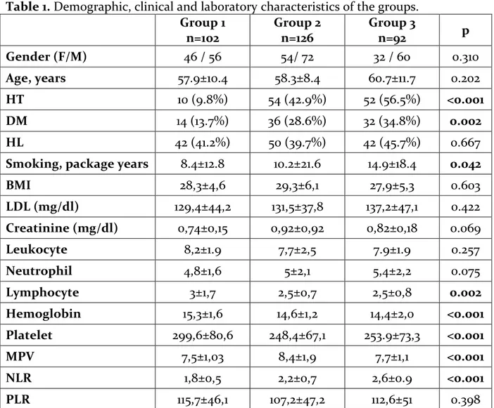
Table 1. Demographic, clinical and laboratory characteristics of the groups. Group 1 n=102 Group 2 n=126 Group 3 n=92 p Gender (F/M) 46 / 56 54/ 72 32 / 60 0.310 Age, years 57.9±10.4 58.3±8.4 60.7±11.7 0.202 HT 10 (9.8%) 54 (42.9%) 52 (56.5%) <0.001 DM 14 (13.7%) 36 (28.6%) 32 (34.8%) 0.002 HL 42 (41.2%) 50 (39.7%) 42 (45.7%) 0.667
Smoking, package years 8.4±12.8 10.2±21.6 14.9±18.4 0.042
BMI 28,3±4,6 29,3±6,1 27,9±5,3 0.603 LDL (mg/dl) 129,4±44,2 131,5±37,8 137,2±47,1 0.422 Creatinine (mg/dl) 0,74±0,15 0,92±0,92 0,82±0,18 0.069 Leukocyte 8,2±1.9 7,7±2,5 7.9±1.9 0.257 Neutrophil 4,8±1,6 5±2,1 5,4±2,2 0.075 Lymphocyte 3±1,7 2,5±0,7 2,5±0,8 0.002 Hemoglobin 15,3±1,6 14,6±1,2 14,4±2,0 <0.001 Platelet 299,6±80,6 248,4±67,1 253.9±73,3 <0.001 MPV 7,5±1,03 8,4±1,9 7,7±1,1 <0.001 NLR 1,8±0,5 2,2±0,7 2,6±0.9 <0.001 PLR 115,7±46,1 107,2±47,2 112,6±51 0.398
HT: Hypertension, DM: Diabetes Mellitus, HL: Hyperlipidemia, BMI: Body Mass Index, LDL: Low Density Lipoprotein, MPV: Mean Platelet Volume, NLR: Neurophil to Lymphocyte Ratio, PLR: Platelet to Lymphocyte Ratio
The demographic data and laboratory parameters were summarized in Table 1. The gender distribution did not differ among groups (p>0.05). There were no significant differences in the creatinine and low density lipoprotein (LDL) levels, leukocyte and neutrophil counts between the groups (p>0.05). The smoking rate was significantly
55
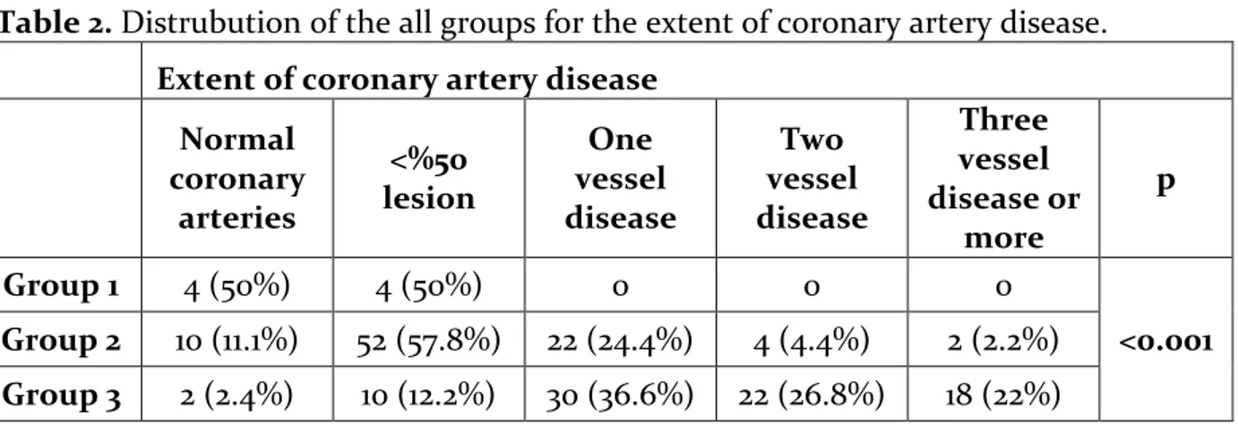
higher in group 3 (p=0.042). Hemoglobin, lymphocyte and platelet counts were significantly higher in group 1 (p<0.001, p=0.002 and p<0.001, respectively). MPV was significantly higher in group 2 (p<0.001). The NLR was significantly higher in group 3 (p<0.001). The PLR did not differ among groups (p>0.05). The extent of CAD, which was defined as total number of affected coronary arteries, was detected to increase significantly from group 1 to group 3 (p<0.001) (Table 2).
Table 2. Distrubution of the all groups for the extent of coronary artery disease. Extent of coronary artery disease
Normal coronary arteries <%50 lesion One vessel disease Two vessel disease Three vessel disease or more p Group 1 4 (50%) 4 (50%) 0 0 0 <0.001 Group 2 10 (11.1%) 52 (57.8%) 22 (24.4%) 4 (4.4%) 2 (2.2%) Group 3 2 (2.4%) 10 (12.2%) 30 (36.6%) 22 (26.8%) 18 (22%)
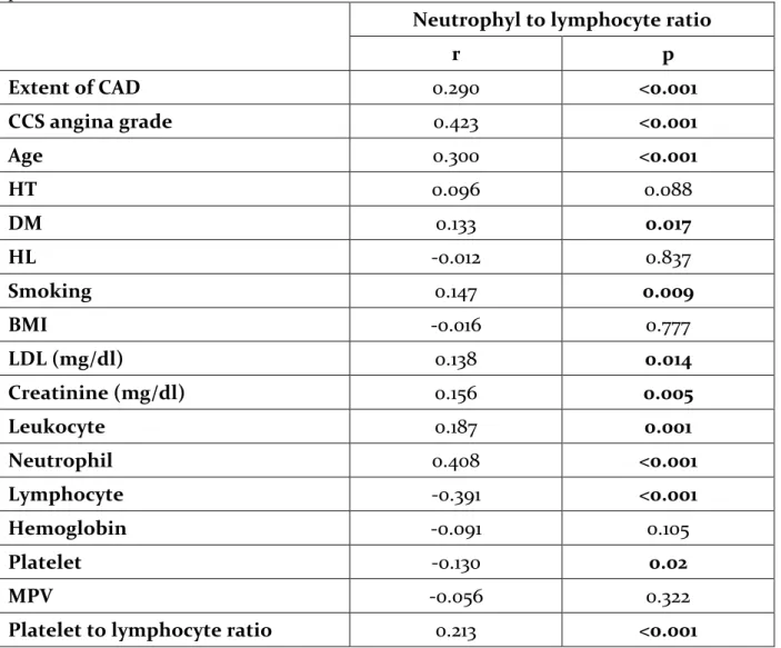
The results of the correlation among parameters with NLR are presented in Table 3. The NLR was positively correlated with extent of CAD, angina grade, diabetes mellitus, age, smoking, creatinine, leukocyte, neutrophil, LDL and PLR. The NLR was negatively correlated with platelet and lymphocyte numbers.
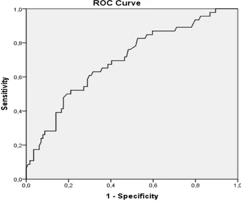
To test the power of NLR for distinguishing group 1 and group 2+3, ROC analysis was performed. The NLR had AUC values of 0.714 (p<0.001), which demonstrate its sufficiency to distinguish patients in group 1 from individuals in group 2+3 (Figure 1). To test the power of NLR for distinguishing group 1+2 and group 3, ROC analysis was performed. The NLR had AUC values of 0.698 (p<0.001), which demonstrate its sufficiency to distinguish patients in group 1+2 from individuals in group 3 (Figure 2). Discussion
In our study, we have demonstrated that NLR has a significant relationship with CCS angina grade of the patients. Patients with higher CCS angina class have higher NLR and those with noncardiac chest pain and atypical angina have smaller NLR values. This finding can be useful in the outpatient setting in evaluation of chest pain especially in patients who are unable to define their symptoms clearly. However, we did not see the similar relationship with PLR. In previous studies, the NLR and PLR, indicating the level of inflammation, was found to be related with severity and prognosis of CAD.2,15-17 The inflammation plays an important role in the
pathophysiology of atherosclerosis and chronic diseases such as cancers, diabetes mellitus, hypertension, connective tissue disease, and chronic kidney disease.1,16,19,20 In
the early phase of coronary artery disease and during acute coronary syndromes, inflammatory molecules play crucial role leading endothelial damage and formation of thrombosis after plaque rupture.1,5,21 Although some inflammatory molecules such as
CRP, tumor necrosis factor (TNF) alpha, interleukin (IL) -1, and IL-6 are considered to be the primary indicators of inflammatory process, WBC count and its subtypes were
56
reported to be useful also in predicting the inflammatory process in cardiovascular diseases.22 NLR has a considerable role in determination of risk, severity, complexity
and mortality of CAD in both stable and unstable presentations.2,4,5,23 Another
inflammatory marker in atherosclerosis is PLR.24 It was reported that PLR is related to
poor prognosis in CAD, an independent predictor of no-reflow in patients with ST elevation myocardial infarction (STEMI), and predicts long term mortality in patients with non-STEMI.16,25 Higher PLR values were found to be an indicator of poor coronary
collateral circulation and cause more frequent limb ischemia in patients with peripheral artery disease.17,26 In some malignancies, PLR was shown to be associated
worse clinico-pathologic results.27 In our study, although NLR had significant
correlation with grade of angina, we could not demonstrate any significant relationship between PLR and angina grade of the patients.
Table 3. Relationship of neutrophil lymphocyte ratio with the other characteristics of patients.
Neutrophyl to lymphocyte ratio
r p Extent of CAD 0.290 <0.001 CCS angina grade 0.423 <0.001 Age 0.300 <0.001 HT 0.096 0.088 DM 0.133 0.017 HL -0.012 0.837 Smoking 0.147 0.009 BMI -0.016 0.777 LDL (mg/dl) 0.138 0.014 Creatinine (mg/dl) 0.156 0.005 Leukocyte 0.187 0.001 Neutrophil 0.408 <0.001 Lymphocyte -0.391 <0.001 Hemoglobin -0.091 0.105 Platelet -0.130 0.02 MPV -0.056 0.322
Platelet to lymphocyte ratio 0.213 <0.001
CCS angina grading system is a useful tool in outpatient setting in determination of severity of angina pectoris.11 Although this grading system has several imperfections
57
generally relevant and practical and used throughout the world for more than 40 years in cardiology practice.12 In the present study, we have evaluated the chest pain
symptoms including noncardiac pain plus atypical angina, CCS angina grade 1 plus 2 and CCS grade 3 plus 4. The analysis between groups yielded significant differences in NLR. The patients in noncardiac plus atypical angina group had the lowest NLR, and the ones in the grade 3 plus 4 group had the higher NLR values. This result may support the fact of lower grade of inflammation in the first group of patients. In the light of these findings, we can conclude that in patients who are unable to define their symptoms, NLR, as a practical test in outpatient clinic, can aid us in evaluation of chest pain and grading of angina pectoris. Our results showed that the extent of CAD, stated as number of diseased coronary vessels, in patients who had undergone coronary angiography, was significantly getting higher from group 1 to group 3. This condition can support the consistency of symptom evaluation with coronary angiographic evidence of CAD. NLR was also found to be positively correlated with extent of CAD similar to the previous studies.2,4 Although NLR has a positive correlation with PLR, we
could not demonstrate any significant relationship between PLR and angina grade of the patients which can be due to relatively smaller sample size of our study group. The presence of significant correlation of NLR with diabetes mellitus, age, smoking, creatinine levels, leukocyte, neutrophil, LDL and PLR can indicate the previously defined strong relationship of NLR with inflammation.21 Unlike many other
inflammatory markers and bioassays, NLR is an inexpensive and readily available marker and provides an additional data about the chest pain symptoms in outpatient setting.
58
Figure 1. ROC analysis of the neutrophil lymphocyte ratio, curves for individual levels and its cooperative power to discriminate two sets of patients in group 1 and group 2+3.
Figure 2. ROC analysis of the neutrophil lymphocyte ratio, curves for individual levels and its cooperative power to discriminate two sets of patients in group 1+2 and group 3.
Study Limitations
This is an observational, single-institution study with a relatively small sample size and was thus subject to various unaccounted confounders inherent in such an analysis. This study did not evaluate the prognostic value of the NLR and PLR and we did not evaluate the inflammatory markers including TNFalpha, IL-1 beta, IL-6 and high sensitive CRP in our study population because of lack of data. Other cardiovascular risk factors including lifestyle, atherosclerosis in the arteries other than coronary circulation, carotid intima media thickness, and ankle-brachial index of patients were not evaluated in our patients. These factors may also contribute to increased inflammatory process. Further studies in larger cohorts are needed for the validation of these findings to better define the role of NLR and PLR in evaluation of chest pain symptoms in the outpatient setting.
Conclusion
NLR is a useful diagnostic marker that can be helpful in differential diagnosis of patients with stable angina pectoris, atypical angina and noncardiac chest pain. NLR, but not PLR, has a significant correlation with CCS angina grading scale.
59
The authors declared no potential conflicts of interest with respect to the research, authorship, and/or publication of this article and received no financial support for the research, authorship, and/or publication of this article.
References
1. Libby P, Ridker PM, Hansson GK. Inflammation in atherosclerosis: from pathophysiology to practice. J Am Coll Cardiol 2009;23:2129-38.
2. Kaya A, Kurt M, Tanboga IH, et al. Relation of Neutrophil to Lymphocyte Ratio with the Presence and Severity of Stable Coronary Artery Disease. Clin Appl Thromb Hemost 2014;5:473-7.
3. Hatmi ZN, Saeid AK, Broumand MA, Khoshkar SN, Danesh ZF. Multiple inflammatory prognostic factors in acute coronary syndromes: a prospective inception cohort study. Acta Med Iran 2010;1:51-7.
4. Angela Papa, Michele Emdin, Claudio Passino, Michelassi C, Battaglia D, Cocci F. Predictive value of elevated neutrophil–lymphocyte ratio on cardiac mortality in patients with stable coronary artery disease. Clinica Chimica Acta 2008;1-2:27–31.
5. Guasti L, Dentali F, Castiglioni L, et al. Neutrophils and clinical outcomes in patients with acute coronary syndromes and/or cardiac revascularisation. A systematic review on more than 34,000 subjects. Thromb Haemost 2011;4:591-9.
6. Uthamalingam S, Patvardhan EA, Subramanian S, et al. Utility of the neutrophil to lymphocyte ratio in predicting long-term outcomes in acute decompensated heart failure. Am J Cardiol 2011; 3:433-8.
7. Thaulow E, Erikssen J, Sandvik L, Stormorken H, Cohn PF. Blood platelet count and function are related to total and cardiovascular death in apparently healthy men. Circulation 1991;2:613–7. 8. Nikolsky E, Grines CL, Cox DA, et al. Impact of baseline platelet count in patients undergoing
primary percutaneous coronary intervention in acute myocardial infarction (from the CADILLAC trial). Am J Cardiol 2007;8:1055–61.
9. Ommen SR, Hammill SC, Gibbons RJ. The relative lymphocyte count predicts death in patients receiving implantable cardioverter defibrillators. Pacing Clin Electrophysiol 2002;10:1424–8. 10. Arruda-Olson AM, Reeder GS, Bell MR, Weston SA, Roger VL. Neutrophilia predicts death and
heart failure after myocardial infarction: a community-based study. Circ Cardiovasc Qual Outcomes 2009;6:656–62.
11. Campeau L. Letter: Grading of angina pectoris. Circulation 1976;3:522-3.
12. Campeau L. The Canadian Cardiovascular Society grading of angina pectoris revisited 30 years later. Can J Cardiol 2002;4:371-9.
13. Ryan TJ, Faxon DP, Gunnar RM, et al. Guidelines for percutaneous transluminal coronary angioplasty. A report of the American College of Cardiology/American Heart Association Task Force on Assessment of Diagnostic and Therapeutic Cardiovascular Procedures (Subcommittee on Percutaneous Transluminal Coronary Angioplasty). Circulation 1988;78:486-502.
14. Gibbons RJ, Chatterjee K, Daley J, et al. ACC/AHA/ACP–ASIM guidelines for the management of patients with chronic stable angina: executive summary and recommendations: a report of the American College of Cardiology/American Heart Association Task Force on Practice Guidelines (Committee on Management of Patients With Chronic Stable Angina). Circulation 1999;99:2829–48.
15. Sönmez O, Ertaş G, Bacaksız A, et al. Relation of neutrophil-to-lymphocyte ratio with the presence and complexiety of coronary artery disease: an observational study. Anadolu Kardiyol Derg 2013;13:662-7.
16. Yildiz A, Yuksel M, Oylumlu M, et al. The Utility of the Platelet-Lymphocyte Ratio for Predicting No Reflow in Patients With ST-Segment Elevation Myocardial Infarction. Clin Appl Thromb Hemost 2015;21: 223-8.
17. Acar G, Kalkan ME, Avci A, et al. The Relation of Platelet-Lymphocyte Ratio and Coronary Collateral Circulation in Patients with Stable Angina Pectoris and Chronic Total Occlusion. Clin Appl Thromb Hemost 2013 (doi: 10.1177/1076029613508599).
18. Sunbul M, Gerin F, Durmus, E et al. Neutrophil to lymphocyte and platelet to lymphocyte ratio in patients with dipper versus non-dipper hypertension. Clin Exp Hypertens 2014;36:217-21.
60
19. Lee S, Choe JW, Kim HK, Sung J. High-sensitivity C-reactive protein and cancer. J Epidemiol 2011;21:161-8.
20. Okyay GU, Inal S, Oneç K, et al. Neutrophil to lymphocyte ratio in evaluation of inflammation in patients with chronic kidney disease. Ren Fail 2013;35:29-36.
21. Tamhane UU, Aneja S, Montgomery D, Rogers EK, Eagle KA, Gurm HS. Association between admission neutrophil to lymphocyte ratio and outcomes in patients with acute coronary syndrome. Am J Cardiol 2008;102:653-7.
22. Horne BD, Anderson JL, John JM, et al. Intermountain Heart Collaborative Study Group. Which white blood cell subtypes predict increased cardiovascular risk? J Am Coll Cardiol 2005; 45:1638-43.
23. Sahin DY, Elbasan Z, Gur M, et al. Neutrophil to Lymphocyte Ratio is Associated with the Severity of Coronary Artery Disease in Patients With ST-Segment Elevation Myocardial infarction. Angiology 2013;64:423-9.
24. Sari I, Sunbul M, Mammadov C, et al. Relation of neutrophil to lymphocyte and platelet to lymphocyte ratio with coronary artery disease severity in patients undergoing coronary angiography. Kardiol Pol 2015 (doi: 10.5603/KP.a2015.0098).
25. Azab B, Shah N, Akerman M, McGinn JT Jr. Value of platelet/lymphocyte ratio as a predictor of all-cause mortality after non-ST-elevation myocardial infarction. J Thromb Thrombolysis 2012; 34:326-34.
26. Gary T, Pichler M, Belaj K, et al. Platelet-to-lymphocyte ratio: a novel marker for critical limb ischemia in peripheral arterial occlusive disease patients. PloS One 2013;8:e67688.
27. Asher V, Lee J, Innamaa A, Bali A. Preoperative platelet lymphocyte ratio as an independent prognostic marker in ovarian cancer. Clin Transl Oncol 2011;13:499-503.