Ultrasonographic and Color Doppler Ultrasonographic
Parameters to Discriminate Thyroid Nodules
Tiroid Nodüllerinde Ultrasonografi ve Renkli Doppler Ultrasonografi Bulguları Birlikteliğinde Malignite Saptanması
Ayșegül Gürsoy Çoruh
1, Zehra Akkaya
1, Derya Öztuna
2, Suat Kemal Aytaç
1, Serdar Akyar
11 Ankara Üniversity, School of Medicine, Department of Radiology
2 Ankara University, Scholl of Medicine, Department of Biostatistics Objective: To determine parameters that can be used to predict malignant thyroid nodules by using
gray-scale ultrasonography and color Doppler ultrasonography
Materials-methods: Gray-scale ultrasonography and color Doppler ultrasonography findings were
retro-spectively analyzed in 60 nodules with known histopathology. Of 60 nodules, 12 nodules were malignant. The evaluation criteria for gray-scale ultrasonography were: size, echotexture, internal morphology, con-tour, presence of microcalcification, and presence of halo sign. The evaluation criteria for color Doppler ultrasonography were: vascular flow pattern and resistive index. Vascular flow patterns were classified as Types I, II, III, and IV.
Results: Irregular contour and type IV flow patterns were the most significant independent predictors of
malignancy in malignant thyroid nodules. Resistive index was significantly higher in malignant nodules. The cut off value was established as resistive index ≥0.69; 91% sensitivity and 97% specificity were calculated in the identification of malignant nodules. A combination of the absence of a halo sign, type IV flow pattern, and irregular contour had the highest specificity and positive predictive value (66.7% sensitivity, 100% specificity, 100% positive predictive value, 92.3% negative predictive value).
Conclusion: We believe that by the integration gray-scale ultrasonography with color Doppler
ultrasonog-raphy findings, it is possible to determine which nodule is malign/benign without performing fine needle aspiration biopsy (FNAB) and unnecessary operations could be prevented.
Key Words: Thyroid Nodule, Ultrasonography, Doppler Ultrasonography, Resistive Index, Vascularization
Amaç: Tiroid nodüllerinde ultrasonografi bulgularına ek olarak renkliDoppler ultrasonografi bulguları ile
birlikte malign - benign ayrımını yapabilmek
Materyal - Metod: Çalıșmamızda total yada subtotaltiroidektomi sonucunda patolojileri bilinen 60 nodül
incelenmiștir. Retrospektif olarak 60 nodülün 12’si malign karakterdeydi. Nodüllerde ultrasonografi bulguları olarak; boyut, ekojenite, natür, kenar yapısı, mikrokalsifikasyon varlığı, periferikhalo varlığı değerlendirilme-ye alındı. Renkli Doppler ultrasonografi bulguları olarak kanlanma desenlerine ve rezistif indeks değerlerine bakıldı. Kanlanma desenleri 4 sınıfta gruplandırıldı; tip I, tip II, tip III, tip IV .
Bulgular: Düzensiz kenar yapısı ve tip IV kanlanma deseni malignnodül saptamada en özgül bağımsız
pa-rametreler olarak saptandı. Rezistif indeks değeri malignnodüllerde daha yüksek bulundu. Rezistif indeks için cutoff değeri % 91 duyarlılık ve % 97 özgüllük ile 0,69 olarak hesaplandı. Düzensiz kenar yapısı, perife-rikhalo kaybı ve tip IV kanlanma deseni kombinasyonu malignnodül saptamada en özgül ve en yüksek pozi-tif tahmini değere sahip bulgu olarak bulundu (% 66.7 duyarlılık, %100 özgüllük, %100 pozipozi-tiftahminideğer, %92.3 negatif tahmini değer).
Sonuç: Ultrasonografik ile renkli Doppler ultrasonografi bulguları birlikte malign – benign nodül ayrımı
yapılabilir ve gereksiz cerrahi ișlem engellenilebilir.
Anahtar Sözcükler: Tiroid Nodülü, Ultrasonografi, Renkli Doppler Ultrasonografi, Rezistif İndeks, İnternal Kanlanma
Thyroid nodules are the most common thyroid disease with increasing prevalence, especially in iodine deficient areas (1). Ultrasound (US) is the primary and the most effective imaging modality in screening thyroid disease. Incidental thyroid nodules detected by US at an approximate rate of 70% of the population and detected by autopsy at a rate of approximately 50% in adults.(2). A great majority of
thyroid nodules are benign. Approximately 4-14% nodules are malignant.(3).
In the literature, several studies reported that the most efficient method in discriminating between benign and malignant nodules is fine needle aspiration biopsy (FNAB) with a sensitivity of 65-98% and specificity of 72-100%.(4). Even in large,
Received: April 16,2016 Accepted: Aug 22,2016 Corresponding author
Aysegul Gursoy Coruh, M.D. E-posta: [email protected], [email protected] Phone: +90 505 400 77 16
Ankara University, School of Medicine, İbni Sina Hospital, Department of Radiology, Sıhhiye/Ankara/ Turkey
experienced centers inadequate specimen and non-diagnostic cytology rate is up to 15-20% (5,6). Furthermore, false negative results are reported at a range of 3-21%.(7). Consequently, a non-invasive, safe, and low cost diagnostic procedure is required, with high accuracy, to discriminate malignant and benign thyroid nodules. Several gray-scale US and color Doppler ultrasonography (CDUS) features have been reported to be highly suggestive of malignancy such as hypoechogenicity, presence of microcalcification, irregular contour, hypervascular central flow, and high Resistive Index (RI) (3,8,9). On the contrary, several authors claim that there are no correlations with the central flow, echogenicity, shape, and malignancy (9,11).
The purpose of this study is to evaluate the capabilities of gray-scale US and CDUS alone and in combination, in detecting malignant thyroid nodules. Furthermore, this study also aimed to determine which thyroid nodule should receive surgery using the US and CDUS parameters without performing FNAB.
Materials and Methods
The researchers of the current study retrospectively reviewed the records of patients who were underwent surgical intervention between the years 2004 and 2009. Thirty patients’ (median age: 42.37±10.10 years; range: 25-61), consisting of 24 women (80%) and 6 men (20%), gray-scale US and CDUS data were evaluated by one radiologist. Sixty nodules were included in the study. Each nodule was reported as benign or malignant nodule based on histologic classification. Out of 60 nodules, 48 (80%) nodules were benign and 12 (20%) nodules were malignant. All US and CDUS were conducted by the same radiologist with more than ten years of experience in thyroid ultrasound. Sonography was performed using the Sonoline Antares system (Siemens, Washington, USA) using 4–9 mHz and 5–13 mHz linear array transducers.
The evaluation criteria for gray-scale US included: size, echotexture, internal morphology, contour, presence of
microcalcification, and the presence of a halo sign. The size of the nodule was defined as the largest size in any of the three dimensions, which were measured in transverse and longitudinal planes. Microcalcification was defined as hyperechoic spots below 2 mm with/without acoustic shadowing and without comet artifact. Internal morphology was categorized as solid, mixed, or cystic according to the ratio of solid and cystic components. A solid nodule was defined when a nodule had a cystic component, compromising less than 25% of it and a cystic nodule was defined when a nodule consisted of more than 75% cystic spaces. Echogenicity of the nodule was identified by comparing the nodule with the thyroid parenchyma and strap muscles, and qualified as hypoechoic, hyperechoic, and isoechoic. The contour of the nodule was categorized as well-defined or spiculated/irregular. The absence or discontinuity of a peripheral halo sign was evaluated. On CDUS, vascular flow patterns were
classified as Types I, II, III, and IV.
Type I: No visible flow
Type II: Peri-nodular (peripheral) flow Type III: Peri-nodular flow (peripheral)
with minimal internal flow
Type IV: Marked internal flow (intrinsic
hypervascularity) or marked internal flow with peripheral flow. Marked internal flow was qualified as flow in the central part of the tumor greater than that in the surrounding thyroid parenchyma.
Resistive index (RI) was recorded, which was performed on one to three arteries of greater caliber in a vascularized nodule. Nodules consisting of central and peripheral flow, the measurements were taken in both vascular portions. Resistive Index was automatically calculated in the software, and for all nodules, average values were obtained. Statistical analyses were performed using
SPSS 11.5. Frequency (percent) for categorical variables, mean±standard deviation for metric variables was given as descriptive statistics. In
order to compare two independent groups in terms of categorical and metric variables, the chi-square test and Mann-Whitney U-test were used, respectively. The area under receiver operating characteristics (ROC) curve was evaluated for the examination of diagnostic performance of RI. Additionally, the sensitivity, specificity, positive predictive value (PPV), and negative predictive value (NPV) were calculated. P<0.05 was considered statistically significant.
Results
In this study, there were 48 (80%) benign and 12 (20%) malignant nodules. Out of 12 malignant nodules, 11 were papillary carcinomas, 1 was a follicular carcinoma. All malignant nodules were solitary. Overall nodule size ranged from 3 mm to 50 mm with a mean value 14.87±10.54 mm. Out of 60 nodules, 58 (96.7%) were solid and 2 (3.3%) were mixed morphology. All malignant nodules and 46 (95.8%) benign nodules had a solid appearance. None of the nodules were in cystic in nature. There were no statistically significant differences between benign and malignant nodules regarding size (p=0.3) and internal morphology (p=1).
Hypoechogenicity was a common gray-scale US feature in malignant nodules (91.7%), whereas for benign nodules, all three categorized echogenicity were seen. A total of 15 (31.3 %) of 48 benign nodules were hypoechoic, 19 (39.6%) nodules were isoechoic, and 14 (29.2%) nodules were hyperechoic. For echogenicity, statistically significant difference was found between benign and malignant nodules (p=0.001).
Of 12 malignant nodules, 11 nodules (91.7%) had an absent or discontinu-ous peripheral halo. Ten (83.3%) ma-lignant nodules showed irregular con-tour, whereas irregular contour was detected in 3 (6.3%) out of 48 benign nodules. Microcalcifications were de-tected in 7 of 12 (58.3%) among the malignant nodules (Figure 1) and 4 of 48 (8.3%) among the benign nodules.
There was a statistically significant difference between benign and ma-lignant nodules regarding the pres-ence of microcalcifications, irregular contour, and the absence of a periph-eral halo (p<0.001). Table 1 shows the distribution of the gray-scale ul-trasonographic features related to the histology, which were found statis-tically significant for indicating
ma-lignant thyroid nodules in this study. The gray-scale US features of hy-poechogenicity, irregular contour, presence of microcalcification, and the absence of a peripheral halo were more common in malignant nodules. Comparing the gray-scale US find-ings, the feature with the highest specificity and positive predictive value was irregular contour (93.8%
and 76.9%, respectively). Table 2 shows the comparative intragroup analysis of gray-scale US features and CDUS features between benign and malignant nodules.
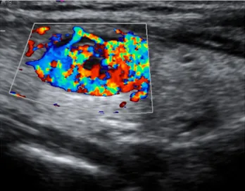
A great majority of malignant nodules had internal vascular flow. A total of 8 of 12 (66.7%) malignant nodules were categorized as Type IV in
Figure 1. Microcalcifications in a hypoechoic thyroid nodule Figure 2. Type IV vascular flow pattern in papillary carcinoma with a
pathological diagnosis of papillary carcinoma
Table 1: Gray-scale ultrasonographic features related to histology
Echogenicity Peripheral halo Contour Microcalsification Total
Hyperechoic Isoechoic Hypoechoic Presence Absence Well-defined Irregular Presence Absence Benign (29.2%) 14 19 (39.6%) (31.3%) 15 41 (85.4%) 7 (14.6%) 45 (93.8%) (6.3%) 3 (8.3%) 4 44 (91.7%) 48 Malignant (8.3%) 1 (0%) 0 (91.7%) 11 (8.3%) 1 11 (91.7%) 2 (16.7%) 10 (83.3%) 7 (58.3%) 5 (41.7%) 12
Table 2: Diagnostic data for gray-scale ultrasound and color Doppler ultrasound features of malignant nodules
Malignant % Benign % Sensitivity % Specificity % Positive predictive value % Negative predictive value %
Hypoechogenicity 91.7 31.3 91.6 68.7 42.3 97
Absence of peripheral halo 91.7 14.6 91.7 85.4 61.1 97.2
Irregular contour 83.3 6.3 83.3 93.8 76.9 95.7
Presence of microcalcification 58.3 8.3 58.3 91.7 63.6 89.8
Type IV flow pattern 66.7 16.7 66.7 83.3 50 90.9
Table 3: Distribution of the types of flow patterns among benign and malignant nodules
Benign Malignant Total
Type I flow pattern 6 (12.5%) 0 (0%) 6 (10%)
Type II flow pattern 15 (31.3%) 0 (0%) 15 (25%)
Type III flow pattern 19 (39.6%) 4 (33.3%) 23 (38.3%)
Type IV flow pattern 8 (16.7%) 8 (66.7%) 16 (26.7%)
Figure 3. Spectral analysis of arterial vascularity shows a high resistance waveform with a RI
value of 0.75. This nodule had a diagnosis of papillary carcinoma.
CDUS (Figure 2). In the current study, none of the malignant nodules showed Type I or Type II flow pat-tern. Table 3 shows the distribution of the types of flow patterns among benign and malignant nodules. Among the other types of flow pat-tern, Type IV presented highest spec-ificity and sensitivity (83.3% and 66.7%, respectively) with a 50% posi-tive predicposi-tive value and a 90.9% negative predictive value.
RI was significantly higher in malignant nodules (Figure 3). The mean value of RI was 0.80±0.15 (range: 0.50-1) in
ma-lignant nodules and 0.52±0.06 (range: 0.45-0.77) in benign nodules. Among the benign nodules, the highest RI (RI=0.77) was detected in follicular ad-enoma. The best results were calculated for a RI≥0.69 cut off value, with 91% sensitivity and 97% specificity in dis-criminating malignant nodules. We also evaluated a combination of
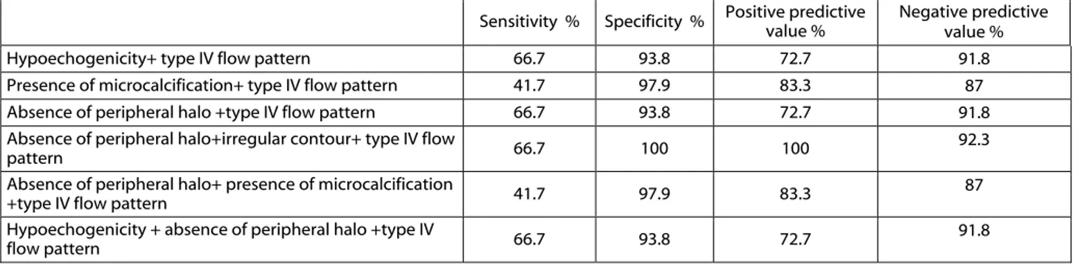
gray-scale US features and CDUS features for discriminating malignant nodules (Table 4). The absence of a peripher-al hperipher-alo, irregular contour, and Type IV flow pattern indicated the highest specificity (100%) and positive pre-dictive value (100%) to predict
ma-lignancy with a 66.7% sensitivity and 92.3% negative predictive value.
Discussion
Nodular thyroid disease is very common and a great majority of nodules are benign. Thyroid cancer prevalence ranges from 1.5% to 10% (12). Alt-hough thyroid malignancy is rare, it is important to discriminate malignant from benign nodules because it is one of the most curable malignancy. Fur-thermore, early and accurate diagnosis could alter the therapeutic approach (13). Thyroid US is the most prevalent method to detect and characterize the thyroid nodules. There are a great number of studies discussing the so-nographic and Doppler findings for thyroid nodules in the literature, yet there is no common consensus. The current study demonstrated that the integration of CDUS and gray-scale ultrasound findings improved the specificity and positive predictive val-ue in identifying malignant nodules. In the current study, no statistically
signifi-cant difference was found in predicting malignancy according to nodule size and morphology (p=0.3, p=1, respec-tively). Twelve (100%) malignant nod-ules and 46 (95.8%) benign nodnod-ules had a solid appearance. Likely for the benign nodules, the mean nodule size was 14.67±11.07 mm, and for the ma-lignant nodules the mean nodule size was 15.68±8.4 mm. The results of the current study supported the findings in the literature. Previous researchers simi-larly declared that nodule size and in-ternal morphology were not predictive of malignancy (14,18).
Table 4:Gray-scale ultrasound and color Doppler ultrasound combination features related to malignant histology in thyroid nodules
Sensitivity % Specificity % Positive predictive value % Negative predictive value %
Hypoechogenicity+ type IV flow pattern 66.7 93.8 72.7 91.8
Presence of microcalcification+ type IV flow pattern 41.7 97.9 83.3 87
Absence of peripheral halo +type IV flow pattern 66.7 93.8 72.7 91.8
Absence of peripheral halo+irregular contour+ type IV flow
pattern 66.7 100 100 92.3
Absence of peripheral halo+ presence of microcalcification
+type IV flow pattern 41.7 97.9 83.3 87
Hypoechogenicity + absence of peripheral halo +type IV
Hypoechogenicity was a common gray-scale US feature in malignant nodules (12,18,19). In the current study, a to-tal of 11 of 12 malignant nodules were hypoechoic. Benign nodules’ echogenicities were variable. In this study hypoechogenicity is a valuable gray-scale US marker with a sensitivi-ty of 91.6% and specificisensitivi-ty of 68.7%. Hypoechogenicity was also frequently seen in benign nodules; therefore its specificity was the lowest in the gray-scale ultrasonographic criteria. There was a statistically significant
diffe-rence between benign and malignant nodules regarding the presence of microcalcifications (p< 0.001). Kim et al. (17) demonstrated that the pre-sence of microcalcification is the most sensitive gray-scale US criteria in their study. Presence of microcalci-fication has a high specificity of 91.7%, which was the second highest specific finding in gray-scale US, sen-sitivity however was relatively lower compared to other findings that sug-gest malignancy such as contour ab-normality and hypoechogenicity which may have resulted from lownumber of patients included in the study. Similarly, Moon et al. (12) declared that the presence of micro-calcification can be considered as a highly specific finding for malig-nancy. The current findings are in concordance with their study.
In previous studies, a great majority of authors reported the association be-tween the absence of a complete pe-ripheral halo and malignancy (15,16,20,21). A peripheral halo is composed of compressed thyroid pa-renchyma and prominent vessels. In agreement with these reports, in the current study of 12 malignant nod-ules, 11 nodules (91.7%) had absent or discontinuous peripheral halo. An absent halo sign is the most sensitive parameter in the current study with a sensitivity of 91.7%, but it is less spe-cific. In predicting malignancy with gray-scale ultrasound features, irregu-lar/spiculated contour is the best sin-gle parameter with a specificity of 93.8%, sensitivity of 83.3%, positive predictive value of 76.9%, and nega-tive predicnega-tive value of 95.7%.
CDUS examination has become a consi-derable imaging technique for discri-minating malignant and benign thy-roid nodules. Regarding CDUS fin-dings, some vascular patterns were classified to differentiate benign and malignant nodules. In previous stu-dies, many authors have shown the association between the increased intranodular blood flow and malig-nancy [3,9,19,22,23]. According to the data of the current study, increa-sed intranodular blood flow (Type IV) was a statistically significant crite-rion to suggest malignancy (p=0.001). Tamsel et al. [24] and Algin et al. [25] did not find any relationship between high intranodular blood flow and ma-lignancy .It is assumed that technical issues (settings of a wall filter and pulse repetition frequency) and pati-ents lack of cooperation (breathing-swallowing motion artifacts) may ha-ve hidden the possible relationship between intranodular vascularity and malignancy in these studies.
In addition to vascular patterns, there are several studies that have shown the relationship with the histology of the thyroid nodules and vascular resis-tance [3,5,13,19,26]. RI is a good spectral Doppler parameter for eva-luating thyroid nodules because it is not dependent on the angle of inso-nation. In the literature, many aut-hors declared that RI values in malig-nant nodules were higher than in be-nign nodules [3,5,19,23,26]. Ivanac et al. [3] calculated the optimal cut-off value as RI≥0.70 to distinguish ma-lignant nodules from benign nodules with a sensitivity of 80% and specifi-city of 92%. De Nicola et al [5], Hol-den [19], and Cerbone et al. [13] re-ported similar results and found mean RI values for carcinomas of 0.75, 0.76, and 0.75, respectively. These studies suggested that large quantities of stenosis and occlusions in the neovascularization of differen-tiated thyroid carcinomas lead to an increase in vascular resistance. Simi-larly, in the current study, RI was sig-nificantly higher in malignant nodules (0.80±0.15 vs. 0.52±0.06) and RI≥0.69 was highly suggestive of ma-lignancy with a sensitivity of 91% and specificity of 97%. However, Tamsel
et al. [24] reported poor sensitivity and specificity values of 11% and 4%, respectively, for an RI cut-off of 0.75. The study of Tamsel et al. [24] was based on cytological results acqu-ired by FNAB. False negative results could arise in some cases, which might have potentially altered the re-sults of this study.
In the literature, they also analyzed gray-scale US and CDUS features in com-bination and reported increased spe-cificity by combining CDUS and US in the detection of malignancy in thy-roid nodules [9,15,16]. In the current study with the combination parame-ters (Table 4), there was an impro-vement in specificity and positive predictive value. On the contrary, Stacul et al. [8] did not report an imp-rovement in diagnostic performance of malignant thyroid nodules. This disagreement may be related, using the cytology as the reference stan-dard, and therefore the results could be deviated. In the current study, among the associations of several US patterns with CDUS; the absence of a peripheral halo, irregular contour, and Type IV flow pattern is the most valuable combination with a specifici-tiy of 100%, sensitivity of 66.7%, po-sitive predictive value of 100%, and negative predictive value of 92.3%. There are some limitations in this study.
Firstly, the number of the patients that were included in the study was limited. Secondly, a great majority of the malignant nodules were papillar carcinoma. Additional studies should be performed with a larger popula-tion including other types of thyroid malignancies. The strength of this study was that all nodules in this study had histological reports ob-tained from surgical specimens. In conclusion, the results of present study
indicate that CDUS gave an incremen-tal value to gray-scale US in the diag-nosis of malignant thyroid nodules. Due to the high specificity and positive predictive value, the findings of com-bined CDUS and gray-scale US fea-tures can determine malignant thyroid nodules and patients with such nodules may undergo surgery without the need of FNAB.
REFERENCES
1. Belfiore A, Rosa GL, Giuffrida D, et al. The management of thyroid nodules. J Endocrinol Invest. 1995; 18: 155-158 2. Alexander EK, Hurwitz S, Heering JP, et
al. Natural history of benign solid and cystic thyroid nodules. Ann Intern Med. 2003; 138: 315-318
3. Ivanac G, Brkljacic B, Ivanac K, et al. Vascularisation of benign and malignant thyroid nodules: CD US evaluation. Ult-raschall Med. 2007;28:502-506.
4. Gharib H. Fine-needle aspiration biopsy of thyroid nodules: advantages, limitati-ons, and effect. Mayo Clin Proc 1994;69:44-49.
5. De Nicola H, Szejnfeld J, Logullo AF, et al. Flow pattern and vascular resistive in-dex as predictors of malignancy risk in thyroid follicular neoplasms. J Ultrasound Med. 2005;24:897-904.
6. Gharib H, Goellner JR. Fine-needle aspiration biopsy of the thyroid: an app-raisal. Ann Intern Med. 1993;118:282-289.
7. Wu HH, Jones JN, Osman J. Fine-needle aspiration cytology of the thyroid: ten ye-ars experience in a community teaching hospital. Diagn Cytopathol. 2006;34:93-96.
8. Stacul F, Bertolotto M, De Gobbis F, et al. US, colour-Doppler US and fine-needle aspiration biopsy in the diagnosis of thyroid nodules. Radiol Med. 2007;112:751-762.
9. Rago T, Vitti P, Chiovato L, et al. Role of conventional ultrasonography and color flow-doppler sonography in predicting malignancy in 'cold' thyroid nodules. Eur J Endocrinol. 1998;138:41-46.
10. Iannuccilli JD, Cronan JJ, Monchik JM. Risk for malignancy of thyroid nodules as assessed by sonographic criteria: the need for biopsy. J Ultrasound Med. 2004;23:1455-1464.
11. Shimamoto K, Endo T, Ishigaki T, et al. Thyroid nodules: evaluation with color Doppler ultrasonography. J Ultrasound Med. 1993;12:673-678.
12. Kim SJ, Moon WK, Cho N. Sonographic criteria for fine-needle aspiration cytology in a Korean female population undergo-ing thyroid ultrasound screenundergo-ing. Acta Radiol. 2010;51:475-481.
13. Cerbone G, Spiezia S, Colao A, et al. Power Doppler improves the diagnostic accuracy of color Doppler ultrasonog-raphy in cold thyroid nodules: follow-up results. Horm Res. 1999;52:19-24. 14. Ginat DT, Butani D, Giampoli EJ, et al.
Pearls and pitfalls of thyroid nodule so-nography and fine-needle aspiration. Ult-rasound Q. 2010;26:171-178.
15. Phuttharak W, Somboonporn C, Hong-domnern G. Diagnostic performance of gray-scale versus combined gray-scale with colour doppler ultrasonography in the diagnosis of malignancy in thyroid nodules. Asian Pac J Cancer Prev. 2009;10:759-764.
16. Appetecchia M, Solivetti FM. The asso-ciation of colour flow Doppler sonog-raphy and conventional ultrasonogsonog-raphy improves the diagnosis of thyroid carci-noma. Horm Res. 2006;66:249-256. 17. Kim EK, Park CS, Chung WY, et al. New
sonographic criteria for recommending fine-needle aspiration biopsy of nonpal-pable solid nodules of the thyroid. AJR Am J Roentgenol. 2002;178:687-691.
18. Frates MC, Benson CB, Doubilet PM, et al. Prevalence and distribution of carci-noma in patients with solitary and multip-le thyroid nodumultip-les on sonography. J Clin Endocrinol Metab. 2006;91:3411-3417. 19. Holden A. The role of colour and duplex
Doppler ultrasound in the assessment of thyroid nodules. Australas Radiol. 1995; 39:343-349.
20. Koike E, Noguchi S, Yamashita H, et al. Ultrasonographic characteristics of thy-roid nodules: prediction of malignancy. Arch Surg. 2001;136:334-337.
21. Moon WJ, Jung SL, Lee JH, et al. Benign and malignant thyroid nodules: US diffe-rentiation-multicenter retrospective study. Radiology 2008;247:762-770.
22. Chammas MC, Gerhard R, de Oliveira IR, et al. Thyroid nodules: evaluation with power Doppler and duplex Doppler ultrasound. Otolaryngol Head Neck Surg. 2005;132:874-882.
23. Bianek-Bodzak A, Zaleski K, Studniarek M, et al. Color Doppler sonography in malignancy of thyroid nodules. J Ultraso-und Med. 2003;22: 758
24. Tamsel S, Demirpolat G, Erdogan M, et al. Power Doppler US patterns of vascu-larity and spectral Doppler US parameters in predicting malignancy in thyroid no-dules. Clin Radiol. 2007;62:245-251. 25. Algin O, Algin E, Gokalp G, et al. Role
of duplex power Doppler ultrasound in differentiation between malignant and benign thyroid nodules. Korean J Radiol. 2010;11:594-602.
26. Frates MC, Benson CB, Doubilet PM, et al. Can color Doppler sonography aid in the prediction of malignancy of thyroid nodules? J Ultrasound Med.2003;22:127-131.