Traumatic Diaphragmatic Hernia as a Rare Cause of Colonic
Obstruction
Kolon Obstrüksiyonunun Nadir Görülen Bir Nedeni: Travmatik Diafragmatik Herni
Kürșat Karadayı
1,
Șule Karadayı
2,
Erkan Hazar
3,
Ekber Șahin
4,
Mustafa Turan
11 Cumhuriyet University Faculty of Medicine, Department of General
Surgery
2 Cumhuriyet University Faculty of Medicine, Department of Emergency
Medicine
3Sivas State Hospital Department of General Surgery
4 Cumhuriyet University Faculty of Medicine, Department of Thoracic
Surgery
Travmatik diafragma hernileri nadirdir, tanı ve tedavisinde bir takım güçlükler vardır. Bu yazıda künt travmadan üç yıl sonra tanı konulan ve splenik fleksuranın herniasyonu nedeniyle kolon obstruksiyonu gelișen sol diafragma hernili 43 yașındaki hastamızı sunduk. Hastamıza subtotal kolektomi ve primer iliokolik anastomoz yapıldı ve diafragmatik defekt tamir edildi. Akut kolon obstrüksiyonu gelișen hastalarda travma öyküsü varsa diafragma hernisinden șüphelenilmelidir.
Anahtar Sözcükler: Travma, Diafragma Hernisi, İnkarserasyon.
Traumatic diaphragmatic hernia (TDH) is uncommon and presents a diagnostic and therapeutic challenge. Herein, we present the case of a 43-year-old patient with left-sided TDH and colonic obstruction due to herniation of the splenic flexura of the colon, which was diagnosed three years after the initial blunt trauma. A subtotal colectomy and primary ileocolic anastomosis were performed and the diaphragmatic defect was repaired. TDH should be suspected in patients with an acute colonic obstruction, particularly if the patient has a history of trauma.
Key words: Trauma, Diaphragmatic Hernia, Incarceration. Diaphragmatic rupture occurs in 0.8–
3.6% of patients after blunt thoracoabdominal trauma, and the preoperative diagnosis is often difficult. Patients with an undiagnosed diaphragmatic rupture can develop symptoms after a delay of weeks, months, or even years (1). A traumatic diaphragmatic hernia (TDH) following traumatic diaphragmatic rupture is uncommon and presents a diagnostic and therapeutic challenge. Here, we present the case of a patient with a left-sided TDH and colonic obstruction due to herniation of the splenic flexura, which was diagnosed three years after the initial blunt trauma.
Case
A 43-year-old man was admitted to the emergency service complaining of sudden onset cramping abdominal pain, vomiting,
abdominal distension, and constipation. He had been previously hospitalised for a rib fracture and received medical treatment for a traffic accident three years ago.
Abdominal distension with tenderness and muscle guarding was found on physical examination. Initial laboratory results were as follows: haemoglobin, 13 g/dL; and white blood cell count, 17800 K/uL. Plain radiographs of the abdomen demonstrated the classic inverted ‘U’ sign of the sigmoid volvulus
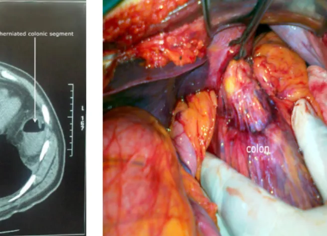
(Fig. 1). A computed tomography
(CT) scan revealed a distended transverse and right colonic segments and a herniated left colonic segment into the thorax
(Fig. 2). Laparotomy was performed. The section of the left colon was herniated into the chest from the left diaphragmatic defect. The transverse descending colon
Ankara Üniversitesi Tıp Fakültesi Mecmuası 2013, 66 (1)
DOI: 10.1501/Tıpfak_000000840
CERRAHİ BİLİMLER/SURGICAL SCIENCES
Case Report/Olgu Sunumu
Received: Feb 03,2012 Accepted: Feb 14,2014 Correspoding Author
Dr.Șule Karadayı GSM: 0505 502 51 86 Fax: 0346 327 12 74
E-mail: [email protected]
Baskent University Adana Teaching and Research Hospital, Department of Thoracic Surgery, Yüreğir/ADANA
Ankara Üniversitesi Tıp Fakültesi Mecmuası 2013, 66 (1)
Traumatic Diaphragmatic Hernia as a Rare Cause of Colonic Obstruction 50
and caecum were dilated and volvulated (Fig. 3). A herniated ischaemic colonic segment and omentum were reduced into the abdominal cavity. A subtotal colectomy and a primary ileocolic anastomosis with a stapler were performed. The diaphragmatic defect was repaired with interrupted non-absorbable sutures. A chest tube was placed in the left side of the thorax. The operating time was 180 minutes. The thoracic drain was removed on the third postoperative day, and the patient was discharged on postoperative day 10.
Discussion
TDH is characterised by displacement of the internal abdominal organs into the chest cavity through a pathological diaphragmatic aperture due to trauma (2). Diaphragmatic injuries are mainly left-sided (79.4%), and the most commonly injured abdominal
organs are the stomach, colon, spleen, small bowel, and liver (3,4). In our case, the herniated segment was the splenic flexure of the colon. The proximal colonic segment was dilated and volvulated.
The diagnosis of TDH may be delayed, particularly if diaphragmatic damage is not established during the acute trauma period. Severe diagnostic problems are caused by the development of post-traumatic diaphragmatic hernia strangulation and acute bowel occlusion (2).
The approach of choice is heavily influenced by the surgeon’s speciality; units that are managed by thoracic surgeons report much higher rates of thoracotomy than series reported by general surgeons (5). A diaphragmatic defect is closed with interrupted unabsorbable sutures, and, when possible, a two-layer closure should be performed. Synthetic grafts are
generally not required and pleural drainage should be performed. The use of laparoscopic techniques for TDH is still rare (2). We preferred a laparotomy in these cases because of excessive abdominal distension. The diaphragmatic defect was small, so we performed primary repair of the TDH with non-absorbable sutures. Thus, the risk of infection due to a synthetic graft and the overall operation time and cost were minimised. We resected the colon since the herniated colonic segment was ischaemic and the proximal colonic segment was volvulated. If the volvulated segment is not resected, the probability of recurrenct volvulus is high (6).
TDH should be suspected in patients with an acute colonic obstruction, particularly if the patient has a history of trauma. Early diagnosis of TDH is very important for appropriate surgical management.
Figure 1: Plain radiograph of the abdomen
demonstrate the classic inverted ‘U’ sign of a sigmoid volvulus.
Figure 2: Computed tomography scan
revealed a distended transverse and right colonic segment and a herniated left colonic segment into the thorax.
Figure 3: The herniated left colon was
Journal Of Ankara University Faculty of Medicine 2013, 66 (1)
Kürșat Karadayı, Șule Karadayı, Erkan Hazar, Ekber Șahin, Mustafa Turan 51
REFERENCES
1. Wirbel RJ, Mutschler W. Blunt
rupture of the right hemi-diaphragm with complete dislocation of the right hepatic lobe: Report of a case. Surg Today 1998;8:850-852.
2. Andreev AL, Protsenko AV, Globin
AV. Laparoscopic repair of a posttraumatic left-sided diaphragmatic hernia complicated by strangulation and colon obstruction. JSLS. 2010;14: 410-413.
3. Chughtai T, Ali S, Sharkey P, Lins M,
Rizoli S. Update on managing diaphragmatic rupture in blunt trauma: a review of 208 consecutive cases. Can J Surg 2009; 52: 177-181.
4. Turhan K, Makay O, Cakan A et al.
Traumatic diaphragmatic rupture: Look to see. Eur J Cardiothorac Surg 2008;33:1082-1085.
5. Morgan BS, Watcyn-Jones T. Garner
JP. Traumatic diaphragmatic injury. JR Army Med Corps 2010;156:139-149.
6. Yildiz SY, Berkem H, Yuksel BC, et
al. Isolated intrathoracic hiatal herniation of the twisted sigmoid colon: Report of a case. Dis Colon Rectum. 2009;52:740-741
Ankara Üniversitesi Tıp Fakültesi Mecmuası 2013, 66 (1)
Traumatic Diaphragmatic Hernia as a Rare Cause of Colonic Obstruction 52