Resuscitation complications encountered in forensic
autopsy cases performed in Muğla province
Halil Beydilli, M.D.,1 Yasemin Balcı, M.D.,2 Şahin Işık, M.D.,2 Melike Erbaş, M.D.,3 Ethem Acar, M.D.,1 Bülent Savran, M.D.3
1Department of Emergency Medicine, Muğla Sıtkı Koçman University Faculty of Medicine, Muğla 2Department of Forensic Medicine, Muğla Sıtkı Koçman University Faculty of Medicine, Muğla 3Ministry of Justice, The Forensic Branch Manager of Forensic Medicine Institute of Muğla, Muğla
ABSTRACT
BACKGROUND: The purpose of this study was to determine complications of resuscitation seen during autopsies and evaluate the effectiveness of basic life support training.
METHODS: Autopsy case reports conducted in The Forensic Branch Manager of Muğla were retrospectively examined. Demo-graphic data of the patients with resuscitation complications such as age, gender, manner of death, and kinds and features of the complications were recorded.
RESULTS: In total, seventy-fourof the 100 cases with resuscitation complications were males. The autopsies in most of these cases were performed during the summer season. Among the patients, 68% died for non-traumatic reasons. Rib fractures were detected in seventy-one patients and sternum fractures in thirty-two patients. Moreover, damage to the pericardium (2%) and lung parenchymal (4%), heart lesions (4%), and liver lacerations (2%) were detected. Regarding rib fractures, fractures were found between the first and eighth ribs on both sides, with the highest numbers occurring in the fourth rib.
CONCLUSION: Resuscitation complications are important since they can be presumed to have carried out for traumatic reasons. Resuscitation complications seen in autopsy cases with non-traumatic causes can be perceived as traumatic events. They can be as-sumed incorrectly as trauma symptoms. These complications can be reduced with a good resuscitation training of the health personnel. Key words: Autopsy; basic life support; closed-chest compression; resuscitation; resuscitation complications.
the last 10 years. The training mentioned above, in-service training, and certification courses have been provided espe-cially to personnel working in emergency services, intensive care, and 112 units. In order to maintain the required quality standards in hospitals, in-service training is provided every 6 months in the form of BLS or cardiopulmonary resuscitation courses. These training sessions are also monitored by the education and quality units of the hospitals.[2,3]
Airway patency establishment and closed-chest compression techniques are taught in first aid and BLS training courses. Training is provided according to the current guidelines pub-lished by the American Heart Association (AHA) and the European Resuscitation Council (ERC). Much of the infor-mation about closed-chest compression, chest compression physiology, the effects of various compression rates, ventila-tion/compression ratios, compression and relaxation times have been obtained from animal studies. This information has also been updated and strengthened by human studies. Finally, clear information about chest compressions was provided in
the 2010 AHA and ERC guidelines.[4,5]
Address for correspondence: Halil Beydilli, M.D.
Muğla Sıtkı Koçman Üniversitesi Tıp Fakültesi, Acil Tıp Anabilim Dalı, Muğla, Turkey
Tel: +90 252 - 211 48 35 E-mail: [email protected]
Qucik Response Code Ulus Travma Acil Cerrahi Derg 2015;21(6):463–468
doi: 10.5505/tjtes.2015.66169 Copyright 2015
TJTES
INTRODUCTION
Today, many people worldwide have taken courses in first aid and basic life support (BLS) or have undergone diverse train-ing in this area. In Turkey, BLS traintrain-ing is provided as part of first aid courses. Such first aid and BLS training is widely avail-able to anyone and provided by expert associations within regulations from the Ministry of Health or at certain cen-tres approved by the ministry.[1] In this regard, the Ministry of
Effective chest compression is an early procedure during BLS used to provide blood flow. Chest compressions should be strong and effective. At least 100 compressions should be performed per minute, and their depth should be at least 5 cm for adults. After each compression, the chest should be allowed to pull back, and compression and decompression times should be approximately equal. There should not be any
interruption between chest compressions.[4,5] Application of
closed-chest compression itself is traumatic. Unwanted com-plications caused by chest compressions, particularly rib and sternum fractures, can be encountered. Additionally, injuries to internal organs, such as the heart, lungs, liver, or stomach,
may occur.[6–10] Complication rate can vary depending on the
individual performing the chest compression (medical per-sonnel vs. lay people), the surface on which it is performed (soft vs. hard surface), the location where it is performed (in or outside a hospital), and the quality of the education and ability of the individual applying it.[6–10]
After Kouwenhoven and Baringer provided two separate de-scriptions of closed-chest massage, which was the basis of cardiopulmonary resuscitation in hospitals in the 1960s, stud-ies aimed at reducing complications, morbidity, and mortal-ity gained more attention. In 1976, Enarson and colleagues published the first studies on resuscitation complications.[9,10]
The recognition of various complications caused by resuscita-tion attempts in a series of autopsies gained great importance from a forensic viewpoint as well. Additionally, the develop-ment of cardiopulmonary resuscitation created a need for multidisciplinary post-mortem studies to help prevent
com-plications. Little research has been done in this field in our country. In our study, we analysed post-mortem autopsy findings in patients to whom BLS was provided by medical personnel in or outside the hospital.
The aim of this study was to analyse in detail the findings of autopsy reports from patients who received BLS from medi-cal personnel to determine the frequency of complications, evaluate the effectiveness of the training provided, and inves-tigate the subjects in the relevant literature.
MATERIALS AND METHODS
In this study, autopsy reports from cases who received BLS performed between 2011 and 2013 in the Forensic Branch Manager of Muğla were analysed retrospectively. Complica-tions related to closed-chest compressions were recorded in the cases who underwent BLS. Moreover, they were analysed in terms of socio-demographic characteristics, cause of death and the location where BLS was performed. Patients with thorax trauma were excluded from the study.
Complications related to closed-chest compressions per-formed during resuscitation attempts, rib fractures, sternum fractures, chest wall ecchymosis, haemothorax, cardiac con-tusion, pulmonary concon-tusion, liver laceration, pericardial inju-ries, and soft tissue damage, such as marks left on the skin by defibrillator pads were included in the evaluation.
For statistical analyses, the SPSS software was used. Percent-Table 1. Complications related to chest compressions
Complication All patients Male Female p
Skin Defibrillator pads skin marks or burn marks 16 13 3 0.471
Ecchymosis and bleeding in skin/subcutaneous tissue 9 5 4 0.034
Upper airway Endotracheal ecchymosis 1 1 – –
Thorax Rib fracture 71 52 19 0.786
Haematoma around rib fractures 8 7 1 0.364
Ecchymosis around rib fractures 17 14 3 0.799
Sternal fracture 32 24 8 0.876
Ecchymosis around sternal fractures 4 2 2 0.264
Haematoma around sternal fractures 4 3 1 0.963
Anterior mediastinal hemorrhage 1 1 – –
Pneumothorax 1 1 – –
Lung Lung contusion 4 3 1 0.963
Pleural rupture 2 1 1 0.464
Heart Pericardial injury/haematoma 2 1 1 0.464
Endomyocardial hemorrhage 1 1 – –
Atrioventricular hemorrhage 1 1 – –
ages for the classified variables and averages for the
con-tinuous variables were calculated routinely. We used χ2 and
Fisher’s exact test for comparisons. P values <0.05 were con-sidered to indicate statistical significance.
RESULTS
Among the cases who underwent BLS and whose autopsy was performed in the Forensic Branch Manager of Muğla be-tween 2011 and 2013, resuscitation complications were de-tected in one hundred (Table 1) patients. Of these one hun-dred patients, there were 74 males and 26 females. In Table 2, the distribution of rib fractures according to gender is shown. The average age of the patients was 54.57 (range, 0–93) years. Autopsies were performed in sixty-eight cases for non-trau-matic reasons. The resuscitation process was performed on seventy-four patients in the hospital and twenty-six patients at the scene of an accident by 112 service ambulance person-nel. There was no statistically significant difference in terms of resuscitation complications according to the scene of the event or practitioner (p>0.05).
The autopsy results identified rib fractures in seventy-one patients; 61 of these fractures were located in the ribs of the right thorax area and another 66 in the ribs of the left thorax. In fifty-nine cases, there were rib fractures on both sides. On each side, rib fractures were detected between the first and eighth ribs. The highest number of fractures on each side occurred in the third (right: 52%, n=52, left: 56%, n=56), fourth (right: 54%, n=54, left: 64%, n=64), and fifth ribs (right:
44%, n=44, left: 57% n=57). The distribution of rib fractures according to their location is shown in Table 2.
In total, ten rib fractures were parasternal (10 males [M]). There were two axillary rib fractures (1 M/1 Famles [F]). Moreover, twenty-three rib fractures (15 M/8 F) were local-ized at the midclavicular line. There were haematomas around eight rib fractures (6 M/2 F). Ecchymosis was found around twelve rib fractures (7 M/5 F). Superficial skin ecchymosis/ haematomas were detected in nine cases (5 M/4 F).
In thirty-two (32%) cases, sternum fractures were detected at the levels of the second, third, and fourth ribs, with the highest number at the level of the third rib (n=14) (Table 3). There were both rib fractures as well as sternum fractures in thirty-one (31%) cases (8 females and 23 males).
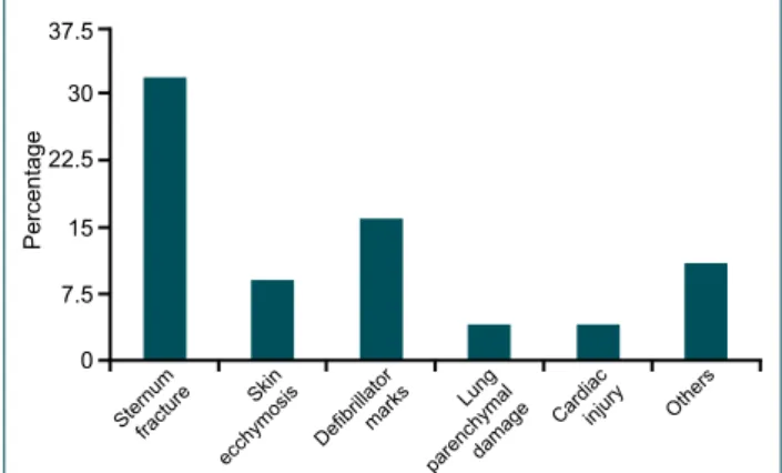
Apart from rib fractures, pericardium and heart injuries were detected in four cases, lung parenchymal damage in four, and rupture of the pleura in two. From the external examinations, defibrillator pad marks were found in sixteen patients, and skin and/or subcutaneous soft tissue ecchymosis and bleeding were detected in nine patients (Fig. 1).
Regarding chest injuries related to CPR, there was no statis-tically significant difference between the traumatic and non-traumatic groups in terms of rib or sternum fractures, age, or gender (p>0.05). Although more cases occurred during summer, no statistically significant difference by season was found (Fig. 2; p>0.05).
Table 2. Distribution of rib fractures according to location
and gender
Fracture location Men Women Total
Parasternal 10 10 –
Midclavicular 23 15 8
Axillar 2 1 1
Not specified 36 26 10
Table 3. Distribution of sternum fractures according to
location and gender
Fracture location Men Women Total
1 Level of 2nd rib 6 6 0
2 Level of 2nd–3rd intercostal space 6 5 1
3 Level of 3rd rib 14 9 5
4 Level of 3rd–4th intercostal space 2 1 1
5 Level of 4th rib 5 4 1
Total 33* 25 8
*Had broken in two locations in a patient’s sternum.
Figure 1. Rates of complications other than rib fractures. 37.5 22.5 7.5 15 30 Percentage
Sternumfracture Skin ecchymosis Defibrillator
marks Lung Cardiacinjury Others parenchymaldamage 0 Figure 2. Seasonal distribution of the cases. 7.8 15.5 23.3 31. 38.8 Percentage Winter 22.4 21.5 30.1 25.8
Spring Summer Autumn
DISCUSSION
If closed-chest compressions are not performed properly during BLS, they can be traumatic. After examination of the patients, complications related to thorax compression such as rib and sternum fractures, were observed. Additionally, injuries to internal organs such as the heart, lung, liver, and
stomach, occur frequently.[6–10] Complication rate can vary
depending on the individual performing the chest compres-sion (e.g., medical personnel vs. lay people at the scene), the surface on and location at which it is performed (in the hos-pital vs. outside), and the education level and capability of the
individual applying it.[6–10] Thus, we believe that continuous
development of BLS training strategies and high-quality train-ing in cardiopulmonary resuscitation can reduce resuscita-tion-related complications.
When analysing complications caused by closed-chest com-pressions, methodological issues must be resolved first. Besides autopsies, computed tomography (CT) and X-ray equipment were used in this research. In this study, 86% of rib fractures detected by autopsy were not detected by AP chest
X-rays.[11] CT is much more sensitive than X-ray radiography,
especially for rib fracture detection. Regardless, in general, an autopsy is the best method for detecting resuscitation
com-plications.[6,11,12] According to the above mentioned studies,
detailed autopsy is still the most sensitive method.
The incidences of rib and sternum fractures, the most of-ten encountered complications of chest compressions, were 4–97% according to autopsy and 8-32% according to X-ray. For sternum fractures, the incidences were 1–43% by autopsy
and 0–26% by X-ray.[6–13] However, in a prospective CT study
performed in patients who were resuscitated as a result of successful BLS, the incidence of rib fractures amounted to
31% and that of sternum fractures to 4%.[6] The third, fourth,
and fifth ribs, especially on the left side of the sternum, were fractured most often. In our autopsy study, the rates of rib and sternum fractures were similar and amounted to 71% for rib fractures and 32% for sternum fractures. Fractures resulting from complications related to resuscitation were observed mostly on the left side (66%) in the third, fourth, and fifth ribs. Although the studies were heterogeneous, it is clear that complications appearing during cardiopulmonary resuscitation are still very common.
Having analysed the distribution of rib fractures according to location (Table 2), most fractures occurred at the midcla-vicular line. Kricher and colleagues have stated that most ribs
fracture at the sterno-condral junction.[10] However, Baubin
and colleagues have claimed in their study using cadavers that most ribs fractures result from chest compressions occur-ring at the axillary line.[14] However, according to the study of
Buschmann and Tsokos and many researches performed, frac-tures usually occur at the midclavicular line.[6–9,11–13] In three
studies conducted in Turkey in 2003, 2008, and 2010, most
rib fractures were reported to occur at the midclavicular line.
[7,8,15] The results of our study were consistent with the
lit-erature reports, in that most rib fractures occurred at the midclavicular line.
The localization of sternal fractures has been reported to be between the second and fourth ribs. Most fractures occur at the sternum at the level of the third intercostal space. Similar results have been reported in domestic and foreign research studies.[7,8,10–12,15,16]
Having analysed the results in terms of skin complications and internal organ damage, in the cases of distant organs (e.g., retinal haemorrhage and subarachnoid haemorrhage) and internal organ damage, their occurrence is not common. When compressing the thorax, stress created by increased thoracic pressure affects many organs. Lacerations of diverse
organs related to this issue have been described.[11] In our
research, injury to internal organs (lung, heart, and liver) was seen at a low rate (10%). The rates of internal organ injury have not changed much since the reports of Krischer and col-leagues and Paaske and colcol-leagues on such injuries and other life-threatening complications related to closed-chest com-pressions. Krischer and colleagues have reported in detail on ten complications, mentioned above, among 705 cases, while Paaske and colleagues have noticed three complications
among 268 cases.[10–18] Life-threatening complications were
rare in these studies.
Due to closed-chest compressions, damage to intrathoracic organs is often encountered. They usually occur in relation to rib and sternum fractures. However, this is not always the case. Organ injuries can also occur without fracture. The most important factors leading to this situation include misapplication and external chest compressions that are too strong, fast, deep, and long. Multiple injuries such as lung con-tusions, pleural rupture, anterior mediastinal bleeding, peri-cardial injury, cardiac injury, pneumothorax, haemothorax, and mediastinal emphysema have been observed. When the heart is tightened between the anterior chest wall and spine during chest compressions, epicardial petechiae and
myocar-dial haemorrhage can occur.[6–18] In addition, in our autopsy
reports, cases with pulmonary contusion, pleural rupture, pericardial injury/bleeding, and endomyocardial bleeding were observed. These findings were similar to those of the litera-ture reports.
Intra-abdominal injuries are rare resuscitation complications. However, many organs may be affected. Liver and spleen lac-eration, gastric dilatation and gastric perforation are encoun-tered. Moreover, intestinal trauma, intraperitoneal bleeding, and retroperitoneal haematomas have been recorded as complications. In the literature, liver damage is seen at an incidence of ~0.6 3% and is the most frequent intra-abdom-inal complication related to CPR.[10,19–26] Liver laceration was
the left lobe. The most important factor in this is the close anatomical relationship between the left lobe of the liver and the sword-shaped lower end of the sternum. A similar lacera-tion was seen in our cases. Two other important factors that can increase the risk of liver laceration are hepatic ischaemia
and liver distension due to venous obstruction.[10,19–26] The
reason for liver laceration in our case was thought to be simi-lar. Moreover, treatments with antiplatelet, antithrombotic, and thrombolytic agents in patients with myocardial infarc-tion may cause such lacerainfarc-tions.[21,22,26]
No significant gender influence on rib fractures has been detected by the majority of studies. However, rib fractures generally occur more frequently in elderly women with high osteoporosis rates and in both genders of more advanced age.[9–13] However, according to research conducted in our
country by Şam and colleagues in 2003, more complications were detected in men and younger patients (average age of
35 years).[15] In the studies of Boz and colleagues from 2008,
the rate of resuscitation complications was found to be high
in men and the elderly.[7] Similarly, in a study conducted in
2010 by Özer and colleagues, the rate of resuscitation
com-plications was high (71.9%) in men and the elderly.[8] In our
study, similar to others conducted in Turkey, more than half of the cases (55%) were 55 years and over, and most (75%) were male.
Hoke et al. and Black et al. have reported an incidence of rib cage fractures of less than 50% and of sternum fracture of less than 30%. In our study, we detected 71% rib cage frac-tures and 32% sternum fracfrac-tures, higher than the average in-cidences reported in the literature. These results emphasise the importance of the education provided on resuscitation, and significant effort should be given to revise this to lower the complication rates.
A limitation of our study is the lack of data regarding sur-face type on which the chest compressions were performed, the depth of application, and the duration of application. Moreover, to obtain more detailed results on resuscitation complications, more in-depth reports on chest compressions cases should be prepared based on the following: a sufficient number of cases, homogenized study populations, exclusion of associated conditions, and stratification by age, gender, du-ration of application, depth of application, and soft or hard surfaces.
Our research was conducted with approval from our local ethics committee. There are no conflicts of interest to re-port.
Conclusion
In BLS, resuscitation is a procedure performed to bring pa-tients back to life and to increase their chance of survival. However, at the same time, it can also cause complications
that may even lead to the patient’s death. Resuscitation per-formed in the correct manner will cause less organ injury and prevent potentially fatal complications. It should be noted that chest compressions applied with enough strength to break the ribs or sternum can harm the thorax and many organs and may decrease the patient’s chance of survival. Awareness of the complications occurring with BLS is vital in autopsy investigations. In patients in whom the major cause of death is trauma, these complications may be interpreted as additional trauma symptoms. However, in patients who died due to non-traumatic reasons, autopsy may be misinterpret-ed by these complication findings, suggesting that the cause of death involved trauma. Thus, detailed information about the application of BLS and its possible complications are cru-cial for forensic medical specru-cialists to distinguish them from actual trauma symptoms. Moreover, it is also important for doctors, who usually perform resuscitation, and other medi-cal personnel to be aware of these complications in terms of comprehension and a reduction in medical errors. Thus, BLS training should be repeated at appropriate intervals based on current approaches.
Note: The English in this document has been checked by at least two professional editors, both native speakers of Eng-lish. For a certificate, please see:
http://www.textcheck.com/certificate/zzLYdk Conflict of interest: None declared.
REFERENCES
1. http://www.saglik.gov.tr/TR/belge/1-552/ilkyardim-yonetmeligi.html. Access date:15.01.2014.
2. http://www.kalite.saglik.gov.tr/content/files/mevzuat/saglikta_perfor-mans_ve_kalite_yonergesi_yeni/hkskitap.pdf. Access date:15.01.2014. 3.
http://www.kalite.saglik.gov.tr/content/files/mevzuat/saglikta_per-formans_ve_kalite_yonergesi_yeni/3_2ek4112son.pdf. Access date:15.01.2014.
4. Berg RA, Hemphill R, Abella BS, Aufderheide TP, Cave DM, Hazinski MF, et al. Part 5: adult basic life support: 2010 American Heart Asso-ciation Guidelines for Cardiopulmonary Resuscitation and Emergency Cardiovascular Care. Circulation 2010;122(18 Suppl 3):685–705. 5. Koster RW, Baubin MA, Bossaert LL, Caballero A, Cassan P, Castrén M,
et al. European Resuscitation Council Guidelines for Resuscitation 2010 Section 2. Adult basic life support and use of automated external defibril-lators. Resuscitation 2010;81:1277–92.
6. Kim MJ, Park YS, Kim SW, Yoon YS, Lee KR, Lim TH, et al. Chest injury following cardiopulmonary resuscitation: a prospective computed tomography evaluation. Resuscitation 2013;84:361–4.
7. Boz B, Erdur B, Acar K, Ergin A, Türkçüer I, Ergin N. Frequency of skeletal chest injuries associated with cardiopulmonary resuscitation: forensic autopsy. [Article in Turkish] Ulus Travma Acil Cerrahi Derg 2008;14:216–20.
8. Özer E, Şam B, Tokdemir MB, Çetin G. Complications of cardiopul-monary resuscitation. Cumhuriyet Tıp Dergisi Cumhuriyet Tıp Dergisi 2010;32:315–22.
OLGU SUNUMU
Muğla ilinde yapılan ot
opsi olgu sonuçlarında karşıl
aşılan resüsitasyon komplikasyonları
Dr. Halil Beydilli,1 Dr. Yasemin Balcı,2 Dr. Şahin Işık,2 Dr. Melike Erbaş,3 Dr. Ethem Acar,1 Dr. Bülent Savran31Muğla Sıtkı Koçman Üniversitesi Tıp Fakültesi, Acil Tıp Anabilim Dalı, Muğla 2Muğla Sıtkı Koçman Üniversitesi Tıp Fakültesi, Adli Tıp Anabilim Dalı, Muğla 3Adalet Bakanlığı, Muğla Adli Tıp Şube Müdürlüğü, Muğla
AMAÇ: Bu çalışmanın amacı, otopsiler sırasında görülen resüsitasyon komplikasyonların belirlenmesi ve temel yaşam desteği eğitimi etkinliğini değerlendirmektir.
GEREÇ VE YÖNTEM: Muğla Adli Tıp Şube Müdürlüğü’nde gerçekleştirilen otopsi olgu raporları geriye dönük olarak incelendi. Resüsitasyon komp-likasyonları olan hastaların yaş, cinsiyet, ölüm şekli ve çeşitleri gibi demografik verileri ve kompkomp-likasyonların özellikleri kaydedildi.
BULGULAR: Toplam, resüsitasyon komplikasyonlara 100 olgunun 74’ü erkektir. Bu olguların çoğunda otopsileri yaz sezonunda yapıldı. Hastalar arasında %68 travmatik olmayan nedenlerle öldü. Kaburga kırıkları 71 hastada, sternum kırıkları 32 hastada tespit edilmiştir. Ayrıca, perikart (%2) ve akciğer parankim (%4), kalp lezyonları (%4) ve karaciğer laserasyon (%2) hasarı tespit edildi. Kaburga kırıkları ile ilgili olarak, meydana gelen kırıklar en yüksek sayılarda dördüncü kaburga, her iki tarafta ilk ve sekizinci kaburga arasında bulundu.
TARTIŞMA: Resüsitasyon komplikasyonları önemli, çünkü bu komplikasyonlar otopside travmatik nedenlerle yapılmış sanılabilir. Travmatik olmayan nedenlerle yapılan otopsi durumlarında görülen resüsitasyon komplikasyonları travmatik olaylar olarak algılanabilir. Onlar, yanlış olarak travma be-lirtileri sanılabilir. Bu komplikasyonlar sağlık personelinin iyi resüsitasyon eğitimi ile azaltılabilir.
Anahtar sözcükler: Kapalı-göğüs sıkıştırma; otopsi; resüsitasyon; resüsitasyon komplikasyonlar; temel yaşam desteği. Ulus Travma Acil Cerrahi Derg 2015;21(6):463–468 doi: 10.5505/tjtes.2015.66169
ORİJİNAL ÇALIŞMA - ÖZET
9. Black CJ, Busuttil A, Robertson C. Chest wall injuries following cardio-pulmonary resuscitation. Resuscitation 2004;63:339–43.
10. Krischer JP, Fine EG, Davis JH, Nagel EL. Complications of cardiac re-suscitation. Chest 1987;92:287–91.
11. Lederer W, Mair D, Rabl W, Baubin M. Frequency of rib and sternum frac-tures associated with out-of-hospital cardiopulmonary resuscitation is un-derestimated by conventional chest X-ray. Resuscitation 2004;60:157–62. 12. Kim EY, Yang HJ, Sung YM, Cho SH, Kim JH, Kim HS, et al. Multi-detector CT findings of skeletal chest injuries secondary to cardiopulmo-nary resuscitation. Resuscitation 2011;82:1285–8.
13. Buschmann CT, Tsokos M. Frequent and rare complications of resuscita-tion attempts. Intensive Care Med 2009;35:397–404.
14. Baubin M, Sumann G, Rabl W, Eibl G, Wenzel V, Mair P. Increased fre-quency of thorax injuries with ACD-CPR. Resuscitation 1999;41:33–8. 15. Şam B, Saka E, Süner Ç. Adli otopsilerde resusitasyon komplikasyonları.
Adli Tıp Bülteni 2003;8:5–8.
16. Hoke RS, Chamberlain D. Skeletal chest injuries secondary to cardiopul-monary resuscitation. Resuscitation 2004;63:327–38.
17. Paaske F, Hansen JP, Koudahl G, Olsen J. Complications of closed-chest cardiac massage in a forensic autopsy material. Dan Med Bull 1968;15:225–30.
18. Hellevuo H, Sainio M, Nevalainen R, Huhtala H, Olkkola KT, Ten-hunen J, et al. Deeper chest compression - more complications for cardiac arrest patients? Resuscitation 2013;84:760–5.
19. Kapłon-Cieślicka A, Kosior DA, Grabowski M, Rdzanek A, Huczek Z,
Opolski G. Coronary artery dissection, traumatic liver and spleen injury after cardiopulmonary resuscitation - a case report and review of the lit-erature. Arch Med Sci 2013;9:1158–61.
20. Rosen J, Tuchek JM, Hartmann JR. Liver laceration in the hemodynami-cally unstable post-cardiac massage patient: early recognition and man-agement-case report. J Trauma 1999;47:408–9.
21. Adams HA, Schmitz CS, Block G, Schlichting C. Intra-abdominal bleeding after myocardial infarction with cardiopulmonary resusci-tation and thrombolytic therapy. [Article in German] Anaesthesist 1995;44(8):585–9. [Abstract]
22. Pezzi A, Pasetti G, Lombardi F, Fiorentini C, Iapichino G. Liver rupture after cardiopulmonary resuscitation (CPR) and thrombolysis. Intensive Care Med 1999;25:1032.
23. Druwé PM, Cools FJ, De Raedt HJ, Bossaert LL. Liver rupture after car-diopulmonary resuscitation in a patient receiving thrombolytic therapy. Resuscitation 1996;32:213–6.
24. Meron G, Kurkciyan I, Sterz F, Susani M, Domanovits H, Tobler K, et al. Cardiopulmonary resuscitation-associated major liver injury. Resuscita-tion 2007;75:445–53.
25. Camden JR, Carucci LR. Liver injury diagnosed on computed tomog-raphy after use of an automated cardiopulmonary resuscitation device. Emerg Radiol 2011;18:429–31.
26. Ziegenfuss MD, Mullany DV. Traumatic liver injury complicating car-dio-pulmonary resuscitation. The value of a major intensive care facility: a report of two cases. Crit Care Resusc 2004;6:102–4.