ORIGINAL
ARTICLE
/Interventional
imaging
Central
venous
occlusion
in
hemodialysis
access:
Comparison
between
percutaneous
transluminal
angioplasty
alone
and
nitinol
or
stainless-steel
stent
placement
S.
Gür
a,∗,
L.
O˘
guzkurt
b,
M.
Gediko˘
glu
caDepartmentofRadiology,DivisionofInterventionalRadiology,IzmirKatipCelebiUniversity,
FacultyofMedicine,IzmirTurkey
bDivisionofInterventionalRadiology,DepartmentofRadiology,KocUniversity,Facultyof
Medicine,IstanbulTurkey
cDivisionofInterventionalRadiology,DepartmentofRadiology,BaskentUniversity,Faculty
ofMedicine,AdanaTurkey
KEYWORDS Centralvein occlusion; Angioplasty; Hemodialysis; Centralvenous occlusion; Stenting Abstract
Purpose:Thepurposeofthisstudywastocomparetheprimaryandsecondarypatencyrates ofpercutaneoustransluminalangioplasty(PTA)alonewiththoseofmetallicstentplacement inpatientswithhemodialysisaccessandcentralvenousocclusion(CVO)andtocomparethe respectiveeffectsofnitinolandstainless-steelstentsonpatency.
Materıalsandmethods:Atotalof150consecutivepatientswithhemodialysisaccesswho under-wentendovascular treatmentforsymptomaticCVOwithipsilateralfunctioning hemodialysis accesswereevaluated.Therewere67menand83womenwithameanageof56.2±15.2(SD) years(range:15—86years).TheprimaryendovasculartreatmentofCVOwasPTAalone.Stent placementeitherwithnitinolorstainless-steelstentswasperformedasabailoutprocedure. Theresultswereanalyzedonaperpatientbasis.
Results:Technicalsuccesswasachievedin141/150patients(94%).Ofthe141patients,109 (77%) underwentPTAaloneand32 (23%)underwent stentplacement.The meannumber of interventionsinthestentgroup[4.3±2.5(SD)]wassignificantlyhigherthanthatinthePTA alonegroup[2.6±2.8(SD)](P=0.002).Theprimarypatencyratesat12,24,and60months forthestentgroup(58.7%,41.9%,and27.9%,respectively)weresignificantlyhigherthanthose inthePTAalonegroup(42.4%,36.3%,and20.2%,respectively)(P=0.036).Secondarypatency ratesat12,24,and60monthsforthestentgroup(87.6%,80.7%,and50.3%,respectively)were
∗Correspondingauthorat:DivisionofInterventionalRadiology,DepartmentofRadiology,IzmirKatipCelebiUniversity,FacultyofMedicine,
KarabaglarIzmir,35360,Turkey.
E-mailaddress:[email protected](S.Gür).
https://doi.org/10.1016/j.diii.2019.03.011
(P=0.011).
Conclusıon:AlthoughPTAaloneisaneffectiveinterventionaltreatmentstrategyofCVOinshort term,stentplacementyieldsgreaterprimaryandsecondarypatencyratesinthelong-term.But themeannumberofinterventionsperveinafterstentingissignificantlyhigher.Closefollow-up andmultiplere-interventionsarenecessarytoensurelong-termpatency.
©2019Soci´et´efranc¸aisederadiologie.PublishedbyElsevierMassonSAS.Allrightsreserved.
Central venous occlusion (CVO) is a common and serious complicationofhemodialysisaccess.TheincidenceofCVO ranges between 25% and 40% [1,2]. Previous history of venouscatheterization isthe most commoncauseof CVO inhemodialysisaccess[3].Central venousinjury and sub-sequentinflammatory responsetrigger intimalhyperplasia andleadtoCVO[4].Fibrinsheathformationafterchronic central venous catheterization is common and leads to catheterdysfunctionandCVO[5].Furthermore,angioplasty canaggravatethevenousintimalresponseandthus accel-eratestenoticprocess[4].Theprimarystandardtreatment forCVOinhemodialysisaccessispercutaneoustransluminal angioplasty(PTA)[6].However,additionalstentplacement may be needed in patients who have insufficient blood flowduringinterventionor earlypostoperativeperiod[7]. Nitinol stents and stainless steel stents have been used for stenting [8—10]. Primary patency rates of stents and PTA are poor and secondary patency can be maintained withrepeatedinterventions. Treatment withconventional PTA and stenting paradoxically triggers the development ofvenous neointimalhyperplasia inlong-term. Therefore, effortstopreventobstructioncausedbyneointimal hyper-plasiatriggeredbyinterventionaltreatmentareincreasing. Studies have focused on covered stents and paclitaxel-coated balloons (PCBs) to improve long-term patency of recurrent hemodialysis access vascular stenosis and CVO causedbyneointimalhyperplasia[11—15].
Inthe present study,itwashypothesized that stenting mayhavebetterprimaryandsecondarypatencyratesthan PTAaloneandthepatencyofnitinolstentsmaybelonger thanthatofstainless-steelstentsinthetreatmentofCVO inhemodialysisaccess.
Thepurposeofthisstudywastocomparetheprimaryand secondarypatencyratesofPTAalonewiththoseofmetallic stentplacementin patients with hemodialysisaccessand CVOandto comparethe respectiveeffectsof nitinoland stainless-steelstentsonpatency.
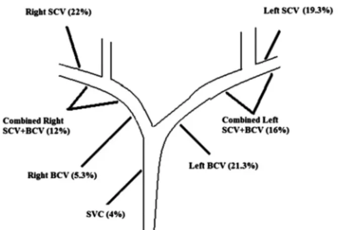
Figure 1. Diagram shows all central vein lesion sites in 150 patientswithcorresponding prevalencein percentage.SCV indi-cates subclavian vein, BCV indicates brachiocephalic vein, SVC indicatessuperiorvenacava.
Materials
and
methods
Patients
Thisstudywasapprovedbytheinstitutionalreviewboardof ourlocalethicscommittee.Therecordsof150consecutive hemodialysis patients whounderwent endovascular treat-ment of symptomatic CVO ipsilateralhemodialysis access between 2000 and 2014 were retrospectively reviewed. There were 67 men and 83 women with a mean age of 56.2±15.2 (SD) years (range: 15—86 years). CVO was locatedintherightsubclavianvein(SCV)in33/150patients (22%), left brachiocephalic vein (BCV) in 32/150 patients (21.3%), left SCV in 29/150 patients (19.3%), left SCV and BCV in 24/150 patients (16%), right SCV and BCV in 18/150 patients (12%), right BCV in 8/150 patients (5.3%)andsuperiorvenacava(SVC)in6/150patients(4%) (Fig.1).
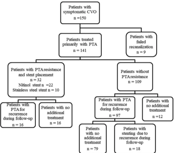
Figure2. Flowchartofthestudy.CVOindicatescentralvenousocclusion.PTAindicatespercutaneoustransluminalangioplasty.
A total of 141 patients who underwent successful endovascular treatment were included in the study. Nine patientsinwhomtheCVOcouldnotbetraversedduringthe primaryinterventionwereexcludedfromthestudygroup. Additionally,patientswithpriorepisodeofsevereallergyto iodinatedcontrastmaterialandclinicallyunstablepatients werenottreatedandalsoexcludedfromthestudy.Patients werereferredtoourinterventionalradiologyclinicwith pro-longedbleedingfollowinghemodialysis,elevationofstatic venouspressure,andipsilateralarmorneckswelling.
Demographicdataofpatientsandmedicalrecordswere obtainedfromthehospitalinformationsystem.Detailswere obtainedfrominterventionalradiologyreportsand venogra-phyimagesstoredindigitalmedia.
Procedure
details
AdiagnosisofCVOwasconfirmedbasedonvenography.All procedureswereperformedby3interventionalradiologists withatleast5yearsofexperienceandwithnationalboard certificationapprovedbytheCardiovascular and Interven-tionalRadiologicalSocietyofEuropa(S.G.,10years;L.O., 25 years;M.G., 5 years).All venogramsand endovascular interventions were performed using a digital subtraction angiography unit (Innova® 3100, General Electric Health-care, or Multistar®, Siemens Healthineers). All endovas-cularinterventionswereoutpatientproceduresperformed
during the same session with diagnostic venography. The lesionstreatedwithPTAorstentplacementwerethesame lesionsand the results were considered on a per patient basis.
Accesstotheveinwasachievedwithasingle-wall punc-tureunder ultrasound guidance.We initially placeda 5-F vascular sheath into the access vein. The basilic or the brachial veins were favored for the diagnostic venogram andendovascularintervention;however,thecephalicvein was also used if these vessels were not suitable. When CVOwasconfirmedbyvenography,the5-Fvascularsheath wasreplacedby an 8-F to 10-Fvascular sheathand 5000 IUofheparinwasadministeredintravenously.Thestenotic vessel segment was passed by a guidewire and PTA was performed. When occlusion waspresent, then it was tra-versed withan angled or a straight hydrophilic guidewire (Glidewire,Terumo,or Roadrunner,Cook)withthehelpof anangledcatheter(Kumpe,Cook,).Highpressureballoons (Atlas/Conquest,Bard)wereusedforPTA-resistantlesions. Thediametersoftheballooncathetersrangedfrom10-to 18-mm(mean,12.8±1.5mm[SD]).Stentdiametersranged from10—18mm[mean,11.2±4.7mm(SD)]andstentlength rangedfrom40—90mm.Stentswereonlyusedasabailout procedureandallthestentsusedwereself-expandingand eitherstainless-steel(Wallstent®,BostonScientific)or niti-nol(Protégé®, ev3,or Smart®,Cordis,). Theflow chartof thestudyisgiveninFig.2.
Female 59 20 83
Age(year)±SD 54.7±15.9[15—78] 60.7±12.6[30—86] 56.2±15.3[15—86]
PTA:percutaneoustransluminalangioplasty;SD:standarddeviation.Numbersinbracketsarerange.
Follow-up
Repeated venography was performed in patients with recurrent symptoms.Follow-up wasfinished when loss of follow-up, patient death, abandoning of the ipsilateral hemodialysisaccess,orrenaltransplantationoccurred.
Outcome
definitions
CVOwasdefinedasastenosisgreaterthan50%orocclusion ofthecentralvenoussystemthatincludedSCV,BCVandSVC. Technical failurewasdefinedasthe impossibilitytocross vascularlesions.Technicalsuccesswasdefinedas restora-tion of flow without significant residual stenosis<30%. A significant decrease in the pressure gradient in patients with persistent collateral vein filling was also accepted assuccess. Post-interventionprimarypatencywasdefined astheintervalof uninterruptedpatencyafter endovascu-lar intervention within a dialysis circuit to thrombosis or repeatedendovascularinterventioninthePTAalonegroup and stent groups. Post-intervention secondary patency of PTA was defined as the interval from the initial proce-dureuntilpermanentocclusionorinterventionnecessitating stent placement. Post-intervention secondary patency of stentgroupwasdefinedastheintervalfromtheplacement offirststentuntilpermanentocclusionof thelesion, sur-gicalligation,renaltransplant,and/orlosstofollow-upor death[16].
Statistical
analysis
Astatisticssoftwarepackage(SPSS,version19.0,SPSS)was used for statistical analysis. Quantitative variables were expressed as mean±standard deviation (SD) and range, or median, first (Q1) and third quartiles (Q3). Qualita-tivevariableswereexpressedasrawnumbers,proportions andpercentages.Survival curvesforthe primaryand sec-ondaryveinpatencyofthedifferentinterventiontypesand differentstenttypesweregeneratedusingKaplan-Meier sur-vivalanalysis,and thelog-rank test wasusedtocompare luminal patency. Differences between means for contin-uous variables were evaluated using the Student t-test.
The Mann-Whitney U test was used to compare the out-comeof patients treatedwithstainless steel withthatof patients treated with nitinol stent. Pearson’s correlation testwasusedtoinvestigatetherelationshipbetweenlesion lengthandsecondarypatencyresults.Significancewassetat
P-value<0.05.
Results
Themostcommonpatientcomplaintswereipsilateralarm swelling (145/150 patients; 97%), additional face or neck swelling (57/150 patients; 38%), breast swelling (5/150 patients;3.3%),andinabilitytopuncturethevascularaccess (19/150 patients; 12.6%). There wasno significant differ-ence between the PTA and stent groups in terms of age and sex(Table 1).Twenty-five patients (25/150;17%) had radiocephalicarteriovenousfistula(AVF),and125patients (125/150;83%) hadbrachiocephalicAVF.Onehundredand thirty-five patients (135/150; 90%) had aprevious ipsilat-eralcentralvenouscatheterplacement.Eighty-fivepatients (85/150; 57%) had a history of a single catheter place-ment, andthe remaining50 patients (50/150;33.3%) had a history of more than one catheter placement. Eighty-four patients (84/150; 56%) had an internal jugular vein (IJV) catheter, and 51 patients (51/150; 34%) had a SCV catheter. All of SCV catheters were implanted in outside institutions.
Ninety-onepatients(91/150; 61%)had stenosis greater than 50%, and 59 patients (59/150; 39%) had occlusion. Forty-twopatients(42/150;28%)hadmultiplestenosesand twenty-fourofthem(24/150;18%)wereontheleftside.All of SCV-BCVcombinedlesionsin 42patients (42/150; 28%) acceptedasasinglelesionandtheresultswereconsidered onaperpatientbasis.Thelocationsandtypesoflesionsare presentedinTable2.Technicalsuccesswasachievedin141 patients(141/150;94%).TheCVOcouldnotbere-canalized inninepatients(9/150;6%).Ofthe141patients,14(14/141; 10%) had additionalacute centralvenous thrombosis that was found initially. Catheter-directed thrombolysis in 4 patients(4/141;2.8%)andmanualaspirationthrombectomy in10(10/141;7%)patientswasperformed.Allpatientswith acuteorsubacutecentralvenousthrombosishadan under-lyingchronicstenosis or occlusion,andweretreatedwith PTA alonein 12patients (12/141; 10.6%)or withPTA and bailoutstentingin2patients(2/141;1.4%).
Stentswereplacedin32patients(22nitinolstentsand10 stainlesssteel stents)withPTA-resistant lesions.However, PTAwasagaintheprimarytreatmentforin-stentocclusive lesions,andrepeatstentingwasperformedinPTA-resistant lesions.In6/141patients(4%),fourstenosis(4/141;2.8%) andtwoocclusions(2/141;1.4%)werepresentintheSVC.
The mean follow-up interval for all patients was 36.5±21.9 (SD)months (range:1—93months). The mean follow-upintervalwas36.6±21(SD)months(range:1—93 months) for the PTA group and 37.6±27.3 (SD) months (range: 6—93 months) for the stent group. According to
Table2 Locationsandtypesoflesionsin141treatedpatients.
Lesionsites PTAgroup(n=109) Stentgroup(n=32) Total(n=141)
Typeoflesions Typeoflesions
Stenosis Occlusion Stenosis Occlusion
SCV 31(31/141;21.9) 19(19/141;13.4) 2(2/141;1.4) 3(3/141;2.1) 55(55/141;39) BCV 19(19/141;13.4) 5(5/141;3.5) 9(9/141;6.3) 4(4/141;2.8) 37(37/141;26.2) SCV+BCV 19(19/141;13.4) 11(11/141;7.8) 3(3/141;2.1) 10(10/141;7) 43(43/141;30.4)
SVC 4(4/141;2.8) 1(1/141;0.7) 0(0/141;0) 1(1/141;0.7) 6(6/141;4.2)
Total 73(73/141;51.7) 36(36/141;25.5) 14(14/141;10) 18(18/141;12.7) 141(141/141;100)
SCVindicates subclavian vein;BCV indicates brachiocephalicvein;SVC indicates superiorvena cava.Numbers inparentheses are proportionsfollowedbypercentages.
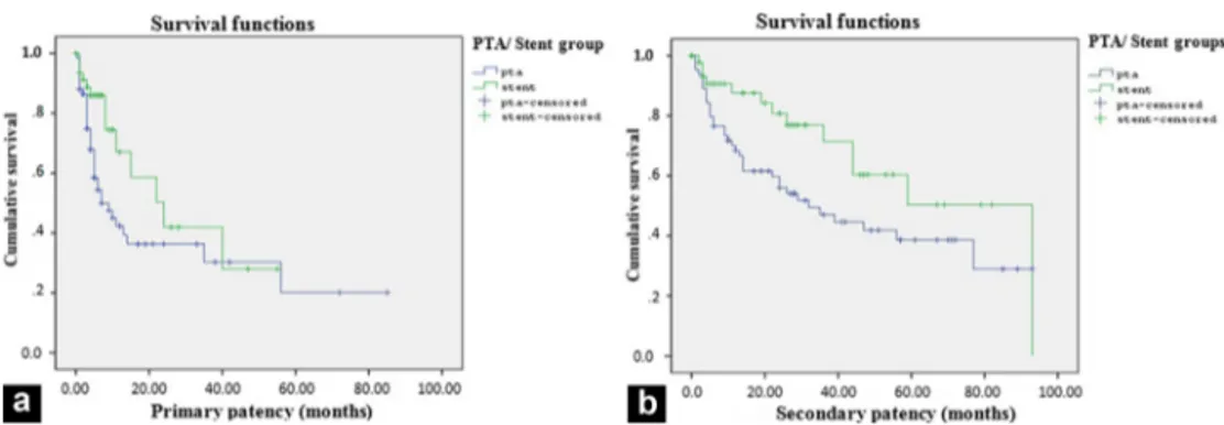
Figure3. Graphsshowpatencyofcentralveinafterprimarypercutaneoustransluminalangioplasty(PTA)in109patientsandafterstenting in32patients.Graphsshowprimarypatency(a)andsecondarypatency(b)ofcentralveins.Thereissignificantdifferenceinprimaryor secondarypatencyratesbetweenthePTAandstentgroup(P=0.036andP=0.046,respectively)byKaplan-Meieranalysis.PTA=percutaneous transluminalangioplasty.
Figure4. Graphsshowpatencyofcentralveinafterplacementof22nitinolstentsandplacementof10stainless-steelstents.Graphs showprimarypatency(a)andsecondarypatency(b)ofcentralvein.Primaryandsecondarypatencyratesinthenitinolstentgroupare significantlygreatercomparedtothestainless-steelstentgroup(P=0.024and0.011,respectively)byKaplan—Meieranalysis.
Kaplan-Meier analysis, the primary patency rates at 12, 24, and60 months weresignificantly greater in the stent group (58.7%,41.9%, and27.9%, respectively)than in the PTA alone group (42.4%, 36.3%, and 20.2%, respectively) (P=0.036)(Fig.3).Secondarypatencyratesat12,24,and60 monthsweresignificantlygreaterinthestentgroup(87.6%, 80.7%,and50.3%,respectively)thaninthePTAalonegroup (68.4%, 56%, and 38.6%, respectively) (P=0.046) (Fig. 3). Furthermore,theprimarypatencyratesat6and12months were significantly greater in the nitinol stent group (89% and 80.9%, respectively) than in the stainless-steel stent
group(78.8% and 38.4%, respectively)(P=0.007) (Fig.4). Thesecondarypatencyratesat6,12and24monthswere sig-nificantlygreaterinthenitinolstentgroup(92.8%,87.7%and 65.8%,respectively)thaninthestainless-steelstentgroup (85.7%,76.2%and65.3%,respectively)(P=0.011)(Fig.4).
Themeannumberofinterventionsperveinwas2.9±2.8 (SD)forallpatients(range,1—17).ForthePTAgroup,the mean number was 2.6±2.8 (SD) (range, 1—10), and the meannumberwas4.3±2.5(SD)(range,1-17)forthestent group.The meannumberofinterventionspervein follow-ingstenting(4.3±2.5 [SD])wassignificantlygreater than
LesionSites SCV 4(1—8) 8(6.2—22.2) 9(1—8) 24(6—51) 22(11—55) 29.5(6—51) BCV 8(5—13.5) 4(2—6.5) 6(5—13.5) 24.5(12.5—51.7) 24(3.5—46.5) 28(12.5—51.7) SCV-BCV 5(3—8.5) 5(3—9.5) 6(3—8.5) 19(5.7—39.7) 24(6—37.5) 27(5.7—39.7) LesionTypes Stenosis 5(3—12) 6(3.7—10.2) 6(3—11) 25(10—41.2) 28(8.5—50.5) 31(10—42) Occlusion 4(3—9) 4(2.5—9.5) 6(3—9) 11.5(4.7—56.2) 17(3.5—44) 24(4—47)
PTA:percutaneoustransluminalangioplasty;PTS:percutaneoustransluminalstenting;SCV:subclavianvein;BCV:brachiocephalicvein.
that after management with PTA alone (P=0.002). In all patients with stenting, neointimal hyperplasia developed in the stent walls during the follow-up period. Ninety-sevenpatients (97/109; 89%)in thePTA group underwent 176repeatPTAprocedures.In16 patients(16/32;50%)of thestent group,150 re-interventions were performed for recurrentlesions.Seventy-nineofthe recurrentlesionsin seventy-nine patients (79/109; 72.4%) responded well to PTAalone,butadditionalstentswereneededin18patients (18/109;16.5%).
Therewasnosignificant differenceinprimaryand sec-ondarypatencyratesofPTAandstentingforocclusionversus stenosis(P=0.6,P=0.3,P=0.2,P=0.1).Medianprimaryand secondarypatencyforthePTAgroup,thestentgroup,and thewholestudy group arereportedaccording to obstruc-tionsite(BCV,SCV, orcombined)andlesiontype(stenosis or occlusion) in Table 3. A poor negativecorrelation was found betweenlesion length andsecondary patencyafter PTA(r=−0.35;P=0.004).
Fourpatients(4/141;2.8%)hadextravasationfollowing PTA. After temporal balloon tamponade, the extravasa-tion was resolved in all patients. Two patients required placement of additional stents due to shortening of the stainless-steelstentsinbothSCVs.Complicationswere min-imalanddidnotrequirehospitalizationaccordingtoSociety ofInterventionalRadiologyStandardsofPracticeCommittee
[16].Duringfollow-up,twonitinolstentsthatwereplaced intheSCVwerefracturedandonenitinolstentintheleft BCVcollapsedbecauseofextrinsiccompression.Thesethree patientsweretreatedwithstent-in-stentplacement.There wasnofracturewiththestainless-steelstents.Therewere noperiproceduralmortalities.
Discussion
Inthisstudy,wehave comparedthepatencyrates ofPTA alonetothoseofbailoutstentinginpatientswithCVOand hemodialysisaccess.Wefoundthatstentingyieldedlonger primaryandsecondarypatencyratesthanPTA.Inthestent group,thefrequencyof re-interventionswasgreaterthan thatinthePTAalonegroup.Thecomplicationratesinboth PTA alone and stent groups were very low. Additionally,
nitinol stents yield significantly longer primary and sec-ondarypatencyratescomparedtostainless-steelstents.
One-yearprimarypatencyafterPTAhavebeenreported between 20% and50% in the literatureand themaximum one-yearprimarypatencywas77%inthestudythatwas con-ductedbyOzyeretal. [10,17—20].Theresults ofpresent study areconsistentwiththoseof priorstudies. One-year primary and secondary patency after primary stenting or stentingafterafailedPTAhavebeenreportedbetween14% and76%aswellas33%and100%,respectively[9,21—23].In recentliterature,itwasreportedthatPCBsseemtoimprove patencyrates of centralveins[24].However,further ran-domizedcontrolledtrialsareneededtofullydeterminethe efficacyofthistreatment.
This present study showed that bailout stenting had higher primary and secondary patency results compared to PTA alone. This result is different from those of pre-vious studies [10,17,20]. Although our study showed that patency results of stenting were better than PTA, stent-ing has been recommended as a salvation procedure by theSociety of InterventionalRadiologyandin theK/DOQI guidelines[7,16].Whilestentingmayhavesuperiorpatency, therearedrawbackssuchasjailingotherveinsand poten-tiallyincreasingneointimalhyperplasia.Probablyduetothe developmentofneointimalhyperplasia,themeannumberof re-interventionsfollowing stentingwassignificantlyhigher thanthatafterPTA[20].
Neointimalhyperplasiaisthemostimportantfactorthat decreases long-term patency in the subsequent period of endovascular treatment of CVO. Over the last ten years, clinicalinvestigationshavefocusedonovercoming neointi-mal hyperplasia[25]. Covered stents andPCBshave been used to improve long-term patency of central veins and hemodialysisaccess stenosis[5,11—15,26]. The advantage of covered stents include providing relative inert stable intravascular matrix for endothelization and a barrier for thedevelopmentofneointimalhyperplasia[27].Verstanding etal.reportedthatcentralvenouscoveredstentplacement isassociatedwithprolongedaccesspatency;butcoverage ofmajorvenousconfluencesisdisadvantage[12].Itshould beavoidedifitispossible.Althoughtherearemany stud-iesthatemphasizetheeffectofPCBsonlong-termpatency inthehemodialysisaccessstenosis,theusageofthese bal-loons in CVOis notclear. A studyshowed thatPCBs yield
longerpatencycomparedtostandardballoonangioplastyin theendovasculartreatmentofcentralveinrestenosis[28]. ThemostcommoncauseofCVOinhemodialysispatientsis previoushistoryofvenouscatheterization[3].SCV catheter-izationis an importantrisk factor of CVOandif itcauses CVO,itinhibits subsequentAVFcreation.Thus,theuseof SCV catheterhasbeen notrecommendedin patientswith kidney disease,accordingtoK/DOQIguidelines[7]. Inour study,44%ofpatientshadaSCVcatheterization.However, noneofthemwereplacedinourclinic.
Inthepresentstudy,itwasfoundthatnitinolstentshad significantlylongerprimaryand secondarypatency results comparedtothestainless-steelstents.Thisfindingmaybe interested withstentstructure andcomposition. Todate, this association hasnot been clearlydemonstrated in the literature.Accordingtotheliterature,inadditiontostent typeandcomposition,recanalizationtypeandotherfactors can beeffective onthe outcomes[29,30]. Self-expanding stents have generally been used for CVO. The stainless-steelstentisa first-generation,self-expandablestentand the advantages of this stent include a low profile, flexi-bility,andradiopacity.Thedisadvantagesofstainless-steel stentinclude an unpredictable amount of shortening dur-ingdelivery.Inaddition,itscapacityfor changingposition and concentricnarrowing asa result of eccentric loading and decreased radial strength [31]. Shortening is espe-cially observed in regions exposed tocontinuous pressure andmovement, suchasthecostoclavicular spaceand the tortuous vein that occurs in the left BCV. Nitinol stents are second generation, self-expandable stents. In addi-tiontosuperelasticity,nitinolstentshaveseveralphysical characteristics that mayconfer longer patency compared to stainless-steelstents [32]. Some previous studies have reportedhigherpatencyratesfornitinolstentscomparedto stainless-steelstents,whileothersfoundnosignificant dif-ferencebetweenthepatencyratesofthetwostenttypes
[10,20,23,24,33,34].
Therearesomelimitationsinthestudy.First,itisa retro-spectivestudy.Randomizedcontrolledstudiesmaybemore informativetoevaluatethemosteffectivetreatmentofCVO in hemodialysis patients. Currently there areno such tri-als.Secondly,thenumberofpatientsinthestentgroupwas smallerthanthatinthePTAgroup.Inaddition,stentingwas notaprimarytreatmentandwasselectivelyperformedin PTA-resistant lesions.Finally, there wasnouniformity for choosingbetweenbaremetalstenttypesasthisdepended onoperator’spreference.
In conclusion, the endovascular approach is an effec-tive treatment in CVO of hemodialysis access. However, systematic follow-up and re-interventions are necessary to maintain access patency in the long-term. Stenting offers higher primary and secondary patency rates than PTA alone. Stent placement should not be performed as the primary treatment for CVO; stents should be recom-mendedinonlybailouttreatment.Althoughwefoundthat nitinol stents have longerprimary and secondary patency resultsthanstainless-steelstentsandbailoutstentingthan PTAalone,moreprospective,randomizedcontrolledtrials shouldbeperformedtoevaluatetheeffectivenessof differ-entendovasculartreatmentmodalities.
Human
and
animal
rights
Theauthorsdeclarethattheworkdescribedhasbeen car-riedoutinaccordancewiththeDeclarationofHelsinkiofthe WorldMedicalAssociation revisedin 2013for experiments involvinghumans.
Informed
consent
and
patient
details
Theauthors declarethatthis reportdoes notcontainany personalinformationthatcouldleadtotheidentificationof thepatient(s).The authors declare that they obtained a written informed consent from the patients and/or volunteers included inthe article. The authorsalso confirm thatthe personaldetailsofthepatientsand/orvolunteershavebeen removed.
Funding
Thisworkdidnotreceive anygrantfromfundingagencies inthepublic,commercial,ornot-for-profitsectors.
Author
contributions
AllauthorsattestthattheymeetthecurrentInternational Committeeof Medical JournalEditors (ICMJE)criteria for Authorship.
Credit
author
statement
Serkan Gür, M.D.:Formal analysis; Investigation; Method-ology;Projectadministration;Resources;Software; Super-vision; Validation; Visualization; Writing - original draft; Writing-review&editing.
LeventOguzkurt,M.D.:Methodology;Project administra-tion,Supervision;Validation,Writing-originaldraft;Writing -review&editing.
Murat Gedikoglu,M. D.: Investigation,Resources; Soft-ware.
Disclosure
of
interest
Theauthorsdeclarethattheyhavenocompetinginterest.
References
[1]Lumsden AB, MacDonald MJ, Isiklar H, Martin LG,KikeriD, HarkerLA, et al. Centralvenous stenosisinthe hemodialy-sispatient:incidenceandefficacyofendovasculartreatment. CardiovascSurg1997;5:504—9.
[2]GlanzS,GordonDH,LipkowitzGS,ButtKM,HongJ,Sclafani SJ.Axillaryandsubclavianveinstenosis:percutaneous angio-plasty.Radiology1988;168:371—3.
parison between catheter exchange alone and catheter exchangewithfibrinsheathangioplasty.DiagnIntervImaging 2019;100:157—62.
[6]KunduS.CentralVenousObstructionManagement.JVascSurg 2009;26:115—21.
[7]Gilmore J. KDOQI clinical practice guidelines and clini-calpracticerecommendations-2006updates.NephrolNursJ 2006;33:487—9.
[8]HaageP,VorwerkD,PirothW,SchuermannK, GuentherRW. Treatmentofhemodialysis-relatedcentralvenousstenosisor occlusion:resultsofprimaryWallstentplacementand follow-upin50patients.Radiology1999;212:175—80.
[9]Gray RJ, Horton KM, Dolmatch BL, Rundback JH, Anaise D, Aquino AO, et al. Use of Wallstents for hemodialysis access-related venous stenoses and occlusions untreatable withballoonangioplasty.Radiology1995;195:479—84.
[10]MayaID,SaddekniS,AllonM.Treatmentofrefractorycentral veinstenosisinhemodialysispatientswithstents.SeminDial 2007;20:78—82.
[11]Anaya-AyalaJE,SmolockCJ,ColvardBD,NaoumJJ,Bismuth J,LumsdenAB,etal.Efficacyofcoveredstentplacementfor central venousocclusivedisease inhemodialysis patients. J VascSurg2011;54:754—9.
[12]Verstandig AG, Berelowitz D, Zaghal I, Goldin I, Olsha O, ShamiehB,etal.Stentgraftsforcentralvenousocclusive dis-ease inpatientswithipsilateralhemodialysisaccess.JVasc IntervRadiol2013;24:1280—7.
[13]LaiCC,FangHC,TsengCJ,LiuCP,MarGY.Percutaneous angio-plastyusingapaclitaxel-coatedballoonimprovestargetlesion restenosisoninflowlesionsofautogenousradiocephalic fistu-las:apilotstudy.JVascIntervRadiol2014;25:535—41.
[14]ZhengJ,CuiJ,QingJM,IraniZ.Safetyandeffectivenessof combinedscoringballoonandpaclitaxel-coatedballoon angio-plasty for stenosis inthehemodialysis access circuit. Diagn IntervImaging2019;100:31—7.
[15]KarunanithyN,MesaIR,DorlingA,CalderF,KatsanosK,Semik V, et al. Paclitaxel-coated balloon fistuloplasty versus plain balloon fistuloplastyonly to preservethepatencyof arteri-ovenousfistulaeusedforhaemodialysis(PAVE):studyprotocol forarandomisedcontrolledtrial.Trials2016;17:1—12.
[16]DariushniaSR,WalkerTG,SilberzweigJE,AnnamalaiG, Krish-namurthyV,MitchellJW,etal.Qualityimprovementguidelines for percutaneous image-guided management of the throm-bosed or dysfunctional dialysis circuit. JVasc Interv Radiol 2016;27:1518—30.
[17]BakkenAM,ProtackCD,SaadWE,LeeDE,WaldmanDL,Davies MG.Long-termoutcomesofprimaryangioplastyandprimary stentingofcentralvenousstenosisinhemodialysispatients.J VascSurg2007;45:776—83.
[18]SurowiecSM,FegleyAJ,TanskiWJ,SivamurthyN,IlligKA,Lee DE,etal.Endovascularmanagementofcentralvenousstenoses
F.Long-termresultsof angioplastyand stentplacementfor treatmentofcentralvenousobstructionin126hemodialysis patients:a10-yearsingle-centerexperience.AmJRoentgenol 2009;193:1672—9.
[21]MickleyV,GörichJ,RilingerN,StorckM,AbendrothD.Stenting ofcentralvenousstenosesinhemodialysispatients:long-term results.KidneyInt1997;51:277—80.
[22]Aytekin C, Boyvat F, Ya˘gmurdur MC, Moray G, Haberal M. Endovascular stent placement in the treatment of upper extremitycentralvenousobstructioninhemodialysispatients. EurJRadiol2004;49:81—5.
[23]RajanDK,SalujaJS.Useofnitinolstents following recanal-izationofcentralvenousocclusionsinhemodialysispatients. CardiovascInterventRadiol2007;30:662—7.
[24]Hongsakul K, Bannangkoon K, Rookkapan S, Boonsrirat U, KritprachaB.Paclitaxel-coatedballoon angioplastyfor early restenosisofcentralveinsinhemodialysispatients:a single-centerinitialexperience.KoreanJRadiol2018;19:410—6.
[25]KitrouP,SpiliopoulosS, KarnabatidisD,KatsanosK. Cutting balloons,covered stents and paclitaxel-coated balloons for thetreatmentofdysfunctionaldialysisaccess.ExpertRevMed Devices2016;13:1119—26.
[26]Kitrou PM, SpiliopoulosS, PapadimatosP, Christeas N, Pet-sas T, Katsanos K, et al. Paclitaxel-coated balloons for thetreatmentof dysfunctionaldialysis access:resultsfrom a single-center, retrospective analysis. Cardiovasc Intervent Radiol2017;40:50—4.
[27]Kundu S. Review of central venous disease in hemodialysis patients.JVascIntervRadiol2010;21:963—8.
[28]MassmannA, Fries P,Obst-Gleditsch K, MinkoP, Shayesteh-KheslatR,BueckerA.Paclitaxel-coatedballoonangioplastyfor symptomaticcentralveinrestenosisinpatientswith hemodial-ysisfistulas.JEndovascTher2015;22:74—9.
[29]Kovalik EC, NewmanGE, Suhocki P,Knelson M, Schwab SJ. Correctionofcentralvenousstenoses:useofangioplastyand vascularWallstents.KidneyInt1994;45:1177—81.
[30]HuangY,ChenB,TanG,ChengG,ZhangY,LiJ,etal.The fea-sibilityandsafetyofathrough-and-throughwiretechniquefor centralvenousocclusionindialysispatients.BMCCardiovasc Disord2016;16:1—11.
[31]Kundu S. Central venous disease in hemodialysis patients: prevalence, etiology and treatment. J Vasc Access 2010;11:1—7.
[32]ClarkTWI.Nitinolstentsinhemodialysisaccess.JVascInterv Radiol2004;10:1037—40.
[33]VogelPM,PariseC.ComparisonofSMARTstentplacementfor arteriovenousgraftsalvageversussuccessfulgraftPTA.JVasc IntervRadiol2005;16:1619—26.
[34]Honnef D, Wingen M, Günther RW, Haage P. Sharp central venousrecanalizationbymeansofaTIPSneedle.Cardiovasc InterventRadiol2005;28:673—6.