Analysis of cyst fluid antigens from different hosts for the
serodiagnosis of hydatidosis
Ahmet DOĞANAY1, Bahadır GÖNENÇ1, H. Oğuz SARIMEHMETOĞLU1, Hatice ÖGE1, Semih ÖGE1, Ramazan ADANIR2, Hamza AVCIOĞLU3, Mehmet TANYÜKSEL4
1 Ankara University, Faculty of Veterinary Medicine, Department of Parasitology, Ankara; 2Akdeniz University, Faculty of
Veterinary Medicine, Department of Parasitology, Burdur; 3Atatürk University, Faculty of Veterinary Medicine, Department of Parasitology, Erzurum; 4Gülhane Military Medicine Academy, Department of Microbiology and Clinical Microbiology, Ankara,
Turkey.
Summary: The aims of this study were to determine the antigenic differences of hydatid cyst fluid from different animals and to investigate the effect of the origin of antigenic materials on the diagnosis of hydatidosis in humans and mice. For this purpose, the proteins of hydatid cyst fluid (HCF) isolated from the liver of sheep and donkeys and secondary hydatid cysts fluid (SHCF) from albino mice infected with protoscolices isolated from sheep and donkeys were separated by SDS-PAGE. This study was performed at the University of Ankara, Faculty of Veterinary Medicine, Department of Parasitology. For comparing antigenic profiles and detecting specific protein bands of those antigens, 10 infected and 10 non-infected human sera were tested for cystic echinococcosis (CE), and 16 positive and 9 negative mice sera were tested for secondary hydatidosis (SH) by using Western blotting technique. The specific protein band for mice sera was detected as 8 kDa (100%) for sheep’s and donkey’s hydatid cyst fluid. The specific proteins for human sera were detected at 24 kDa (100%) and at 8 kDa (100%) for sheep’s and donkey’s hydatid cyst fluid respectively. No band was detected in negative mice and human sera. It was concluded that SHCF antigen from mice could be considered useful as being easy to provide and give good results for the diagnosis of CE.
Key words: Cyst fluid antigen, diagnosis, donkey, mice, sheep, Western blotting.
Hydatidosisin serodiagnosisinde çeşitli konaklardan elde edilen antijenlerin analizi
Özet: Çalışmanın amacı farklı hayvanların hidatik kist sıvısının antijenik farklılıklarını saptamak ve insan ve farelerde hidatidosisin identifikasyonunda antijenik materyalin orijininin etkisini araştırmaktır. Bu amaçla Ankara Üniversitesi Veteriner Fakültesi Parazitoloji Anabilim Dalında yapılan çalışmada, koyun ve eşek karaciğerlerinden elde edilen hidatik kist sıvılarındaki proteinler ile yine koyun ve eşek orijinli protoskolekslerle enfekte albino farelerden elde edilen sekonder hidatik kist sıvılarındaki proteinler SDS-PAGE yöntemi ile ayrılma işlemine tabi tutulmuştur. Bu antijenik yapıların spesifik protein bantlarının tespiti ve karşılaştırılması için kistik ekinokokoz yönünden 10 enfekte, 10 enfekte olmayan insan serumu ve sekonder hidatidosis için 16 pozitif ve 9 negatif fare serumu Western blot yöntemi ile test edilmiştir. Fare serumlarında koyun ve eşek kist sıvıları için tek spesifik protein bandının 8 kDa (%100) olduğu saptanırken, insan serumları kullanıldığında koyun hidatik kist sıvısı için 24 kDa (%100), eşek hidatik kist sıvısı için 8 kDa (%100) bandın spesifik olduğu tespit edilmiştir. İnsan ve fare negatif serumlarında banda rastlanmamıştır. Sonuç olarak kistik ekinikokozun teşhisinde farelerden elde edilen sekonder hidatik kist sıvılarının iyi sonuç verdiği ve elde edilmesinin kolay olması yönüyle hastalığın tanısında rahatça kullanılacağı ortaya konmuştur.
Anahtar sözcükler: Kist sıvısı antijeni, tanı, eşek, fare, koyun, Western blotting.
Introduction
Cystic echinococcosis is a zoonotic disease (CE) caused by infection with the metacestode stage of the tapeworm Echinococcus granulosus. Cystic echinococcosis has public health importance not only in areas of endemicity but also in regions without endemicity due to the migration of infected people (2, 8, 17) and livestock exchanges (1, 4, 12).
The diagnosis of CE in human is not possible via routine laboratory procedures and clinical symptoms. In particular, it is very difficult to diagnose newly formed
cysts by radiography and ultrasound. Early diagnosis of this disease is very important for successful treatment and the prevention of complications. SDS-PAGE and Western blotting are useful techniques in immunodiagnosis, as they greatly reduce cross-reactions (18, 20).
Immunodiagnosis of CE is commonly performed as a primary screening test with specific antigens (11). Antigenic profiles of protoscolices, fluid and germinal layer of cyst hydatids were studied by Western blotting in many studies and got different results due to host
differences (5, 6, 7, 13, 16). In general hydatid cyst fluid (HCF) is collected from naturally infected intermediate hosts such as sheep, cattle, horses, camels, pigs or moose or from CE patients for preparation of purified native antigens (5, 6, 7, 13, 14). However, the nature and quality of the antigens are variable among the host species (12, 15). In order to standardize the origin of HCF of E.
granulosus, it need to be investigated the antigenic
quality of HCF prepared from experimentally infected animals. For this purpose, mice were infected with protoscolices originating from hydatid cysts isolated from sheep and donkeys and their usefulness was evaluated for the serodiagnosis of CE in humans and secondary hydatidosis in mice in this study.
Materials and Methods
Serum Samples
Mouse sera: In this study, a total of 30, 1 month old, males and females in equal Swiss albino mice were used and divided into three equal groups. In the two experimental groups, mice were intraperitoneally infected with protoscolices recovered from hydatid cysts of ovine livers (n = 10) from sheep slaughtered in a slaughter house in Ankara, and donkey livers (n = 10) from donkeys slaughtered for carnivore animals in Ankara Zoo. The other group (n = 10) injected intraperitoneally normal saline solution served as the negative control group. The viability of protoscolices was verified by morphology, movement, flame cell activity and eosin uptake. For inoculation, a suspension of 3000 viable protoscolices per ml was prepared in physiological salt solution, containing 1000 units/ml of penicillin and 0.001 gr/ml of streptomycine. Ten months after the inoculation 0.5 ml of blood samples were taken directly from the heart of the mice and mice were necropsied. Sera and secondary hydatid cysts fluid (SHCF) were collected and stored at -20°C and -70°C respectively until used. This study was performed at the University of Ankara, Faculty of Veterinary Medicine, Department of Parasitology. During the experiment, all animals were handled in accordance with the Animal Welfare Act. Ethical committee approval was received from Ankara University, Faculty of Veterinary Medicine (2002/58).
Human sera: A total of 20 human sera were kindly provided by the Gülhane Military Medical Academy (Ankara, Turkey). Among them, 10 sera belonged to patients surgically confirmed as hydatid cyst in liver, 10 sera were from patients negative for hydatidosis confirmed by advanced imaging methods and serologically [Kit I: IBL Echinococcus IgG (LOT EG-131, IBL Immuno-biological laboratories) - Kit II: DRG Echinococcus (LOT LN457, DRG International Inc.)] and negative for other parasite infections confirmed by routine parasitic examination. Ethical committee approval was received from Gülhane Military Medicine Academy (1491-16-2003).
Antigen Preparations: The HCF obtained from
ovine and donkey liver and the SHCF obtained from mice were centrifuged at 10.000g for 30 minutes at 4°C. The supernatant fraction was filtered through 0.45 μm membrane filter and dialysed against distilled water at 4°C for 24 hours. HCF and SHCF antigens were stored individually at -70°C until used.
Polypeptid Analysis: Antigens prepared from HCF
and SCHF were separated by SDS-PAGE according to Laemmli, 1970 (9). Proteins were visualized with silver stain technique and their molecular weights were determined in comparison with molecular weight standard (Sigma: M-4038). To determine the most appropriate amount of antigen, a gel (5% stacking + 15% separating) was prepared and 20, 30, 40 and 50µl of antigen were loaded to gel. Forty µl of antigen were the most suitable for all antigen groups in this study.
Western Blotting Analysis: After SDS-PAGE, the
proteins were transferred electrophoretically onto a nitrocellulose membrane using a transfer blot apparatus (BIO-Rad Laboratories). Nitrocellulose containing transferred samples was incubated 30 minutes at room temperature in 3 % skimmed dried milk, and then rinsed in PBS before 30 minutes incubation with sera containing test antibodies. Following three PBS washes to remove unbound antibody, nitrocellulose sheets were incubated 60 minutes in horseradish peroxidase conjugated anti-human-IgG (Sigma: A-6029). Unbound conjugate was removed by three PBS washes before addition of substrate solution containing DAB (3, 3’ – Diaminobenzidine, Sigma D-4418). Bands were visible within 15 minutes and development was stopped by removing substrate with distilled water and air drying the nitrocellulose.
Results
Two mice infected with protoscolices originating from donkeys (group 1); 2 mice infected with protoscolices originating from sheep (group 2), one mouse in uninfected group (group 3) have died for unknown reason during ten months of study. Consequently, 8 mice of group 1, 8 mice of group 2 and 9 mice of group 3 were used for the study.
SDS-PAGE Analysis of HCF and SHCF: In this
study, 11 protein bands in HCF from sheep’s liver (A1); 10 protein bands in HCF from donkey’s liver (A3); 6 protein bands in SHCF originating from sheep (A2); 12 protein bands in SHCF originating from donkeys (A4) were detected between 6.5 kDa - 200 kDa in polyacrylamide gel casted as separating gel (Figure 1).
Western Blotting Analysis: A1, A2, A3 and A4
antigens were separated by SDS-PAGE and transferred nitrocellulose membrane and detected the molecular weight of specific protein bands on nitrocellulose membranes.
Figure 1. Protein Standart (PS). Detected protein bands in HCF from sheep’s (A1) and donkey’s (A3) liver; detected protein bands in SHCF originating from sheep (A2) and donkey (A4) by SDS-PAGE.
Şekil 1. Protein Standardı (PS). SDS-PAGE ile koyun (A1) ve eşek (A3) hidatik kist sıvısında tespit edilen protein bantları; koyun (A2) ve eşek (A4) kökenli sekonder kist hidatik sıvısında tespit edilen protein bantları.
Figure 2. Detected bands in the serum of patients with CE by Western blotting in A1 antigen (strips 1-10) and in A2 antigen (strips 11-20). Negative serum control (strip 21).
Şekil 2. Kistik ekinokokozlu (KE) hasta serumlarında Western blot ile tespit edilen A1 (hat 1-10) ve A2 (hat 11-20) antijen bantları. Negatif serum kontrol (21. hat).
Figure 3. Detected bands in the serum of patients with CE by Western blotting in A3 antigen (strips 1-10) and in A4 antigen (strips 11-20). Negative serum control (strip 21).
Şekil 3. Kistik ekinokokozlu hasta serumlarında Western blot ile tespit edilen A3 (hat 1-10) ve A4 (hat 11-20) antijen bantları. Negatif serum kontrol ( 21. hat).
Figure 4. Detected bands in the sera of mice infected with sheep’s (strips 1-8) and donkey’s (strips 9-16) origin protoscolices by Western blotting in A1 antigen. Detected bands in the sera of mice infected with sheep’s (strips 16-24) and donkey’s (strips 24-32) origin protoscolices by Western blotting in A2 antigen. Negative serum control (strip 33).
Şekil 4. Koyun (hat 1-8) ve eşek (hat 9-16) protoskoleksleri ile enfekte fare serumunda Western blot ile tespit edilen A1 antijen bantları. Koyun (hat 16-24) ve eşek (hat 24-32) protoskoleksleri ile enfekte fare serumunda Western blot ile tespit edilen A2 antijen bantları. Negatif serum kontrol (33. hat).
Figure 5. Detected bands in the sera of mice infected with sheep’s (strips 1-8) and donkey’s (strips 9-16) origin protoscolices by Western blotting in A3 antigen. Detected bands in the sera of mice infected with sheep’s (strips 16-24) and donkey’s (strips 24-32) origin protoscolices by Western blotting in A4 antigen. Negative serum control (strip 33).
Şekil 5. Koyun (hat 1-8) ve eşek (hat 9-16) protoskoleksleri ile enfekte fare serumunda Western blot ile tespit edilen A3 antijen bantları. Koyun (hat 16-24) ve eşek (hat 24-32) protoskoleksleri ile enfekte fare serumunda Western blot ile tespit edilen A4 antijen bantları. Negatif serum kontrol (33. hat).
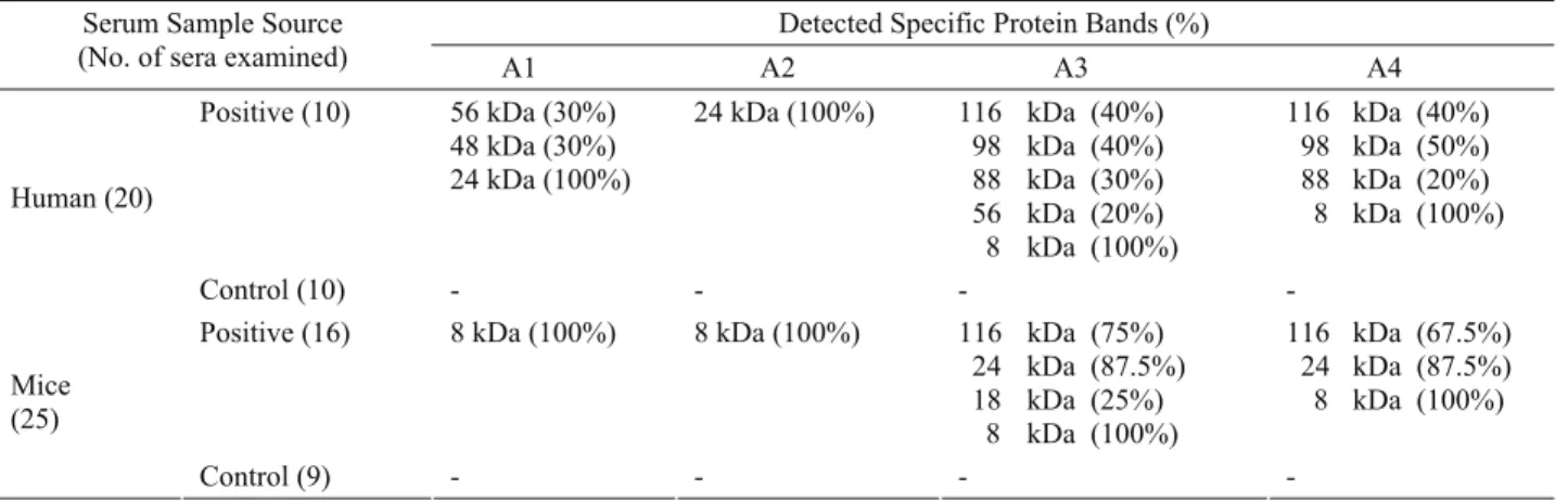
Human sera: Protein bands visualized with human sera were 56 kDa (3/10=30%), 48 kDa (30%) and 24 kDa (100%) for A1, 24 kDa (100%) for A2 (Figure 2) , 116 kDa (40%), 98 kDa (40%), 88 kDa (30%), 56 kDa (20%) and 8 kDa (100%) for A3, 116 kDa (40%), 98 kDa (50%), 88 kDa (20%), 8 kDa (100%) for A4 (Figure 3). There were not any bands on nitrocellulose membranes after testing negative human control sera with these antigens (Table 1).
Mouse sera: Mice infected with protoscolices of ovine origin recognized bands at 8 kDa (8/8=100%) for A1 and 8 kDa (100%) for A2 (Figure 4). Mice infected with protoscolices originating from donkeys recognized bands at 116 kDa (6/8=75%), 24 kDa (87.5%), 18 kDa (25%) and 8 kDa (100%) for A3 and 116 kDa (67.5%), 24 kDa (87.5%) and 8 kDa (100%) for A4 (Figure 5). No band was detected by sera of control group mice (Table 1).
Discussion and Conclusion
In recent years, SDS-PAGE and Western blotting have been widely used in the serological studies. The diagnostic sensitivity and specificity of the tests varied according to the nature and quality of the antigen. Resolution of E. granulosus hydatid cyst fluid by sodium dodecyl sulfate polyacrylamide gel electrophoresis, followed by immunoblotting, resulted in the identification of the arc-5 subunits, including two subunits with relative molecular masses estimated by different laboratories to be between 37 and 38 kDa and 20–22–24 kDa, respectively (11). Diagnostic assessment of these two antigens by immunoblotting performed in different laboratories has resulted in the publication of different sensitivity and specificity parameters (10, 11, 19). The second major parasite antigen in hydatid cyst fluid is a thermostable lipoprotein called antigen B. The major components of antigen B resolve as three bands of 8–12 kDa, 16 kDa, and 23–24 kDa by Western blotting (3, 10,
11). The apparent 8–12 kDa and 23–24 kDa bands were assumed to represent identical antigens (10, 11). Eight and 24 kDa antigens were also detected and accepted as specific in this study. As described in previous reports (7, 14, 15), the smallest subunit of antigen B (8 kDa) is highly sensitive and is expected to be Echinococcus genus specific (7). Our immunoblotting results, obtained with cyst fluid from sheep and donkey indicated that all the sera of mice infected secondary hydatidosis reacted with the antigen B subunit (8 kDa) in cyst fluid.
There were some differences between human CE sera treated with A1, A2 and A3, A4 antigens. However, human CE sera treated with A1, A2 (sheep origin) reacted with 24 kDa, human CE sera treated with A3 and A4 (donkey origin) reacted with 8 kDa. It was thought that the differences of the specificity can be caused by the origin of the antigens.
It was detected some less specific protein bands (18, 48, 56, 88, 98, 116 kDa) than 8 and 24 kDa in human and mice sera and these bands were not evaluated specific for CE. The bands number from mice SHCF antigens was fewer than the bands number from sheep and donkeys HCF. It was determined that it can be caused by sheep and donkeys naturally infection.
In conclusion, primer and seconder HCF antigens of sheep’s origin (A1, A2) and primer and seconder antigens of donkey’s origin (A3, A4) showed very similar patterns of recognition by almost the entire human and mice sera. These results may be provided advantages for standardization of the quality of HCF antigen, when the SHCF antigen from mice maintained in the laboratory under controlled conditions for a standard inoculation period is used for detecting CE in human and animals.
Acknowledgements
This study was supported by Ankara University BAP Directorate, as the project.
Table 1. Detected specific protein bands in A1, A2, A3 and A4 antigens in human and mice sera Tablo 1. İnsan ve fare serumundan elde edilen A1, A2, A3 ve A4 antijenlerine ait spesifik protein bantları
Detected Specific Protein Bands (%) Serum Sample Source
(No. of sera examined) A1 A2 A3 A4 Positive (10) 56 kDa (30%) 48 kDa (30%) 24 kDa (100%) 24 kDa (100%) 116 kDa (40%) 98 kDa (40%) 88 kDa (30%) 56 kDa (20%) 8 kDa (100%) 116 kDa (40%) 98 kDa (50%) 88 kDa (20%) 8 kDa (100%) Human (20) Control (10) - - - -
Positive (16) 8 kDa (100%) 8 kDa (100%) 116 kDa (75%) 24 kDa (87.5%) 18 kDa (25%) 8 kDa (100%) 116 kDa (67.5%) 24 kDa (87.5%) 8 kDa (100%) Mice (25) Control (9) - - - -
References
1. Barıs I, Sahin A, Bilir N, Kalyoncu AF, Emri AS, Akhan O, Barıs B, Copur AS, Selcuk ZT (1998): Hidatik
kist hastalığı ve Türkiye' deki konumu. Türkiye Akciğer
Hastalıkları Vakfı Yayını, No:1, Ankara.
2. Craig PS, Rogan MT, Allan JC (1996): Detection,
screening and community epidemiology of taeniid cestode zoonosis: cystic echinococcosis, alveolar echinococcosis and neurocysticercosis. Adv Parasit, 38,169–250.
3. Gonzales G, Nieto A, Fernandez C, Orn A, Wernstedt C, Hellman U (1996): Two different 8 kDa monomers are
involved in the oligomeric organization of the native Echinococcus granulosus antigen B. Parasite Immunol, 18,
587–596.
4. Güralp N (1981): Helmintoloji. Ankara Üniv. Vet. Fak. Yayınları, No: 368, Ankara.
5. Ioppolo S, Notargiacomo S, Profumo E, Frenchi C, Ortona E, Rigano R, Siracusano A (1996):
Immunological responses to antigen B from Echinococcus granulosus cyst fluid in hydatid patients. Parasite
Immunol, 18, 571– 578.
6. Irabuena O, Nieto A, Ferreira AM, Battistoni J, Ferragut G (2000): Characterization and optimization of
bovine Echinococcus granulosus cyst fluid to be used in immunodiagnosis of hydatid disease by ELISA. Rev Inst
Med Trop Sao Paulo, 42, 255–262.
7. Ito A, Ma L, Schantz PM, Gottstein B, Liu YH, Chai JJ, Abdelhafez SK, Altıntas N, Joshi DD, Lightowlers MW, Pawlowski ZS (1999): Differential serodiagnosis
for cystic and alveolar echinococcosis using fractions of Echinococcus granulosus cyst fluid (antigen B) and Echinococcus multilocularis protoscolex (EM18). Am J
Trop Med Hyg, 60, 188–192.
8. Ito A, Okamoto M, Ishiguro T, Ma L, Suzuki H, Yasui A, Shigeta H, Matsuura T, Hosokawa T, Chai TT (1998): An imported case of cystic echinococcosis in Japan
diagnosed by imaging and serology with confirmation of Echinococcus granulosus-specific DNA sequences. Am J
Trop Med Hyg, 58, 790–792.
9. Laemmli UK (1970): Cleava of structural proteins during
the assembly of the head of bacteriophage T4. Nature, 227,
680.
10. Leggatt GR, Yang W, Mcmanus DP (1992): Serological
evaluation of the 12 kDa subunit of antigen B in Echinococcus granulosus cyst fluid by immunoblot analysis. T Roy Soc Trop Med H, 86, 189–192.
11. Lightowlers M, Gottstein B (1995): Immunodiagnosis of
echinococcosis. 355-410. In: RCA Thompson, AJ
Lymbery (Eds), Echinococcus and Hydatid Disease. CAB International, Wallingford, United Kingdom.
12. Mamuti W, Yamasaki H, Sako Y, Nakaya K, Nakao M, Lightowlers MW, Ito A (2002): Usefulness of hydatid
cyst fluid of Echinococcus granulosus developed in mice with secondary infection for serodiagnosis of cystic Echinococcosis in humans. Clin Diagn Lab Immunol, 9,
573-576.
13. Margutti P, Ortona E, Vaccari S, Barca S, Rigano R, Teggi A, Muhschlegel F, Frosch M, Siracusano A (1999): Cloning and expression of a cDNA encoding an
elongation factor 1beta/delta protein from Echinococcus granulosus with immunogenic activity. Parasite Immunol,
21, 485–492.
14. Meddison SE, Selemanda SB, Schantz PM, Fried JA, Wilson M, Tsang VCW (1989): A specific diagnostic
antigen of Echinococcus granulosus with an apparent molecular weight of 8 kDa. Am J Trop Med Hyg, 40, 377–
383.
15. Poretti D, Felleisen E, Grimm F, Pfister M, Teuscher F, Zuercher C, Reichen J, Gottstein B (1999): Differential
immunodiagnosis between cystic hydatid disease and other cross-reactive pathologies. Am J Trop Med Hyg, 60, 193–
198.
16. Sarkari B, Sadjjadi SM, Abidi H, Izadpanah A,
Kazemian S, Rafati A (2007): Application of Western Blotting Using Native Antigen B for Serodiagnosis of Human Cystic Echinococcosis. Iran J Parasitol, 2, 7-12.
17. Schantz PM, Chai J, Craig PS, Eckert J, Jenkins DJ, Macpherson CNL, Thakur A (1995): Epidemiology and
control of hydatid disease. 233-331. In: RCA Thompson,
AJ Lymbery (Eds), Echinococcus and Hydatid Disease. CAB International, Wallingford, United Kingdom. 18. Sharma SD, Mullenax J, Araujo FG (1987): Western
blot analysis of the antigens of T. gondii recognized by human IgM antibodies. J Immunol, 131, 977-978.
19. Siracuso A, Ioppolo S, Notargiacomo S, Ortona E, Rigano R, Teggi A, Derosa F, Vicari G (1991): Detection
of antibodies against Echinococcus granulosus major antigens and their subunits by immunoblotting. T Roy Soc
Trop Med H, 85, 239–243.
20. Towbin H, Staehelin T, Gordon J (1979):
Electrophoretic transfer of proteins from polyacrylamide gels to nitrocellulose sheets: Procedure and some applications. P Natl Acad Sci USA, 76, 4350.
Geliş tarihi: 08.03.2012 / Kabul tarihi: 17.07.2012
Address for correspondence:
Prof. Dr. Bahadır Gönenç
Ankara University, Faculty of Veterinary Medicine, Department of Parasitology, 06110, Ankara, Turkey. e-mail: [email protected]