Procalcitonine, Nitric oxide and C-Reactive Protein concentrations in
rats with experimentally-induced infectious and noninfectious
inflammation
Pınar Alkım ULUTAŞ, Funda KIRAL, Mürüvvet URAL
Adnan Menderes University, Faculty of Veterinary Medicine, Department of Biochemistry Işıklı/Aydın, TURKEY.
Summary: The aim of this study is to determine the alterations in procalcitonine (PCT), nitric oxide (NO) and C-reactive protein (CRP) levels in infectious and noninfectious inflammation and to evaluate their potential in the differentiation of inflammation. Totally 70 female Wistar rats (Group 1: n=21; Group 2, n=21, Group 3: n=21; Day 0: n=7), weighing 250-300 grams, were used in this study. Before the applications, blood samples were collected in seven rats and the time is accepted as Day 0. Three groups were formed each containing 21 rats. Stapylococcus aureus were administered at a concentration of 10 6 CFU in the infection group (Group 1), subcutaneously. Turbentine oil at a dose of 0.5 mg/kg were injected subcutaneously to twenty-one rats in the noninfectious inflammation group (Group 2). In the control group, 21 rats were injected with saline subcutaneously. Blood samples were collected from randomly selected 7 rats in each group on the 1st, 4th and the 7th days. PCT, NO, and CRP analyzes were performed with commercial test kits in the ELISA reader. According to the obtained results, we determined that serum PCT, NO and CRP levels increased after administrations. The rises of CRP and PCT concentrations were not statistically significant but it was higher in infectious group levels than the other two groups. Based on these results, it can be said that NO shows significant increases during infectious inflammation and can be used for diferential diagnostic purposes in clinics.
Key words: CRP, inflammation, NO, PCT, rat.
Deneysel Olarak İnfeksiyöz ve Noninfeksiyöz Yangı Oluşturulmuş Ratlarda Prokalsitonin, Nitrik
Oksit ve C-Reaktif Protein Konsantrasyonları
Özet: Bu çalışmanın amacı, infeksiyöz ve noninfeksiyöz yangı durumunda prokalsitonin(PCT), Nitrik oksit (NO) ve C-reaktif
protein (CRP) düzeylerini belirlemek ve yangının ayrımının yapılmasında kullanılabilirliğini değerlendirmektir. Bu çalışmada toplam olarak 250-300 gram ağırlığında 70 dişi Wistar rat (Grup 1: n=21, Grup 2: n=21, Grup 3: n=21 ve 0. Gün n=7) kullanıldı. Uygulamalardan önce 7 rattan kan örnekleri alınıp 0. gün olarak kabul edildi. Ayrıca her biri 21 rat içeren 3 grup oluşturuldu. İnfeksiyon grubunda (Grup 1) bulunan 21 rata Staphylococcus aureus 106
CFU derialtı olarak verildi. Noninfeksiyöz yangı grubunda bulunan 21 rata Turbentin yağı 0.5 mg/kg derialtı uygulandı. Kontrol grubundaki 21 rata derialtı fizyolojik tuzlu su verildi. Uygulamalardan sonra her gruptan rastgele seçilen yedişer hayvandan, 1., 4. ve 7. günlerde kan örnekleri toplandı. PCT, CRP ve NO düzeyleri ELISA cihazında ticari test kitleriyle çalışıldı. Elde edilen sonuçlara göre PCT, NO ve CRP düzeylerinin uygulamalardan sonra arttığını belirlendi. CRP ve PCT düzeylerindeki artış infeksiyöz grupta diğer iki gruba göre daha fazlaydı ancak istatistiki önem saptanamadı. Bu çalışmanın sonuçlarına dayanarak, NO’in infeksiyöz yangıda önemli düzeyde arttığı ve kliniklerde ayırıcı tanı amaçlı kullanılabileceği söylenebilir.
Anahtar sözcükler: CRP, PCT, rat, NO, yangı.
Introduction
Early differantiation of bacterial infection and noninfectious inflammation is important due to selection of different treatment models. Sometimes, discrimination of infectious and noninfectious inflammations is difficult because of similar clinical and laboratory findings (17). The differantiation of inflammation and diagnosis of the disease is important for selecting the appropriate treatment and thus preventing complications in the long term (29).
Procalcitonine (PCT) is a polypeptide composed of 116 amino acids that are produced and secreted by thyroid C cells (22). When bacterial infections or septicemia occurs, PCT level increases in circulating blood. The origin of the PCT during infectious conditions are considered to be extrathyroidal such as lung, liver, pancreas, colon and other organs (17). PCT behaves like an acute phase protein similar to other positive reactants, such as CRP, the production of which is also increased by inflammatory stimuli, including infections (23). After
a bacterial stimulus in healthy volunteers, the PCT concentration rise within 4 hr, reaching peak levels after 6 hours, and maintaining a plateau between 8 and 24 hr (29). PCT levels do not increase or slight increase in localised inflammation or infection that no systemic finding (17).
C- reactive protein (CRP) is a marker of nonspesific inflammation and it was used for monitoring the treatment (4, 10). CRP, is reported as major acute phase protein in human and canine (4, 10, 15). CRP is a useful marker of inflammatory clinical situation (12). In the rat CRP has been studied as a marker of inflammation using experimental models (2,12,18). But some researchers investigated that CRP was less sensitive marker of inflammation in the rat than in the human (3, 6).
Nitric oxide (NO), is an endogenous mediator of numerous phsiological processes. NO, produced from L-arginine catalysed by nitric oxide synthase (7). NO, is produced by macrophages and many other cell types in response to inflammation or infection (11). NO levels may be useful to determine inflammation and diseases (17). Some reporters demonstrated NO concentrations can help to use in discrimination of the inflammation (5, 13).
The object of present study was to determine the availibility of use of serum PCT, CRP and NO levels in infectious and noninfectious inflammations induced in rats. Moreoever it was aimed to determine the alterations in these parameters and evaluate their potential usage in differentiation of inflammation.
Materials and Methods
Animals: This study was approved by The
Institutional Animal Ethics Committee of Adnan Menderes University, Turkey (Date:01/03/2011, Number: 2011/18). Experiments were carried out in a semi-acclimatised room at 22°C, with 50-70% humidity and 12/12-hour light/darkcycle. A total of 70 female Wistar albino rats 3 month-old were used in this study. The mean weight of rats was 250-300g. Animals were acclimatised for at least 3 weeks before the experiment.
Study Design: Rats were randomly divided into
three groups for applications.
Group 1: Infectious inflammation group (n=21)
Staph. aureus was given by subcutaneous injection at10 6
CFU/mL (19).
Group 2: Non-infectious inflammation group (n=21) 0.5 mg/kg turbentine oil were administired by subcutaneous injection (27).
Group3: The control group (n=21) received an equal volume of physiologic saline by subcutan way.
Day 0 (n=7): Before the applications, blood samples were collected in rats.
Blood samples were collected via hearth of randomly selected 7 rats from each group at 1, 4 and 7 days after Staphylococcus aureus, turbentine oil and physiologic saline injections. Blood samples were taken under the anaesthesia Serum were separated by centrifugation at 1700 g for 10 min and they were kept frozen-20°C until analysis.
Biochemical analyses: CRP levels were measured
using competative enyzme immunoassay (AssayPro, Assay Max Rat CRP, USA). This assay employs a quantitative sandwich enzyme immunoassay technique that measures rat CRP. The final absorbance of the samples in a microplate reader at 450 nm (Optic Ivyman System 2100C, Spain). The sample concentrations of CRP is determined by using the standart curve and multiply the value by the dilution factor. PCT was measured a with manuel rat spesific commercial ELISA test kits (CUSABIO, rat Procalcitonin ELISA Kit). This assay employs quantitative sandwich enzyme immunuassay. Determine the final absorbance of each well usina a microplate reader set to 450 nm (Optic Ivyman System 2100C, Spain). The results is calculated the calibration curve using Professional ‘Curve Expert 1.3’. NO levels were measured by test combination for determination of NO via nitrate (Nitric Oxide Colorimetric Assay, Roche, USA) on microtiter paltes (Thermo Scientific Multiscan GO, USA). NO, is detected in biological fluids via nitrite. The nitrate present in the sample is reduced to nitrite by reduced NADPH in the presence of the enzyme nitrate reductase. The nitrite formed reacts with sulfanilamde and N-(1-naphtyl) ethylendiamine dihydrochloride to give a red-violet diazo dye. The diazo dye is measured on the basis of its absorbance in the visible range at 550 nm. The result is calculated from the calibration curves using the standart solutions.
Statistically Analyses: SPSS 11.5 package was used
for all computations. All data were checked for normal distribution with Shapiro-Wilk and homogeneity of variance with Levene’s test. If the data were not normally distributed, logarithmic or square root transformation was performed in order to normalize the distribution. Two-way analysis of variance (ANOVA) was conducted to assess the effect of intervention and time on PCT, No and CRP. Post hoc multiple comparisons were performed using Tukey HSD (Honestly significant difference) test. P values <0.05 were considered to be significant. The results were presented as the means ± SE.
Results
All results of this study are shown in Table 1. According to the results of this study, serum PCT levels were not found any statistically significant when compared between groups and days. In spite of PCT
levels were raised 0.175 ng / ml in turbentine group and 0.152 ng / ml in Staph. aureus group in 4 day after application, the levels of increases were not foud statistically significant.
NO levels 10.31 ± 1.83 µM/ml in day 0. This levels were raised to 19.53 ± 6.24 µM/ml in first blood sampling day in Staphylococcus aureus group. This level were significant (p<0,001) when evaluated between groups and days.
CRP levels increased in all groups after the applications and the high CRP levels were remained all sampling days. CRP levels raised at first day of the trial (approximately two fold of before administration day) in
Staph. aureus group. But it was not found any statistical
significance in CRP levels between groups and days. Altough no effects were seen among treatments, time- dependent changes were seen in PCT and CRP leves. It was observed that time and group has significant effects on NO concentrations. Moreover, there was a significant interaction between tretments and time. In entire experimental period, NO levels in Staphylococcus
aureus group were higher when compared to other
treatments. A significant decrease starting from day 4 seen in this group (Table 1).
Discussion and Conclusion
Identifiying infectious and noninfectious inflammation is important because of the treatment procedure differs. Antibiotic treatment is urgent for bacterial infections while immunsuppresive treatment is needed to noninfectious inflammations such as autoimmun disease. Wrong drug choice may cause to deteriorate the patients condition and sometimes the results may be fatal (17). The objective of present study were to evaluate availibility of use of serum PCT, CRP and NO levels in infectious and noninfectious inflammations induced in rats. This study showed that CRP, PCT and NO concentrations were higher in infectious group than the noninfectious inflammation group. However the significant difference was only in NO levels whereas CRP and PCT concentrations were not changed significantly.
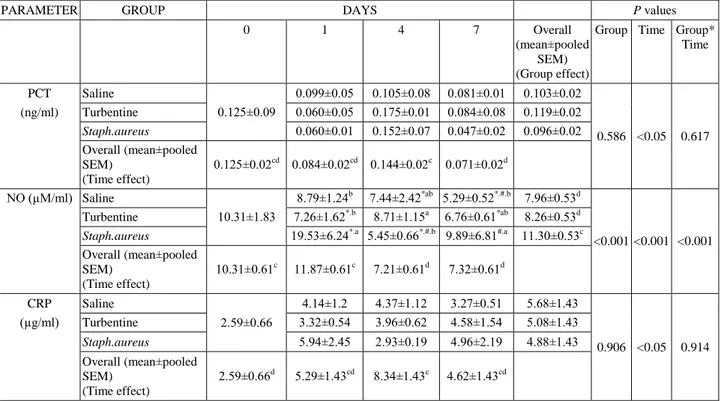
Table 1. Mean serum PCT, NO and CRP concentrations in saline, turbentine and Saphylococcus aureus groups on days of 0, 1, 4 and 7. Tablo 1. Fizyolojik tuzlu su, turbentin ve Staphylococcus aureus gruplarında 0, 1, 4 ve 7. günlerde ortalama serum PCT, NO ve CRP konsantrasyonları.
PARAMETER GROUP DAYS P values
0 1 4 7 Overall
(mean±pooled SEM) (Group effect)
Group Time Group* Time PCT Saline 0.125±0.09 0.099±0.05 0.105±0.08 0.081±0.01 0.103±0.02 0.586 <0.05 0.617 (ng/ml) Turbentine 0.060±0.05 0.175±0.01 0.084±0.08 0.119±0.02 Staph.aureus 0.060±0.01 0.152±0.07 0.047±0.02 0.096±0.02 Overall (mean±pooled SEM) (Time effect) 0.125±0.02cd 0.084±0.02cd 0.144±0.02c 0.071±0.02d NO (µM/ml) Saline 10.31±1.83 8.79±1.24b 7.44±2.42*ab 5.29±0.52*.#.b 7.96±0.53d <0.001 <0.001 <0.001 Turbentine 7.26±1.62*.b 8.71±1.15a 6.76±0.61*ab 8.26±0.53d Staph.aureus 19.53±6.24*.a 5.45±0.66*.#.b 9.89±6.81#.a 11.30±0.53c Overall (mean±pooled SEM) (Time effect) 10.31±0.61c 11.87±0.61c 7.21±0.61d 7.32±0.61d CRP Saline 2.59±0.66 4.14±1.2 4.37±1.12 3.27±0.51 5.68±1.43 0.906 <0.05 0.914 (µg/ml) Turbentine 3.32±0.54 3.96±0.62 4.58±1.54 5.08±1.43 Staph.aureus 5.94±2.45 2.93±0.19 4.96±2.19 4.88±1.43 Overall (mean±pooled SEM) (Time effect) 2.59±0.66d 5.29±1.43cd 8.34±1.43c 4.62±1.43cd
* Statistical difference between output values (Day 0) and other sampling days in same group(same line) (P<0.001).
#
Statistical difference between first day (Day 1) and other sampling days in same group (same line) (P<0.001).
a.b
Statistical difference between groups with different letters in the same day (same column) (P<0.001).
c.d
Statistical difference between overall values with different letters for group and/or time effect (P<0.001). Data represents mean value of experiment ± SE. PCT: Procalcitonine; NO: Nitric oxide; CRP: C-reactive protein. * Aynı grup içinde çıkış günü (0. gün) ile diğer kan alım günleri arasındaki istatistiki fark (Aynı satır)(p<0.01).
# Aynı grup içinde ilk gün(1. gün) ile diğer kan alım günleri arasındaki istatistiki fark (p<0.001) (Aynı satır). ab
: Aynı gün içinde gruplar arası istatistiki fark önemli (aynı sütun). (p<0.001)
cd
: Farklı sütunlardaki tüm değerler arasında grup ve /veya zaman etkisi için istatistiki fark (p<0.001) Veriler ortalama değer±SE olarak verilmiştir. NO. PCT: ProkalsitoninNitrik oksit CRP: C- reaktif protein
PCT has stimulated great interest as a potentially more specific marker for bacterial infection. PCT is produced ubiquitously in response to endotoxin or mediators released in response to bacterial infections that is, interleukin (IL-1β), tumor necrosis factor- (TNF-), and interleukin-6 (IL-6) and strongly correlates with extent and severity of bacterial infections (28). PCT may serve as a useful marker for the detection of systemic bacterial infection in patients with systemic autoimmune disease (9). In this study, PCT levels were showed that nonsignificant increased in infectious and noninfectious group at 4th day after administration. The results of this study indicate that PCT levels were not altered from the cause of inflammation. It is related to PCT levels only rasied significantly during systemic bacterial or fungal infections (7). In healthy subjects, PCT levels are lower than 0.10 ng/ml (7). Many mild increases in PCT concentrations were missed. The results of this study may be related to use of mild inflammation models.
It is reported that CRP is not a major acute phase protein in rats and basal levels of rat CRP is higher than human (24, 25). However, some studies indicated that rat CRP is used for the marker of inflammation (2,12,18). In this study it was determined that CRP concentrations were raised after administrations in three group but this increase was not statistically significant. Connolly et al. (6) reported that plasma CRP levels raised to %138 at 17 days post injection in a rat adjuvant atrhritis model (6). Bürger et al.(3) described an increase at 4 and 15 day of post injection (3). The results of the present study show that CRP levels increased in two fold in Staphylococcus
aureus group in first day after administration. We didn`t
find any significant difference among other treatment groups. Similarly, Myer et al. (21) using subcutanously injected turbentine, determined increased CRP levels in two fold at 24 h post administration (21). Giffen et al.(14) was also determined a slight increase in CRP concentrations (14). This may reflect the difference in models of inflammation used, the route of administration and the method of detection system.
NO, plays an important role in many physiologic functions such as neurotransmission, regulation of immun system and vascular system. In septic shock, endotoxines and cytokines can stimulate NOS in various cells such as macrophages, endothelial cells, vascular smooth muscle cells, leading to NO production (1, 5, 17, 26, 27). NO plays a complex and incompletely understood role in infectious disease and similarly has complex effects on immun system and metabolism. Ahren et al.(1) reported that NO production increased in patients with acute infectious gastroentritis and acute infectious disease such as acute pneumonitis, urinary tract infection and gastroenteritis (26). NO concentration increases as disease severity increases (27). The increase in NO concentrations is a general finding with acute infectious
diseases and may rather be associated with certain pathogens or sites of infection. Charmandari et al. (5) described an significant increase in plasma NO concentrations in infectious or noninfectious diarhea (17). Similarly, in this study, NO concentrations in infectious group showed a statistically significant increase in first day after injection.
According to present study, it can be stated that availability of NO concentrations for discrimination of infectious and noninfectious inflammation is higher than PCT and CRP concentrations. Similar to this, NO levels can be a useful tool for diferential diagnostic purposes in clinics.
References
1. Ahren C, Jungersteb L, Sandberg T (1999): Plasma nitrate as an index of nitricoxide formation in patients with acute infectious disease. Scand J Infect Dis, 31, 405-7. 2. Atli M, Erikoğlu M, Kaynak A, Esen HH, Kurban S
(2012): The effects of selenium and vitamin E on lung tissue in rats with sepsis. Clin Invest Med, 1,35, 48-54. 3. Bürger W, Schade R, Hirschelmann R (1987): The rat
C-reactive protein isolation and response to experimental inflammation and tissue damage. Agents Actions, 21, 97– 93.
4. Ceron JJ, Eckersall PD, Martynez-Subiela S.(2005): Acute phase proteins in dogs and cats: current knowledge and future perspectives. Vet Clin Pathol, 34, 99-85. 5. Charmandari E, Meandows N, Patel M et al (2001):
Plasma nitrate concentrations in children with infectious and noninfectious diarrhea. J Crit Care, 32, 427-423. 6. Connolly KM, Stecher VJ, Kent L (1988): Examination
of interleukin- 1 activity, the acute phase response, and leukocyte subpopulations in rats with adjuvant-induced arthritis. J Lab Clin M, 111, 347–341.
7. Delevaux I, Andre M, Colombier M et al (2003): Can procalsitonin meusurement help in help in differentiating between bacterial infection and other kinds of inflammatory proces? Ann Rheum Dis, 40,1250-6. 8. Eberhard OK, Haubitz M, Brunkhorst FM et al (1997):
Usefulness of procalcitonin for differentiation between activity of systemic autoimmune disease (systemic lupus erythematosus/systemic antineutrophil cytoplasmic antibody- associated vasculitis) and invasive bacterial infection. Arthritis Rheum, 40, 1250-6.
9. Ebersole JL, Machen RL, Steffen MJ et al (1997): Systemic acute-phase reactants, C-reactive protein and haptoglobin in adult periodontitis. Clinical Exp Immunol, 107, 352–347.
10. Eckersall PD, Bell R.(2010): Acute phase proteins: Biomarkers of infection and inflammation in veterinary medicine. Vet J, 185, 27-23.
11. Ellis G, Adatia L, Yazdanpanah M, Makela KS (1998): Nitrite and Nitrate Analyses. A Clin Biochem Pers, 31, 220-195.
12. Faddah ML, Baky NAA, Al-Rasheed NM et al (2010): Role of Quercetin and orjinin in amelioriating nano zinc oxide induced nephrotoxicity in rats. BMC Complem Altern M., 12, 60.
13. Forte P, Dykhuizen RS, Milne E et al (1999): Nitric oxide synthesis in patients with infective gastroenteritis. Gut, 45, 361-355.
14. Giffen PS, Turton J, Andrews C et al (2003): Markers of experimental acute inflammation in the Wistar Han rat with particular referance to haptoglobulin and C- reactive protein. Arch Toxicol, 77, 402-392.
15. Gruys E, Toussaint JM, Niewold TA et al (2005): Acute phase reaction and acute phase proteins. J Zhejiang University Sci, 11, 1056-1045.
16. Jocobson LS, Lobestti RG, Becker F et al (2002): Nitricoxide metbabolites in naturally occuring canine babesiosis. Vet Parasitol, 104, 27-41.
17. Joo K, Park W, Lim MJ et al (2011): Serum procalcitonin for differentiating bacterial infection from disease flares in patients with autoimmune diseases. J Korean Med Sci, 26, 1151-1147.
18. Kahyaoğlu A(2011): Deneysel diabet oluşturulan ratlarda bazı akut faz proteinleri ve iz elementler arasındaki ilişkiler. Yüksek Lisans Tezi, Adnan Menderes Üniversitesi, Sağlık Bilimleri Enstitüsü.
19. McLoughlin Rachel M, Solinga Robert M, Rich JM et al (2006): T cells and CXC chemokines modulate the pathogenesis of Staphylococcus aureus wound infections Proceeding of National Academy of Sciences, 103: 10408-10413.
20. Miranda MK, Espey MG, Wink AD (2001): A rapid simple spectrophotometric method for simultaneous detection of nitrite and nitrate. Nitric Oxide, 5, 71-62. 21. Myers MA, Fleck A (1988): Observations on the delay in
onset of the acute phase protein response. Brıt J Exp Pathol, 69, 176–169.
22. Nahum E, Schiller O, Livni G et al(2012): Procalcitonin level as an aid for the diagnosis of bacterial infections following pediatric cardiac surgery. J Crit Care, 27, 220, 11-16.
23. Nijsten MW, Olinga P, The TH et al (2000): Procalcitonin behaves as a fast responding acute phase protein in vivo and in vitro. Crit Care Med, 28, 458-61. 24. Nunomura W, Takakuwa Y, Higashi T (1994): Changes
in serum concentration and mRNA level of rat CRP. Bıochım. Bıophys. Acta., 1227, 55-70.
25. Podilla ND, Blecker WK, Lubbers Y et al (2003): Rat CRP activates the autologous complement system. Immunology, 109, 564-571.
26. Satoi S, Kamiyama Y, Kitabe H et al (2002): Prolonged decreases in plasma nitrate levels at early postoperative phase after hepato-pancreato-biliary surgery. J Lab Clin Med,131, 342-236.
27. Scotté M, Hiron M. Masson, S et al (1996): Differentıal expression of cytokine genes in monocytes, peritoneal macrophages and liver following endotoxin- or turpentine-induced inflammatıon in rat. Cytokine, 8: 115–120. 28. Schuetz P, Suter-Widmer I, Chaudri A et al (2011):
Prognostic value of procalcitonin in community-acquired pneumonia. Eur Resp J, 37, 392-384.
29. Simon L, Gauvin F, Amre DK et al (2004): Serum procalcitonin and C-reactive protein levels as markers of bacterial infection: a systematic review and meta-analysis. Clin Infect Dis, 39, 206–17.
Geliş tarihi: 10.03.2014 / Kabul tarihi: 14.07.2014 Address for correspondence:
Dr. Pınar Alkım ULUTAŞ Adnan Menderes University, Faculty of Veterinary Medicine, Department of Biochemistry, 09016 Işıklı/Aydın/Turkey e-mail: [email protected]