Clinical, pathological and immunohistochemical findings of bovine
cutaneous papillomatosis
Şule Yurdagül ÖZSOY1, Zafer ÖZYILDIZ2, Murat GÜZEL3
1 Mustafa Kemal University, Faculty of Veterinary Medicine, Department of Pathology, Antakya, 2Harran University, Faculty of
Veterinary Medicine, Department of Pathology, Şanlıurfa, 3Mustafa Kemal University, Faculty of Veterinary Medicine, Department
of Internal Medicine, Antakya, Turkey.
Summary: Bovine cutaneous papillomatosis caused by a bovine papillomavirus, is a common skin disease in Turkey. The aim of the present study was to describe the clinical, histopathological and immunohistochemical aspects of naturally occurring bovine cutaneous papillomatosis. A total of 82 Holstein cattle (9.5%), aged between 5 and 24 months, were diagnosed as cutaneous papilloma by clinical examination. The percentage of papilloma and papillomatosis in male and female was found in 7.3% and 14.8%, respectively. The cauliflower-like growths of varying sizes (0.5-11 cm) were mostly located on the head (63.2%). Histopathology revealed various degrees of acanthosis and hyperkeratosis in all neoplasms. Immunohistochemical (IHC) examination with antibodies against proliferating cell nuclear antigen (PCNA) and Ki-67 were detected in the basal layer of the epidermis and connective tissue. Bovine papillomavirus (BPV-1) antigens were detected in the basal layer. In conclusion, it was decided that the BPV-1, PCNA and Ki-67 antibodies were very useful markers in the diagnosis of bovine cutaneous papilloma.
Key words: Bovine papillomatosis, BPV-1, Ki-67, PCNA.
Sığırlarda deri papillomunda klinik, patolojik ve immunohistokimyasal bulgular
Özet: Bovine papillomavirusun neden olduğu deri papillomatozisi ülkemizde sığırların yaygın bir deri hastalığıdır. Bu çalışmanın amacı doğal olarak oluşmuş sığır deri papillomatozisinde klinik, histopatolojik ve immunohistokimyasal bulguların değerlendirilmesidir. 5 ile 24 ay arasında değişen 82 adet Holstein sığıra (% 9,5) klinik olarak deri papillomu tanısı konuldu. Papilloma ve papillomatozisin erkek ve dişi hayvanlar arasında görülme yüzde oranı sırasıyla %7,3 ve %14,8 olarak belirlendi. Çeşitli boyutlarda (0,5–11 cm) karnabahar görünümündeki üremelerin çoğunlukla baş bölgesinde (%63,2) yerleşim gösterdiği belirlendi. Tümörlerde mikroskobik olarak değişen derecelerde akantozis ve hiperkeratoz saptandı. İmmunohistokimyasal boyamalarda epidermisin bazal katmanında ve bağ dokuda PCNA ve Ki-67 pozitif reaksiyonlar tespit edildi. Bovine papillomavirus antijenleri bazal katmanda belirlendi. Sonuç olarak BPV-1, PCNA ve Ki-67 primer antikorlarının sığır deri papillomunun tanısında önemli markırlar olduğuna karar verildi.
Anahtar sözcükler: Sığır papillomatozis, BPV-1, Ki-67, PCNA.
Introduction
Papillomatosis is seen in many animal species and the disease especially created by host-spesific papilloma viruses (3, 20). Cutaneous papillomatosis is more common in the cattle than other animals (7, 21, 23, 27). In the cattle, six different types of bovine papillomavirus (BPV) have been recognized (BPV-1 to BPV-6). Bovine papilloma may be occurring in all ages (3, 8, 23, 24), but it commonly occurs in young animals and the tumors regress spontaneously due to the animal's immune response without significant scarring (8, 13, 27) The spread of the disease is usually via direct contact, contaminated food and equipment, castration and injections. Inheritance, nutritional and hormonal disorders, sunlight and suppressed immune system may play important roles in pathogenesis of disease (8, 9, 21, 25, 28).
The aim of the present study was to describe the clinical, macroscopic, and histopathological findings of naturally occurring bovine cutaneous papillomatosis. In addition to these findings, the presence and distribution of bovine papilloma virus infection with proliferation cell markers were demonstrated by the immunohistochemical methods.
Materials and Methods
This clinical field study was conducted in six private herds (two dairy and four beef cattle) in Hatay and Osmaniye province of Turkey between March 2008 and October 2009. A total of 865 Holstein cattle (615 males, 250 females), aged between 1 day and 8 years, were involved in this study. The animals had shaped cauliflower-like masses in different part of the body.
Diagnosis was based on presented clinical signs and histopathological findings. The papilloma or papillomatosis classified according to their localization and, the animals were registered according to type of breeding, sex and age. Skin biopsies containing papillomas were obtained under local anesthesia (Lidocaine HCl). Following biopsy, masses were fixed in 10% neutral buffered formalin and embedded in paraffin by routine methods. Sections were cut 5-6 µm in thickness and were stained with hematoxylin and eosin (HE) (19).
Remained tissues were dewaxed and rehydrated by routine methods for immunohistochemical staining as follows. The streptavidin-biotin-peroxidase complex (ABC) technique was performed (Zymed, Histostain Plus Kit, California, USA). Antigen retrieval was facilitated by heating in the citrate buffer (pH 6.0). Endogenous peroxidase activity in tissue sections was blocked by applying 0.3% hydrogen peroxide in 0.01M PBS including 10% methanol and blocked with 5% normal goat serum prior to exposure to primary antisera. The presence of papillomavirus was demonstrated by monoclonal mouse antibody BPV-1-1H8 (Abcam, cat. no: ab2417). Proliferative activity was estimated by the detection of monoclonal antibodies mouse anti-PCNA, clone PC10 (Invitrogen, cat no: PCNA15) and mouse anti-Human Ki-67 antigen clone MIB-1 (Dako, cat no: F7268). The sections were incubated with the rabbit anti mouse biotinylated secondary antibody, and then treated with streptavidin-peroxidase conjugate. Colour labeling was developed by a final incubation step using 3-amino-9-ethyl-carbazole (AEC, Dako/Denmark). Finally, sections were counterstained with Mayer's hematoxylin, rinsed with top water, and mounted with an aqueous mounting medium. For controls, the primary antibody was omitted and replaced by PBS. Following each incubation step, sections were thoroughly washed with phosphate buffered saline (PBS) solution, with the exception of the step after normal goat sera incubation.
All sections were examined using light microscopy (Olympus CX31).
The histopathologic changes in the tumoral masses
was graded as mild, moderate and severe. The number of
immunolabelled cells for 10 microscopic fields at 40X magnification was calculated and scored as none (-) (no
positively staining cells); none or weak (-/+) (<25%); weak (+) (26-50 %); moderate (++) (51-75 %); marked (+++) (>75 %).
Results
The eighty-two of 865 cattles that were ranging between 5 and 24 months (45 male and 37 female) developed clinical papillomatosis. The percentage of papilloma or papillomatosis was found as 54.9% (45/82) in the male and 45.1% (37/82) in the female. (summarized in Table I).
Macroscopically 0.5 to 11 cm in size, grey-white in colour, multiple or soliter, cauliflower-like shaped, sessile or pedunculated, tumoral masses were observed. The eighty-two of 865 (9.5%) cattle were diagnosed within cutaneous papilloma or papillomatosis. Sixty-nine cattle (84.1%) presented solely one tumoral mass on the body; however, 13 cattle (15.9%) had more than one tumoral mass. Papillomatosis were observed both at head (ear and around-around the eyes) and at neck. The masses were found on the head (63.2%), neck (16.80%), interscapular region (12.6%), and thorax (7.4%) of the body. The distribution and location of papillomas are summarized in Table 2.
Table 2. Distribution of bovine cutaneous papillomatosis according to their locations.
Tablo 2. Lokalizasyonlarına göre sığır deri papillomatozisin dağılımı.
Region n %
Head 60 73.2
-Ear and around 21 35.0
-Around the eyes 16 26.7
-Around the noises 15 25.0
-Brow 8 13.3
-Neck 16 19.5
-Interscapular 12 12.6
-Thorax 7 7.4
Total 95* 100
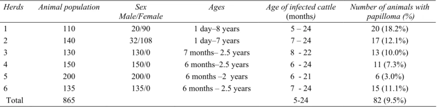
* Some cattle have more than one region. Table 1. Age, sex, breed and population of animals with percentage of the cutaneous papillomatosis.
Tablo 1. Sığırların yaş, cinsiyet ve popülasyonu ile deri papillomatozisin yüzde oranı.
Herds Animal population Sex
Male/Female
Ages Age of infected cattle
(months)
Number of animals with papilloma (%) 1 110 20/90 1 day–8 years 5 – 24 20 (18.2%) 2 140 32/108 1 day–7 years 7 – 24 17 (12.1%) 3 130 130/0 7 months– 2.5 years 8 - 22 13 (10.0%) 4 150 150/0 6 months–2.5 years 6 - 24 11 (7.3%) 5 200 200/0 6 months –2 years 6 - 21 6 (3.0%) 6 135 135/0 6 months – 2.5 years 7 - 24 15 (11.1%) Total 865 5-24 82 (9.5%)
Table 3. Histopathological findings of the tumor Tablo 3. Tümörde gözlenen histopatolojik bulgular
Epidermis Dermis
Hyperkeratosis + (7), ++ (24), +++ (51) Hyperplasia + (43), ++ (39)
Acanthosis ++ (19), +++ (63) Hemorrhage 17
Hydropic degeneration + (2), ++ (18), +++ (62) Lymphocytes infiltration 23 Keratohyalin granules 44
Inclusion bodies 12
(+) Mild number of lesions; (++) moderate number of lesions; (+++) severe number of lesions. Table 4. Immunohistochemical findings of the tumor
Tablo 4. Tümörde gözlenen immunohistokimyasal bulgular Epidermis Dermis BPV-1 - (12), -/+ (32), + (38) - (82) PCNA + (19), ++ (63) -/+ (43), + (39) Ki-67 + (20), ++ (62) -/+ (51), + (31)
(-) None; (-/+) none or weak number of lesions; (+)weak number of lesions; (++)moderate number of lesions; (+++) marked number
of lesions.
Figure 1. Epidermis; hyperkeratosis (H), acanthosis (A) and hydropic degeneration (arrows) in keratinocyts, HE, x 200 µm. Şekil 1. Epidermis, hiperkeratozis (H), akantozis (A) ve keratinositlerde hidropik dejenerasyon (oklar), HE, x 200 µm.
Figure 2. Only some cells in the basal layer of the epidermis have positive reactions for anti-bovine papilloma virus antigen, ABC, x 60 µm.
Şekil 2. Epidermisin bazal katmanında sadece bir kaç hücrede sığır papilloma virus antijenine karşı pozitif reaksiyon, ABC, x 60 µm. Figure 3. PCNA positive reactions in the nuclei of epidermal and mesenchymal cells, ABC, x 60 µm.
Şekil 3.Epidermal ve mezenşimal hücrelerde PCNA pozitif reaksiyon, ABC, x 60 µm.
Figure 4. Ki-67 positive reactions in the nuclei of epidermal and mesenchymal cells, ABC, x 200 µm. Şekil 4. Epidermal ve mezenşimal hücrelerde Ki-67 pozitif reaksiyon, ABC, x 200 µm.
Histopathologically, varying degrees of hyperplasia of the epidermis with irregular papillary projections into the dermis was common and it was seen in all animals. In the epidermis moderate to severe acanthosis, mild to severe hyperkeratosis, hydropic degeneration (Figure1) of keratinocyts and many koilocytes with variably sized keratohyalin granules were present. Also rare presence of intranuclear inclusion bodies were observed only in the basal cells of the epidermis. Dermis showed mild to moderate hyperplasia of the connective tissue that consisted of blood vessels, fibroblasts, focal hemorrhage and mild infiltration of lymphocytes. The main histopathological features are summarized in Table 3.
A few cell of the basal layer of the epidermis was immunostained with BPV-1 (Figure 2). Weak to moderate diffuse nuclear staining of PCNA (Figure 3) and Ki-67 (Figure 4) was detected predominantly in the basal layer of the epidermis. Also superficial layer of hyperplastic dermis showed positive reaction with PCNA and Ki-67. The immunohistochemical lesions are summarized in Table 4.
Discussion and Conclusion
Bovine papillomatosis is a common viral disease of the skin, manifested as benign tumors or warts, caused by bovine papillomavirus (BPV) (23). Papillomavirus may affect all ages of cattle; however, affected cattle were usually younger than 2 years of age (27). In this study, cutaneous papillomatosis was detected in animals aged between 5 and 24 months. Bovine cutaneous papillomas were detected mainly on the head and neck, besides on thorax and in some animals within the other parts of the body (1, 3, 4, 11, 15). In our study most of the lesions were found on the head (63.2%). Papillomatosis may become a significant herd problem when a large group of young, susceptible cattle become infected. The percentage of disease may be increased approximately up to 20-25% (29). In this study, the percentage of bovine papillomatosis was detected between 3.0-18.2% (mean 9.5%) in different herds.
The macroscopic and microscopic findings of the tumor that observed in the present study was similar to described before (1, 11,14). Macroscopically cauliflower-like shaped tumoral masses were observed. The lesions were composed of a generally marked hyperkeratosis of the epidermis with irregular papillary projections into the dermis in the histopathologic examination.
Viral inclusion bodies are rarely reported in naturally occurred skin papillomas (12, 15). Etiology of this disease is connecting to papillomaviruses, and cattle are natural carries of the virus (6, 12, 27). In this study intranuclear viral inclusion bodies were seen in papilloma as demonstrated by previous study (10), specific antigen (BPV-1) in the basal cell layer of epidermis.
The level of cellular proliferation in tumor tissue may be determined by immunohistochemical techniques. With these methods, nuclear antigen associated with cell growth and division, stained and evaluated under the microscope (26). Because of both Ki-67 and PCNA is a useful marker of cell proliferation (5); they used several times in neoplastic skin tissues previously (2, 16, 17). Immunohistochemical studies have shown increased nuclear PCNA and Ki-67 staining at both human and cattle papillomatosis (13, 14, 18, 22). In our study PCNA and Ki-67 immunolabeling was principally present in the epidermis. But as previous study (14) we also observed immunoreactivity in dermis. The immunolabelling of both PCNA and Ki-67 were increased parallel to fibroblastic activity. It seen that Ki-67 immunoreactivity has been shown to bear a close correlation with the PCNA staining.
In this study, the present authors described clinical and pathological findings of cutaneous papillomatosis in the cattle. Specific antigen of BPV-1 was demonstrated by immunohistochemically in bovine cutaneous papillomatosis. The present researchers also decided that the PCNA and Ki-67 antibodies were very useful markers to detection of cellular proliferation in the bovine cutaneous papillomatosis.
Acknowledgements
The author thanks to the Veterinary Surgeons Bünyamin Akin for kindly helps during field studies.
References
1. Atasever A, Cam Y, Atalay O (2005): Bir sığır sürüsünde
deri papillomatosis olguları. Ankara Univ Vet Fak Derg,
52, 197–200.
2. Barrett TL, Smith KJ, Hodge JJ, Butler R, Hall FW, Skelton HG (1997): Immunohistochemical nuclear
staining for p53, PCNA, and Ki-67 in different histologic variants basal cell carcinoma. J Am Acad Dermatol, 37,
430-7
3. Blood DC, Radostits OM (1989): Veterinary medicine. A textbook of the diseases of cattle, sheep, pigs, goats and horses. 7th edn., Bailliere Tindall, London.
4. Borku MK, Atalay O, Kibar M, Çam Y, Atasever A (2007): Ivermectin is an effective treatment for bovine
cutaneous papillomatosis. Res Vet Sci, 83, 360–363.
5. Brugal G (1995): Quantitative microscopy and tumour
cell proliferation. Bul Cancer 82, 511-517.
6. Campo MS (2002): Animal models of papillomavirus
pathogenesis. Virus Res, 89, 249-261.
7. Campo MS (1998): Persistent infection by bovine
papillomavirus. 503-516. In: R Ahmed, ISY Chen, (Eds),
Persistent Viral Infections, Wiley.
8. Campo, MS, Jarrett WF, O’neil W, Baron RJ (1994):
Latent papillomavirus infection in cattle. Res Vet Sci, 56,
151–157.
9. Dinç DA (1995): Papillomatozis. 41-45. In: Evcil Hayvanlarda Memenin Deri Hastalıkları, Dolaşım Bozukluklari ve Operasyonlari. Konya, Ülkü Matbaası.
10. Emhmad AO, Levkut M, Levkutova´ M, Revajova´ V, Ondrejka R, Benı´sˇek Z (1997): Immunohistochemistry
of the progressive and regressive stages of bovine papillomatosis. Acta Veterinaria Brno, 66, 245–248.
11. Goldschmidt MH, Hendrick MJ. (2002). Tumors of the
skin and soft tissues. 47-117.. In DJ Meuten (eds), Tumors
in domestic animals. 4th edn., Iowa State Pres, Ames. 12. Hargis AM (1995): Cutaneous Neoplasia. 504-509. In:
Thomson’s Special Veterinary Pathology. Carlton WW, McGavin MD (eds), 2nd edn., Mosby, Philadelphia, London, Sydney, Tokyo.
13. Hatipoglu F, Ozdemir O, Kıran MM (2009): Detection
of argyrophil nucleolar organizer regions (AgNORs) and proliferating cell nuclear antigen (PCNA) in epithelial skin tumours from domestic animals. Revue Méd. Vét, 160,
477-483.
14. Jelinek F, Tachezy R (2005): Cutaneous papillomatosis
in cattle. J Comp Pathol, 132, 70–81.
15. Jones TC, Hunt RD, King NW (1997): Bovine cutaneous
papillomatosis. 252. Veterinary Pathology, 6th edn.,
Williams & Wilkins, A Waverly Company, Philadelphia, london, Tokyo, Sydney.
16. Karademir N, Güvenç T, Yarım M, Orman MM, Gülbahar MY (1996): Comparison of agnors staining and
pcna immunostaing methods, and mitotic index scores in intracutaneous cornifying epithelioma and squamous cell carcinoma. Ankara Univ Vet Fak Derg, 43, 281-285.
17. Kawahira K (1999): Immunohistochemical staining of
proliferating cell nuclear antigen (PCNA) in malignant and nonmalignant skin diseases. Arch Dermatol Res, 291,
413–418.
18. Lu S, Tiekso J, Hietanen S, Syrjaènen K, Havu VK, Syrjaènen S (1999): Expression of cell-cycle proteins p53,
p21 (WAF-1), PCNA and Ki-67 in benign, premalignant and malignant skin lesions with implicated HPV involvement. Acta Derm Venereol, 79, 268-273
19. Luna LG (1968). Manual of Histologic Staining Methods
of the Armed Forces Institute of Pathology. 32–241.
McGraw-Hill Book Company, New York.
20. Ndarathi CM, Mbuthia PG (1994): Individual
bovine-specific and species-bovine-specific autogenous vaccine in treatment of bovine cutaneous papillomatosis. Indian J
Anim Sci, 64, 218-221.
21. Nicholls PK, Stanley MA (2000): The immunology of
animal papillomaviruses. Vet Immunol Immunopathol, 73,
101–127.
22. Niwa Y (1995):Demonstration of oncogene, E7 region of
18 type HPV in the skin lesions of patients with severe, treatment-resistant atopic dermatitis (abstract, in Japanese). J Jpn Soc Cancer Ther, 30,1226.
23. Olson C (1993): Papillomatosis in cattle. 430–431. In: Howard, J.L. (ed), Current Veterinary Therapy 3 & Food Animal Practice. W. B. Saunders Company, Philadelphia, London, Tokyo.
24. Olson C, Olson RO, Hubbard-Van Stelle S (1992):
Variations of response of cattle to experimentally induced viral papillomatosis. J Am Vet Med Assoc, 201, 56–62.
25. Otter A, Leonard D (2003): Fibropapillomatosis
outbreak in calves. Vet Rec, 1, 570–571.
26. Rosai J (1996): Ackerman's surgical pathology. 47-48. 8th edn., St Louis, C.V. Mosby Co.
27. Smith BP (1996): Papillomatosis (Warts, Fibropapillomas). 1417-1418. In: Large Animal Internal Medicine & Diseases of Horses, Cattle, Sheep, and Goats. Smith BP (Eds). 2nd edn., Mosby-Year Book, Inc. Boston, New York, Philadelphia, London, Tokyo.
28. Theilen G, Wheeldon EB, East N, Madewell B, Lancaster WD, Munn R (1985): Goat papillomatosis. Am J Vet Res, 46, 2519-2526.
29. The Merck Veterinary Manual (2008): Papillomas. http://www.merckvetmanual.com/mvm/index.jsp?cfile=ht m/bc/72214.htm
Geliş tarihi: 16.03.2010 / Kabul tarihi: 11.10.2010
Address for correspondence:
Şule Yurdagül Özsoy Mustafa Kemal University Faculty of Veterinary Medicine
Department of Pathology, Antakya, Turkey E-mail: [email protected]