Yazışma Adresi/Address for Correspondence: Dr. Selin Gaş, Beykent University, School of Dentistry, Department of Oral and Maxillofacial Surgery, Istanbul, Turkey. E-mail: selingas@beykent.edu.tr
ARAŞTIRMA / RESEARCH
Effects of hyaluronic acid and gamma-radiated mineralized allografts on
the healing of rat tibial defects
Hyaluronik asit ve gama radyasyonlu mineralize allogreftlerin sıçan tibial defektlerinin
iyileşmesi üzerine etkileri
Selin Gaş1 , Necat Vakur Olgaç2 , Ahmet Taylan Çebi3 , Çetin Kasapoğlu4 1Beykent University, School of Dentistry, Department of Oral and Maxillofacial Surgery, Istanbul, Turkey. 2Istanbul University, Institute of Oncology, Department of Tumor Pathology, Istanbul, Turkey.
3Karabuk University, School of Dentistry, Department of Oral and Maxillofacial Surgery, Karabuk, Turkey. 4Istanbul University School of Dentistry, Department of Oral and Maxillofacial Surgery, Istanbul, Turkey.
Cukurova Medical Journal 2020;45(2):526-532
Abstract Öz
Purpose: This study aimed to evaluate the effects of
hyaluronic acid (HyA) and gamma-radiated mineralized allografts (Gr-MAs) on the healing of bone defects in rat tibiae.
Materials and Methods: Fifty-two male Sprague Dawley
rats were randomly allocated to four groups: Gr-MA, HyA, Gr-MA combined with HyA (Gr-MA + HyA), and controls with empty defects. The animals were sacrificed on the 7th and 21st postoperative days. The inflammation, necrosis, fibrosis, new bone formation, and bone healing scores were evaluated on the basis of the histopathological findings.
Results: The amount of new bone formation was found
to be significantly greater in the control group than in the experimental groups. In addition, the healing scores were statistically higher in the control and the Gr-MA + HyA groups. Comparisons of the control, graft, and HyA groups indicated that the control group exhibited significantly less necrosis on the 7th day; however, on the 21st day, there were no statistically significant differences among the groups. There were no statistically significant differences among the groups in terms of the inflammation and fibrosis levels on the 7th or 21st days.
Conclusion: Within the limitations of this study, the
application of HyA alone and the addition of HyA to Gr-MA did not improve bone regeneration in rat tibial defects.
Amaç: Hyaluronik asidin ve mineralize allogreftin sıçan
tibiasında oluşturulmuş defektlerde yeni kemik formasyonu ve kemik iyileşme skoru üzerine etkisinin değerlendirilmesi amaçlanmıştır.
Gereç ve Yöntem: 52 adet Spraque-Dawley cinsi sıçan 4
gruba ayrılmıştır: mineralize kemik greft grubu, hyaluronik asit grubu, hyaluronik asit ile kombine olarak uygulanan mineralize kemik greft grubu ve boş defektlere sahip kontrol grubu. Hayvanlar postoperatif 7. ve 21. günlerde sakrifiye edilmiştir. İnflamasyon, nekroz, fibrosis, yeni kemik oluşumu ve kemik iyileşme skoru histopatolojik olarak değerlendirilmiştir.
Bulgular: Yeni kemik oluşumu kontrol grubunda deney
grubuna göre anlamlı oranda daha yüksek bulunmuştur. Ayrıca yalnızca hyaluronik asit ve yalnızca greft gruplarına kıyasla, kontrol grubunda iyileşme skoru daha yüksek bulunmuştur. Greft ve hyaluronik asit grupları kıyaslandığında, 7. gündeki nekroz kontrol grubunda anlamlı oranda düşükken, 21. günde gruplar arasında anlamlı bir farklılık bulunmamıştır. 7. ve 21. günlerdeki inflamasyon ve fibrozis değişkenlerinin oranları gruplar arasında anlamlı bir değişiklik yaratmamıştır.
Sonuç: Hyaluronik asit tek başına veya mineralize kemik
allogrefti ile birlikte uygulandığında, sıçan tibiasında oluşturulmuş kritik boyutta olmayan defektlerde kemik rejenerasyonununda yeterli katkıyı sağlamamıştır.
Keywords: Hyaluronic acid, bone grafting, bone
527
INTRODUCTION
Bone graft materials are commonly used as a void filler for bone defects1-5. Several treatment methods,
such as the application of local and systemic drugs, graft materials, hormones, growth factors, bone morphogenic proteins, physical stimulation, and hyperbaric oxygen, have been used to accelerate the healing of bone defects and fractures2-4. The choice
of bone graft material plays a crucial role in bone grafting augmentation techniques. Bone grafts can be classified as autogenous bone, allografts, xenografts, and synthetic5. Autogenous bone grafts are still
currently accepted as the gold standard for bone augmentation because of their osteogenicity, osteoinductivity, and osteoconductivity. However, because of the limitations of autogenous grafts, several bone substitutes have been introduced. Allografts are tissues that are taken from donors of the same species as the host. Recently, gamma-radiated mineralized allografts (Gr-MAs) have become another preferred treatment that promotes rapid healing and offers complete remodeling5.
Gr-MAs have exhibited better adaptation to the surrounding tissues because of the variations in tissue banks’ allograft processing methods6. Mineralized
allografts and their mineral content provide better volume stability at the grafting site than do other allografts7.
Hyaluronic acid (HyA) is one of the important components of the extracellular matrix of tissues. It is a naturally derived linear high-molecular-weight protein with viscoelastic properties8. HyA
contributes to bone formation and prior osteogenic commitments by regulating cytokines and growth factors9-12. These are some of the basic characteristics
of HyA that can mediate both acute and chronic wound healing. Several studies have demonstrated the effects of bone grafts and HyA on bone healing; however, few have provided comparisons of these materials.
The aim of the present study was to evaluate the histopathological effects of HyA and Gr-MA on the new bone formation and bone healing processes in tibial defects in rats. The null hypothesis tested in the present study is that hyaluronic acid (HyA) and Gr-MA would not contribute to new bone formation and healing when applied together or alone to rat tibial defects.
MATERIALS AND METHODS
Fifty-two male 10–12-week-old Sprague Dawley rats weighing 350–400 g were used in the study. All of the animals were randomly allocated to four groups, each with a different augmentation material for treating the tibial defects. Three experimental groups, each consisting of 16 animals, received the following treatments: The first group received only HyA (HYALOSS™ Matrix; Anika Therapeutics, Padova,
Italy), which was applied to the bone defect. The second group received Gr-MA (Puros® allograft;
Centerpulse Dental Division, Carlsbad, CA, USA), which was applied to the bone defect. The third group received Gr-MA combined with HyA (Gr-MA + HyA). In the control group, which consisted of four animals, the bone defect was irrigated with a sterile saline solution.
Anesthesia and surgical procedure
This study was approved by the Institutional Animal Care and Ethics Committee of Istanbul University Institute for Experimental Medical Research (Project No 2014/92, Date of Ethical Approval 09/10/2014). Experimental protocol has been completed in accordance with the Guide for the Care and Use of Laboratory Animals. The animals were anesthetized by the intraperitoneal injection of a combination of 60 mg/kg ketamine (Ketalar; Eczacibaşi-Warner Lambert, Istanbul, Turkey) and 6 mg/kg xylazine (Rompun® 2%; Bayer, Istanbul, Turkey).
Preoperatively, the skin over the tibia was shaved and disinfected with povidone-iodine. The bone was exposed through a full-thickness skin incision of approximately 2 cm. A right tibial non-critical bone defect with a diameter of 3 mm was prepared with a dental handpiece and a trephine bur under copious saline irrigation (Figure 1). The wounds were closed with 3-0 silk sutures. Half of the animals in each group were sacrificed on the 7th postoperative day,
and the other half were sacrificed on the 21st
postoperative day.
Histopathological evaluation
The tibiae were removed and fixed in 10% phosphate-buffered formaldehyde for 1 week. After fixation, the material was decalcified in a formic acid sodium nitrate solution. Paraffin tissue blocks were then prepared and deparaffinized. After staining with hematoxylin and eosin, the sections were examined under a light microscope (Olympus BX60; Olympus
Optical Co. Ltd., Tokyo, Japan) attached to a color video camera that was connected to a personal computer. The images were captured, and the parameters of interest were measured with analySIS FIVE software (Olympus Optical Co. Ltd.) at 100× magnification. Digital images were obtained from the tissue sections of all the defect areas. These areas were analyzed for inflammation, necrosis, fibrosis, new bone formation, and bone healing. The inflammation and fibrosis were assessed as follows: 0 (–), 1 (1–30%), 2 (30–60%), and 3 (>60%)13.
Micro-abscess formation was also scored as 3. Bone healing was evaluated on the basis of Allen’s14 fracture
healing scores, including the healing stages: non-union—0, incomplete cartilage union—1, complete cartilage union—2, incomplete bony union with phase of ossification—3, incomplete bony union with intermediate phase of ossification—4, incomplete bony union with late phase of ossification—5, and complete bony union—6. The areas occupied by newly formed bone and fibrosis were measured, and the proportion (%) with respect to the total area was determined. Necrosis and inflammation were scored as present (−) or absent (+).
Figure 1. Surgical procedures. (A) Rat tibia after dissection; (B) Defect; (C) Application of gamma-radiated mineralized bone graft; (D) Application of hyaluronic acid.
Statistical analysis
IBM SPSS Statistics for Windows, Version 22 software (IBM Corp., Armonk, NY, USA) was used. Descriptive statistics, such as the median, standard deviation, and frequency, were used. The group variables that were normally distributed were
evaluated with the Shapiro–Wilk test, and those that were not normally distributed were evaluated with the Kruskal–Wallis test. Levene’s test was used for the homogeneity of variances. The Mann–Whitney U test was performed for pairwise comparisons. The Fisher–Freeman–Halton test and Fisher’s exact test were used for the analysis of the categorical variables. The confidence interval was set to 95%, and p < 0.05 was considered statistically significant.
RESULTS Day 7 assessment
In the HyA group, active fibrous connective tissue was observed in the defect area, and in the Gr-MA group, new bone formation was seen around the graft material. In the Gr-MA + HyA group, new trabecular bone was observed to be filling the defect region around the active fibrous connective tissue. In the control group, active fibrous connective tissue was observed around the new bone formation in the defect area (Figure 2).
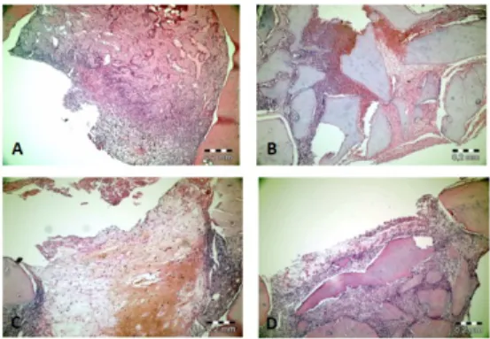
Figure 2. Representative hematoxylin-eosin-stained histopathological sections under 100× magnification on the 7th day. (A) In the control group, new bone formation in and around the active fibrous connective tissue in the defect region; (B) In the gamma-radiated mineralized allograft (Gr-MA) group, new bone trabeculae around the graft material covering large areas of the active connective tissue; (C) In the hyaluronic acid (HyA) group, active fibrous connective tissue around the defect; (D) In the Gr-MA combined with HyA group, the presence of large graft particles in the fibrous connective tissue in the defect region and bone formation around the graft particles.
The necrosis levels were found to be significantly lower in the control group than in the HyA and the Gr-MA groups (p = 0.022). The inflammation levels
529
in the Gr-MA and the control groups were statistically higher on the 7th day than on the 21st day
(p = 0.003).
Figure 3. Representative hematoxylin-eosin-stained histopathological sections under 100× magnification on the 21st day. (A) In the control group, new bone formation filling the defect site and active fibrous connective tissue around the defect; (B) In the gamma-radiated mineralized allograft (Gr-MA) group, new bone formation around the graft material; (C) In the hyaluronic acid (HyA) group, new bone formation in the fibrous connective tissue; (D) In the Gr-MA combined with HyA group, new bone formation covering the defect surface.
Similar to the MA and the control groups, the Gr-MA + HyA group exhibited significantly higher inflammation levels on the 7th day than on the 21st
day inflammation (p = 0.001). The control group had
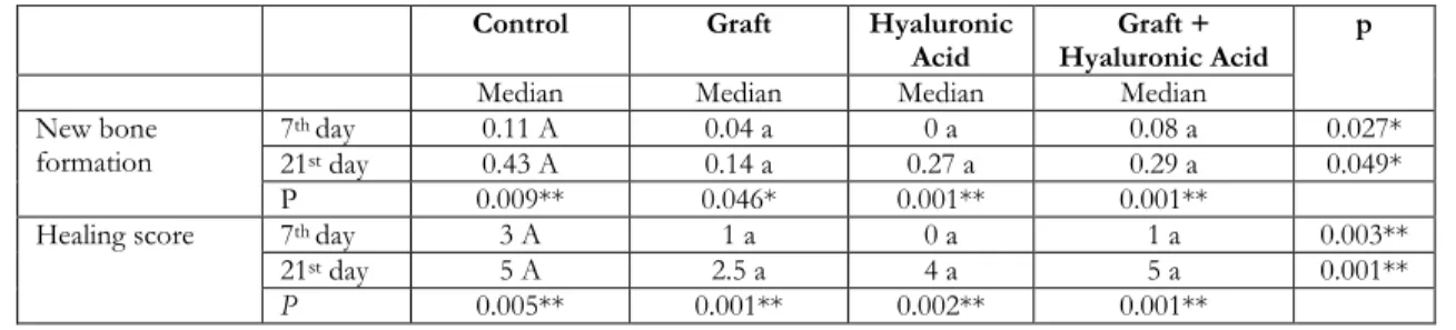
a significantly greater amount of new bone formation than the HyA group (p = 0.025), the Gr-MA group (p = 0.015), and the Gr-MA + HyA group (p = 0.039; Table 1).
The healing scores for the control group were significantly higher than those for the HyA group (p = 0.007), graft group (p = 0.002), and Gr-MA + HyA group (p = 0.004). There were statistically significant differences in the necrosis levels of the groups on the 7th day (p = 0.005).
Day 21 assessment
New bone formation around the graft material was observed in the Gr-MA group, and in the HyA group, bone islands were formed in the fibrous connective tissue. In the Gr-MA + HyA group, new bone formation filled the defect area, and there was residual graft material in the deep tissues. In the control group, fibrous connective tissue around the new bone formation filled the defect area (Figure 3). The control group exhibited a significantly greater amount of new bone formation than the Gr-MA group (p = 0.028) and the Gr-MA + HyA group (p = 0.048). In all the groups, the amount of new bone formation was significantly greater on the 21st day
than on the 7th day. On the 21st day, there were
statistically significant differences in the healing scores (p = 0.001). The control group’s healing scores were significantly higher than those of the HyA and the Gr-MA groups (p = 0.006). For all the groups, the healing scores on the 21st day were significantly
higher than those on the 7th day (Table 1).
Table 1. Healing scores and new bone formation on the 7th and 21st postoperative days Control Graft Hyaluronic
Acid Hyaluronic Acid Graft + p
Median Median Median Median
New bone
formation 7
th day 0.11 A 0.04 a 0 a 0.08 a 0.027*
21st day 0.43 A 0.14 a 0.27 a 0.29 a 0.049*
P 0.009** 0.046* 0.001** 0.001**
Healing score 7th day 3 A 1 a 0 a 1 a 0.003**
21st day 5 A 2.5 a 4 a 5 a 0.001**
P 0.005** 0.001** 0.002** 0.001**
DISCUSSION
This study was conducted to evaluate the effects of HyA and Gr-MA on the healing of bone defects. Allografts, which provide type I collagen and contain
bone morphogenic proteins, are commonly used as void fillers in maxillofacial bone defects15. The
Puros® allograft, a two-piece graft composed of
cortical and cancellous bone, is obtained mostly from the ends of the long bones, such as the humeral head, femoral head, femoral condyles, and tibial plateau.
This graft material receives low-dose gamma irradiation, which ensures biomechanical integrity, preserves protein structure, and inactivates all remaining viruses14. HYAFF®-11, an esterified form
of HyA, forms a scaffold for cell growth16. The
HYALOSS Matrix is composed of bundles of fibers made entirely of HYAFF®-11, a solid derivative of
HyA, a naturally occurring component of the body. Rats were selected as the experimental animal because they are inexpensive, widely available, acceptable to society, and easily housed and maintained17. Because of ethical concerns, the
unilateral defect model was used in this study. Non-critical-sized defects were created because the selected materials were void fillers. The 7th and 21st
days were selected for euthanasia to determine the healing process of the bone defects in relation to the methods used in previous studies12,18.
Osteoconduction is the process that allows bone apposition from existing bone. Osteoconductive graft materials provide an environment that is capable of hosting the mesenchymal stem cells, osteoblasts, and osteoclasts that are essential for the functioning of the bone graft1. Collins et al. showed that there was
no significant difference in the new bone formation after the implantation of biphasic calcium sulfate alone or in combination with gamma-radiated human mineralized allografts in the extraction sockets19. In a
comparison of three bone grafting materials, Zhang
et al. found that biphasic calcium phosphate bone grafts resulted in a greater amount of new bone formation than did demineralized freeze-dried bone allografts and natural bone minerals of bovine origin20. No complications were observed during the
current experiment; therefore, Gr-MA can be safely used for bone formation.
HyA has been shown to play an important role in the healing of bone tissue21. Several studies have
confirmed the efficacy of HyA on the soft tissues 22-25. Studies have more recently focused on the effects
of HyA on bone healing. Demonstrating higher bone volume, Huang et al. asserted that HyA, with an optimal combined administration, could significantly promote the osteogenic and angiogenic activity of bone morphogenic protein 2 (BMP-2) or absorbable collagen sponges (ACSs)25. Mermerkaya et al.
demonstrated the role of hyaluronan-based mesh in promoting osteoblastic function26. In a comparison
of HYAFF®-11 in vitro and HyA in various
molecular weights, Moseley et al. observed that the HYAFF®-11 had superior wound healing and
antioxidant capacity27. Mendes Brazão et al.28
performed a radiological examination of a critical-sized defect in rat calvaria to examine the effects of HyA on bone healing. They concluded that HyA alone or in association with a carrier would not improve bone healing. This was consistent with the findings of the present study. Bezerra et al.29
investigated the effects of HyA on bone healing in critical-sized calvarial defects in rats at 2 months post-surgery. They reported an increased amount of new bone formation in the defects filled with HyA gel and HyA gel + ACS. In the current study, the HyA group exhibited a lower amount of bone regeneration than the control group. This might have been related to the time appointment and the density of the HyA. Bone graft materials can be applied with other kinds of materials to increase their effectiveness. HyA is used in combination with other graft materials to improve wound healing and bone regeneration30.
Arpağ et al. investigated the effects of HyA, xenografts, and autografts on rabbit calvarial defects. They asserted that HyA contributes to xenograft in new bone formation and bone healing by reducing the residual graft volumes31. Koca et al. reported on
the positive contributions of HyA alone or in combination with grafts to healing in critical-sized bone defects in rat jaws32.
Unlike the abovementioned studies, the current study found that HyA alone did not have an effect on new bone formation or the healing scores. Diker et al.33
found that HyA did not adequately improve bone regeneration in rats. However, they reported that bone formation was more noticeable in the graft and HyA + graft defects. Agrali et al. asserted that HyA alone or in combination with a graft and membrane did not significantly contribute to bone regeneration in critical-sized rat calvarial defects34. The current
study obtained similar results.
The contribution of osteoconductive materials is not affected by the size of the defect; thus, a critical-sized defect was not chosen for the current study. The goal was to determine the effectiveness of the materials used as void fillers. The study used rat tibial defects; however, the findings of previous studies were not supported. HyA alone or in combination with grafts did not improve the healing scores.
Aslan et al.35 stated that the amount of the bone
formation in the rat tibial defects filled with HyA + allogenic cancellous bone grafts was greater than that in defects filled with allogenic cancellous bone grafts
531
only. In contrast, the current study found no significant differences in new bone formation in the Gr-MA, HyA, and Gr-MA + HyA groups. One of the reason could be the duration of the different postoperative healing periods in the studies. In our study we only showed the early healing scores, although Aslan et al. analyzed the the 40th
postoperative day of healing. The other reason could be the properties of the HyA used in the study. It had a more stable structure than the equivalents produced in liquid form, it took longer to biodegrade, and it acted as a place holder by turning into a gel after coming into contact with blood. The current study was planned on the basis of the expected effects of the HyA. It is possible that the consideration of the expected effects of the bone graft would have produced different results.
The present study has several limitations. First, the small number of animals limited the number of samples that could be obtained. Second, undecalcified sections, which could have strengthened the study results, were not used. Third, conducting the study over three experiment periods could have facilitated the assessment of the effectiveness of the use of HyA exclusively or in combination with graft materials during the recovery. As a conclusion, hyaluronic acid has a versatile role in tissue repair process from early-stage inflammatory activity to tissue generation granulation. Its effects on process of both soft tissue healing and wound healing are shown in many studies. Within the limitations of the present study, it can be suggested that HyA, used alone or in combination with Gr-MA, is not likely to enhance bone formation in rat tibial defects. There is still much work needed to demonstrate the effects and biological mechanisms of hyaluronic acid in bone healing.
Yazar Katkıları: Çalışma konsepti/Tasarımı: SG, ÇK, NVO; Veri
toplama: SG, ÇK; Veri analizi ve yorumlama: SG, ÇK, MVO, ATÇ; Yazı taslağı: SG, ÇK; İçeriğin eleştirel incelenmesi: SG, ÇK, MVO, ATÇ; Son onay ve sorumluluk: SG, NVO, ATÇ, ÇK; Teknik ve malzeme desteği: -; Süpervizyon:SG, ÇK, MVO; Fon sağlama (mevcut ise): yok.
Etik Onay: Bu çalışma İstanbul Üniversitesi Deneysel Tıbbi Araştırma
Enstitüsü Kurumsal Hayvan Bakım ve Etik Komitesi tarafından onaylanmıştır (Proje No 2014/92, Etik Onay Tarihi 09/10/2014).
Hakem Değerlendirmesi: Dış bağımsız.
Çıkar Çatışması: Yazarlar çıkar çatışması beyan etmemişlerdir. Finansal Destek: Deneysel tıbbi Araştırma Enstitüsü (süreç 2014/92)
ve bu çalışma 49881 numaralı proje ile İstanbul Üniversitesi Araştırma Fonu tarafından desteklenmiştir.
Teşekkür: Bu araştırma, İstanbul Üniversitesi Araştırma Vakfı
tarafından 49881 sayılı hibe ile finanse edilmiştir, İstanbul, Türkiye
Author Contributions: Concept/Design : SG, ÇK, NVO; Data acquisition: : SG, ÇK; Data analysis and interpretation: SG, ÇK, MVO, ATÇ; Drafting manuscript: SG, ÇK; Critical revision of manuscript: SG, ÇK, MVO, ATÇ; Final approval and accountability: SG, NVO, ATÇ,
Technical or material support: -; Supervision: SG, ÇK, MVO; Securing funding (if available): n/a.
Ethical Approval: This study was approved by the Institutional Animal
Care and Ethics Committee of Istanbul University Institute for Experimental Medical Research (Project No 2014/92, Date of Ethical Approval 09/10/2014).
Peer-review: Externally peer-reviewed.
Conflict of Interest: Authors declared no conflict of interest. Financial Disclosure: Institute for Experimental Medical Research
(process 2014/92) and this work was supported by the Research Fund of Istanbul University with the Project Number 49881.
Acknowledgment: This research was funded by Istanbul University
Research Foundation with a Grant Number 49881, Istanbul, Turkey
REFERENCES
1. Fillingham Y, Jacobs J. Bone grafts and their substitutes. Bone Joint J. 2016;98-B:6-9.
2. Blokhuis TJ, Buma P, Verdonschot N, Gotthardt M, Hendriks T. BMP-7 stimulates early diaphyseal fracture healing in estrogen deficient rats. J Orthop Res. 2012;30:720-2.
3. Liu A, Li Y, Wang Y, Liu L, Shi H, Qiu Y. Exogenous parathyroid hormone-related peptide promotes fracture healing in Lepr(-/-) mice. Calcif Tissue Int. 2015;97:581-91.
4. Hoexter D. Bone regeneration graft materials. J Oral Implantol. 2002;28:290-4.
5. Yamada M, Egusa H. Current bone substitutes for implant dentistry. J Prosthodont Res. 2018;62:152-61. 6. Kattz, J. The effects of various cleaning and sterilization processes on allograft bone incorporation. J Long Term Eff Med Implants. 2010;20:271-6.
7. Trajkovski B, Jaunich M, Müller WD, Beuer F, Zafiropoulos GG, Houshmand A. Hydrophilicity, Viscoelastic, and Physicochemical Properties Variations in Dental Bone Grafting Substitutes. Materials (Basel). 2018;30:11:215.
8. Patterson J, Siew R, Herring SW, Lin AS, Guldberg R, Stayton PS. Hyaluronic acid hydrogels with controlled degradation properties for oriented bone regeneration. Biomaterials. 2010;31: 6772-81. 9. Sindel A, Dereci Ö, Toru HS, Tozoğlu S.
Histomorphometric comparison of bone regeneration in critical-sized bone defects using demineralized bone matrix, platelet-rich fibrin, and hyaluronic acid as bone substitutes. J Craniofac Surg. 2017;28:1865-8. 10. Babo PS, Reis RL, Gomes ME. Production and characterization of hyaluronic acid microparticles fort he controlled delivery of growth factors using a spray/dehydration method. J Biomater Appl. 2016;31:693-707.
11. Boyce DE, Thomas A, Hart J, Moore K, Harding K. Hyaluronic acid induces tumour necrosis factor-alpha production by human macrophages in vitro. Br J Plast Surg. 1997;50:362-8.
12. Sasaki T, Watanabe C. Stimulation of osteoinduction in bone wound healing by high-molecular hyaluronic acid. Bone. 1995;16:9-15.
13. Karayürek F, Kadiroğlu ET, Nergiz Y, Coşkun Akçay N, Tunik S, Ersöz Kanay B et al. Combining platelet
rich fibrin with different bone graft materials: an experimental study on the histopathological and immunohistochemical aspects of bone healing. J Craniomaxillofac Surg. 2019;47:815-25.
14. Allen HL, Wase A, Bear WT. Indomethacin and aspirin: effect of nonsteroidal antiinflammatory agents on the rate of fracture repair in the rat. Acta Orthop Scand. 1980;51:595-600.
15. Minichetti JC, D'Amore JC, Hong AY, Cleveland DB. Human histologic analysis of mineralized bone allograft (Puros) placement before implant surgery. J Oral Implantol. 2004;30:74-82.
16. Campoccia D, Doherty P, Radice M, Brun P, Abatangelo G,Williams DF. Semisynthetic resorbable materials from hyaluronan esterification. Biomaterials. 1998;19:2101-27.
17. Mansjur KQ, Kuroda S, Izawa T, Maeda Y, Sato M, Watanabe K et al. The effectiveness of human parathyroid hormone and low-intensity pulsed ultrasound on the fracture healing in osteoporotic bones. Ann Biomed Eng. 2016;44:2480-8.
18. Akyildiz S, Soluk-Tekkesin M, Keskin-Yalcin B, Unsal G, Ozel Yildiz S, Ozcan I et al. Acceleration of fracture healing in experimental model: platelet-rich fibrin or hyaluronic acid? J Craniofac Surg. 2018;29:1794-8.
19. Collins JR, Jiménez E, Martínez C, Polanco RT, Hirata R, Mousa R et al. Clinical and histological evaluation of socket grafting using different types of bone substitute in adult patients. Implant Dent. 2014;23:489-95.
20. Zhang Q, Jing D, Zhang Y, Miron RJ. Histomorphometric study of new bone formation comparing defect healing with three bone grafting materials: the effect of osteoporosis on graft consolidation. Int J Oral Maxillofac Implants. 2018;33:645-52.
21. Necas J, Bartosikova, Brauner P, Kolar J. Hyaluronic acid (hyaluronan): A review. Vet Med. 2008;53:397-411.
22. Hellström S, Laurent C. Hyaluronan and healing of tympanic membran perforations. An experimental study. Acta Otolaryngol Suppl. 1987;442:54-61. 23. Saltı NI, Tuel RJ, Mass DP. Effect of hyaluronic acid
on rabbit profundus flexor tendon healing in vitro. J
Surg Res. 1993;55:411-5.
24. Sonoda M, Harwood FL, Amiel ME, Moriya H, Temple M, Chang DG et al. The effect of hyaluronan on tissue healing after meniscus injury and repair in a rabbit model. Am J Sports Med. 2000;28:90-7.
25. Huang H, Feng J, Wismeijer D, Wu G, Hunziker EB. Hyaluronic acid promotes the osteogenesis of BMP-2 in an absorbable collagen sponge. Polymers (Basel). 2017;4:9:339.
26. Mermerkaya MU, Doral MN, Karaaslan F, Huri G, Karacavuş S, Kaymaz B et al. Scintigraphic evaluation of the osteoblastic activity of rabbit tibial defects after HYAFF 11 membrane application. J Orthop Surg Res. 2016;11:57.
27. Moseley R, Leaver M, Walker M, Waddington RJ, Parsons D, Chen WY et al. Comparison of the antioxidant properties of HYAFF-11p75, AQUACEL and hyaluronan towards reactive oxygen species in vitro. Biomaterials. 2002;23:2255-64.
28. Mendes Brazão MA, de Brito Bezerra B, Casati MZ, Sallum EA, Sallum AW. Hyaluronan does not improve bone healing in critical size calvarial defects in rats-a radiographic evaluation. Braz J Oral Sci. 2010;9:124-7.
29. de Brito Bezerra B, Mendes Brazão MA, de Campos ML, Casati MZ, Sallum EA, Sallum AW. Assocation of hyaluronic acid with a collagen scaffold may improve bone healing in critical-size bone defects. Clin Oral Implants Res. 2012;23:938-42.
30. Casale M, Moffa A, Vella P, Sabatino L, Capuano F, Salvinelli B et al. Hyaluronic acid: Perspectives in dentistry. A systematic review. Int J Immunopathol Pharmacol. 2016;29:572-82.
31. Arpağ OF, Damlar I, Altan A, Tatli U, Günay A. To what extent does hyaluronic acid affect healing of xenografts? A histomorphometric study in a rabbit model. J Appl Oral Sci. 2018;26:e20170004.
32. Koca C, Komerik N, Ozmen O. Comparison of efficiency of hyaluronic acid and/or bone grafts in healing of bone defects. Niger J Clin Pract. 2019;22:754-62.
33. Diker N, Gulsever S, Koroglu T, Yilmaz Akcay E, Oguz Y. Effects of hyaluronic acid and hydroxyapatite/beta-tricalcium phosphate in combination on bone regeneration of a critical-size defect in an experimental model. J Craniofac Surg. 2018;29:1087-93.
34. Agrali OB, Yildirim S, Ozener HO, Köse KN, Ozbeyli D, Soluk-Tekkesin M et al. Evaluation of the effectiveness of esterified hyaluronic acid fibers on bone regeneration in rat calvarial defects. Biomed Res Int. 2018;28:3874131.
35. Aslan M, Simsek G, Dayi E. The effect of hyaluronic acid-supplemented bone graft in bone healing: Experimental study in rabbits. J Biomater Appl. 2006;20:209-20.