See discussions, stats, and author profiles for this publication at: https://www.researchgate.net/publication/335702313
Comparative Genotyping of Echinococcus granulosus Infecting Livestock in
Turkey and Iran
Article in Turkiye parazitolojii dergisi / Turkiye Parazitoloji Dernegi = Acta parasitologica Turcica / Turkish Society for Parasitology · September 2019 DOI: 10.4274/tpd.galenos.2019.6117 CITATIONS 4 READS 281 8 authors, including:
Some of the authors of this publication are also working on these related projects:
Isolation of Toxocara canis and Toxocara cati from dogs and cats from parts of Iran and their molecular characterization by rDNA and mtDNA Genes using PCR-SequencingView project
ELISA AssayView project Afshin Barazesh
Bushehr University of Medical Sciences 67 PUBLICATIONS 383 CITATIONS
SEE PROFILE
Bahador Shahriari (Sarkari) Shiraz University of Medical Sciences 181 PUBLICATIONS 2,484 CITATIONS
SEE PROFILE Galip Sarısu
Mus Alparslan University 1 PUBLICATION 4 CITATIONS
SEE PROFILE
Mehdi Hami
Iran veterinary organization 16 PUBLICATIONS 159 CITATIONS
SEE PROFILE
All content following this page was uploaded by Afshin Barazesh on 18 September 2019.
©Copyright 2019 Turkish Society for Parasitology - Available online at www.turkiyeparazitolderg.org
©Telif hakkı 2019 Türkiye Parazitoloji Derneği - Makale metnine www.turkiyeparazitolderg.org web sayfasından ulaşılabilir.
Der
gisi
PARAZIT
O
L
OJI
Cite this article as:
ÖZ
ABSTRACT
Amaç: Echinococcus granulosus, yaşam döngüsü paterni ve konak tiplerine göre çeşitlilik gösteren farklı suşlara sahiptir. Şimdiye kadar bu parazitin 10 genotipi, moleküler yöntemler kullanılarak tespit edilmiştir. Bu çalışmada, Türkiye ve İran’daki hayvanlarda E. granulosus metasestodlarının genotipik çeşitliliğinin değerlendirilmesi ve karşılaştırılması amaçlanmıştır.
Yöntemler: İran’ın Doğu Azerbaycan eyaletindeki Bonab şehrindeki (30 koyun ve 30 sığır dahil olmak üzere 60 örnek) ve Türkiye’nin Van şehrindeki (15 koyun ve 15 sığır dahil olmak üzere 30 örnek) endüstriyel kesimhanelerinden hidatik kist ile enfekte
Objective: Echinococcus granulosus contains a complex of different strains that represent diversity in the pattern of the life cycle and also their host types. So far 10 genotypes of this parasite have been identified, using molecular methods. The current study aimed to evaluate and compare the genotypic diversity of E. granulosus metacestodes from livestock of Turkey and Iran.
Methods: A total of 90 livestock liver and lung organs infected with hydatid cyst from industrial slaughterhouses of Bonab Province in the East Azerbaijan Province in Iran (60 samples, including 30 sheep and 30 cattle) and Van Province in Turkey (30 samples, including 15 sheep and 15 cattle) were collected. DNA was extracted from the protoscolices or germinal layers and polymerase chain reaction (PCR) were utilized, targeting the partial mitochondrial cytochrome c oxidase subunit 1 (cox1) and NADH dehydrogenase 1 (nad1) genes. PCR products were isolated from the electrophoresis gels and sequenced. The sequences were compared with each other, as well as with those related available sequences in the GenBank, using the BioEdit software and the BLAST algorithm. Finally, the phylogenetic trees were constructed by comparing sequences of cox1 and nad1 fragments, using the MEGA7 software and the maximum likelihood method.
Results: All samples sequenced from Iran corresponded to the genotype G1 (100%). Among the samples from Turkey, 15 samples (78.9%) were identified as G1 while only one sample (5.3%) corresponded to the genotype G3 and 3 isolates (15.8%) were defined as genotypes G1/G3. Five distinct haplotypes were determined within the examined isolates from sheep and cattle in both countries and all isolates clustered in one group. Phylogenetic analysis revealed that the intra-species genetic variations were 0.0-0.6% and 0.0-1.4% for cox1 and nad1, respectively.
Conclusion: The dominant genotype of E. granulosus sensu stricto of livestock in both countries was the G1 (sheep strain) genotype. Our findings indicate that the sheep-dog cycle is the leading cycle of E. granulosus in these two areas. Hence, adopting regional common policies and bilateral cooperation helps to control the disease in livestock as well as in human in these two regions. Further study is required to compare the genetic diversity of human isolates of E. granulosus in these two countries. Keywords: Hydatid cyst, livestock, genotypes, Turkey, Iran
Received/Geliş Tarihi: 09.06.2019 Accepted/Kabul Tarihi: 11.06.2019
Address for Correspondence/Yazar Adresi: Bahador Sarkari, 2Shiraz University of Medical Sciences Faculty of Medicine, Department of
Parasitology and Mycology, Shiraz, Iran
Phone/Tel: +90 987132305291 E-mail/E-Posta: [email protected] ORCID ID: orcid.org/0000-0003-2045-9057
Barazesh A, Sarkari B, Sarısu G, Hami M, Mikaeli F, Aydın A, Ekici A, Ebrahimi S. Comparative Genotyping of Echinococcus granulosus Infecting Livestock in Turkey and Iran. Turkiye Parazitol Derg 2019;43(3): 123-9.
1Bushehr University of Medical Sciences, Faculty of Medicine, Department of Microbiology and Parasitology,
Bushehr, Iran
2Shiraz University of Medical Sciences Faculty of Medicine, Department of Parasitology and Mycology, Shiraz, Iran 3Basic Sciences in Infectious Diseases Research Center, Shiraz University of Medical Sciences, Shiraz, Iran 4Muş Alparslan University, Vocational School of Health Services, Muş, Turkey
5Iran Veterinary Organization, Technical Deputy of East-Azarbaijan Province, Veterinary Directorate, Iran 6Hakkari University, Çölemerik Vocational School, Hakkari, Turkey
7Van Yüzüncü Yıl University Faculty of Medicine, Department of Parasitology, Van, Turkey
Afshin Barazesh1, Bahador Sarkari2,3, Galip Sarısu4, Mehdi Hami5, Fattaneh Mikaeili2,
Abdulalim Aydın6, Abdurrahman Ekici7, Sepideh Ebrahimi2
Türkiye ve İran'da Çiftlik Hayvanlarına Bulaşan Echinococcus
granulosus’un Karşılaştırmalı Genotiplenmesi
Comparative Genotyping of Echinococcus
Türkiye Parazitol Derg 2019;43(3):123-9
124
Barazesh et al. Genotyping of Echinococcus granulosustoplam 90 hayvan karaciğeri ve akciğeri toplanmıştır. Protoskolekslerden veya germinal tabakalardan DNA çıkarıldı ve kısmi mitokondriyal sitokrom C oksidaz subunit 1 (cox1) ve NADH dehidrojenaz 1 (nad1) genlerini hedef alan polimeraz zincir reaksiyonu (PCR) uygulandı. PCR ürünleri elektroforez jellerinden izole edildi ve dizilendi. Diziler, BioEdit yazılımı ve BLAST algoritması kullanılarak Genbank'taki mevcut dizilerin yanı sıra birbirleriyle karşılaştırıldı. Son olarak, filogenetik ağaçlar, Mega 7 yazılımı ve maksimum olasılık yöntemi kullanılarak cox1 ve nad1 parçalarının dizilerini karşılaştırarak inşa edildi.
Bulgular: İran’dan alınan tüm örneklerde (%100) G1 genotipi tespit edildi. Türkiye’den alınan örneklerin 15’inde (%78,9) G1 genotipi, birinde (%5,3) G3 genotipi ve üçünde (%15,8) G1/G3 genotipi tespit edildi. Her iki ülkede de incelenen koyun ve sığır izolatlarında beş farklı haplotip belirlendi ve tüm izolatlar bir grupta kümelendi. Filogenetik analiz, tür içi genetik varyasyonların sırasıyla cox1 ve nad1 için %0,0-0,6 ve %0,0-1,4 olduğunu ortaya koydu.
Sonuç: Her iki ülkedeki hayvanlarda en sık saptanan E. granulosus genotipi G1 genotipi (koyun suşu) idi. Bulgularımız, koyun-köpek döngüsünün bu iki bölgede E. granulosus’un önde gelen döngüsü olduğunu göstermektedir. Bu nedenle, bölgesel ortak politikaların ve ikili işbirliğinin benimsenmesi, bu iki bölgede hayvanlarda olduğu kadar insanlarda da hastalığın kontrol edilmesine yardımcı olacaktır. Bu iki ülkede, E. granulosus’un insan izolatlarının genetik çeşitliliğini karşılaştırmak için daha fazla çalışma gereklidir.
Anahtar Kelimeler: Kist hidatik, hayvan, genotip, Türkiye, İran
INTRODUCTION
Cystic echinococcosis (CE) is one of the most important zoonotic parasitic diseases which is caused by the larval stage of
Echinococcus granulosus (1). The adult form of this parasite lives in
the intestinal tract of canidae as the definite hosts, and humans and herbivores act as intermediate hosts. The intermediate hosts become infected through ingestion of food contaminated with the eggs of these helminthes, passed in the dog’s feces (2). Apart from the great morbidity and mortality of the disease in humans, the disease causes significant economic losses in the livestock (3).
E. granulosus contains a complex of different strains that
represent diversity in the pattern of the life cycle and their host types. Up to now, 10 genotypes of this parasite have been identified, using molecular methods and in particular the sequencing of mitochondrial DNA (mtDNA) (4). E. granulosus has recently been classified in four main groups: sensu stricto (G1-G3 genotypes), equinus (G4), ortleppi (G5), and canadensis (G6-G10) (5). Echinococcus felidis, isolated from South African lions, are classified in a separate group (6). Apart from the G4 genotype, all other strains of E. granulosus have been identified as the cause of human CE. The genotypes G1 and G3 are the most common genotypes identified in livestock and human all over the world (7,8).
The infection has been reported from all of the Middle Eastern countries and in the meantime, Iran and Turkey are considered as hyper-endemic areas for human CE (9-11). About 1% of the surgeries performed in medical centers of Iran are due to hydatid cyst (12). Studies which have been conducted in different areas of Iran reported the seroprevalence rate of 1.2 to 21.4% for human CE and a prevalence of 1.7 to 70% for hydatid cyst among livestock (13). The main transmission pattern of the disease in Iran is involving dogs and sheep, whereas animals such as goats, cattle, wild boars, and camels are also contributing to different degrees to the life cycle of the parasite (14,15).
Both Turkey and Iran are located in the hyperendemic region of CE, and the disease is widespread in these two countries. There are some reports on the genotyping of E. granulosus in various intermediate hosts including humans in different geographical regions of Turkey and Iran. Utuk et al. (16) characterized different isolates of E. granulosus in East and Southeast regions of Turkey, using polymerase chain reaction-restriction fragment length polymorphism analysis of ribosomal ITS1 fragment and DNA sequencing of cox1 gene. They came to the conclusion that the predominant genotype of E. granulosus in Turkey is the common sheep strain (G1 genotype) which is able to infect humans, cattle, sheep, goats, camels as well as the dog as the definitive host. In another molecular study in Turkey, Eryıldız et al. (17) after
collecting 58 E. granulosus isolates from humans and animals in the province of Edirne, they used ITS1 fragments and nad1 genes for characterization and DNA sequencing of cox1 and nad1 genes for genotyping of human and animal E. granulosus isolates. Their study indicated only two genotypes: G1 (sheep strain) and G7 (pig strain) with a predominance G1 strain. Based on their sequence analysis, they identified eight haplotypes of Echinococcus species in their study.
The prevalence of infection in cattle and sheep in Turkey has been reported to be 39.7% and 58.6%, respectively (18,19). During the 2001-2005, about 14.789 human cases of hydatidosis have been recorded by the Ministry of Health and Hospitals in Turkey (17). Eastern regions of Turkey are considered as a high-risk area for CE (20). In a study in Kars’s slaughterhouse, an eastern province in Turkey in the neighborhood of Iran, the rate of infection with hydatid cysts was found to be 31.25%, 63.85% and 25.11% in cattle, sheep, and goats, respectively (21).
To integrate and incorporate information related to morphological taxonomy, molecular genetics, and evolutionary ecology of
E. granulosus, the knowledge and a better understanding of
biodiversity among different genotypes of this parasite are needed. Determination of the dominant genotypes of the parasite in different regions of the world would be necessary for providing an appropriate and effective prevention and controlling measurements (22).
Considering the fact that import and export of livestock have recently been increased between borders of two neighboring countries, Turkey and Iran, as two main foci of both human and animal CE in the Middle East, and given that there has not been a comparative genotyping study of E. granulosus in these regions, the current study aimed to find out and compare the genotypic diversity of E. granulosus of livestock in two neighboring areas, Van Province from Turkey and East Azerbaijan Province from Iran.
METHODS
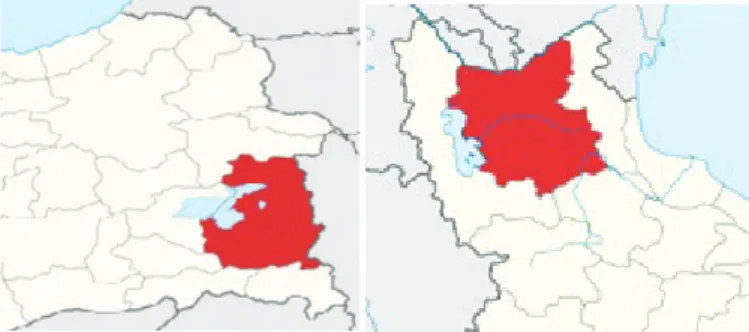
Study AreaThe study was conducted in two regions from two countries with almost similar climatic conditions; Van province from Turkey located in the east of Van Lake which is a part of the coldest region in Turkey, and East Azerbaijan province as a cold area located on the Sahand Mountain range of Iran in the southeast of Urmia Lake (Figure 1).
East Azerbaijan is located in Iranian Azerbaijan, bordering with Armenia and Republic of Azerbaijan with the geographical coordinates of 38° 28’ 45.1020’’ N and 47° 3’ 50.9040’’ Bonab city
is located in the Azerbaijan region. Because of its extensive and large pastures, its livestock numbers are significant compared to other cities in the province. It has a large industrial slaughterhouse and high daily intake capacity, which plays an important role in providing meat and livestock products of the region and also the country.
Van is one of the eastern provinces of Turkey located in neighboring Iran at latitude 38° 29’ 40 N, longitude of 43° 22’ 59 E and altitude of 1.725 meters in Turkey. Van has a harsh continental climate with cold, snowy winters and warm, dry summers. Rainfall occurs mostly during the spring and autumn. Because of Van Lake, the climate of this city can be changed between terrestrial and Mediterranean climate of Central Anatolia and Southeast Anatolia regions (23). Therefore, like the region introduced in Iran, it has similar climate conditions and, is an active and leading province in livestock breeding and production of livestock products in Turkey. A recently described rare sheep breed, Norduz, is mainly raised in a region of the same name in Gürpınar County of Van province (24). The study was approved by the Research Ethics Committee of Shiraz University of Medical Sciences (SUMS, Iran).
Sample Preparation
A total of 90 livestock liver and lung organs infected with hydatid cyst from two areas; Van city of Turkey (30 samples, including 15 sheep and 15 cattle) and Bonab city in East Azerbaijan Province of Iran (60 samples, including 30 sheep and 30 cattle) were obtained. The samples were collected from industrial slaughterhouses of Bonab and Van cities. Protoscolices (PSCs) were collected from the hydatid cyst fluid and after 3 time washes with phosphate buffered saline; the precipitated PSCs were frozen. Also, germinal layers of the cyst were carefully released from the outer host capsules, and were stored at -20 °C until use.
Extraction of Genomic DNA from Isolates
The genomic DNA from either germinal layers or PSCs were extracted, using a DNA extraction kit (YTA, Yekta Tajhiz Azma, Iran), based on the manufactures instructions and also modifications, previously introduced by the authors (25).
Polymerase Chain Reaction and Gel Electrophoresis
For all 90 samples collected from these two countries, polymerase chain reaction (PCR) was performed targeting a 450 bp and 550 bp fragments of cox1 and nad1 of the mitochondrial DNA respectively, using appropriate primers (26,27). The characteristics of the primers used and the genomic regions of the targets are presented in Table 1.
The cycling parameters for the amplification of both genomic pieces was: 1x (5′, 95 °C)+ 40x (45″, 94 °C+35″ 51 °C+45″ 72 °C)+ 1x (10′, 72 °C).
PCR products were separated on a 1.5% agarose gel, and the obtained bands were visualized and recorded by a ultraviolet detector (Bio-Rad, USA).
DNA Sequencing
Of the total 90 available PCR products, 49 samples including 19 samples from Turkey (10 sheep and 9 cattle) and 30 samples from Iran (15 samples from each animal) were selected in terms of the quality of the resulting band on the electrophoresis gel and purified from the gel by EasyPure Quick Gel Extraction Kit (TRANS, TransGen Biotech, South Korea), based on the manufacturer’s instructions. The purified products were sequenced for both cox1 and nad1 fragments from both directions using the same primers which were used in the PCR.
Phylogenetic Analysis
The sequences of E. granulosus isolates from both countries were aligned and compared, using BioEdit and also the BLAST program. Moreover, the obtained sequences were compared with those of available related sequences in the GenBank. Maximum likelihood tree was constructed based on the Tamura-Nei model, using the MEGA 7.0 software. Taenia solium (accession no: AB086256) was
used as the out-group.
RESULTS
All gDNA isolates from collected 90 hydatid cysts from two countries were subjected to molecular analysis targeting both
cox1 and nad1 genomic fragments and the resulting PCR product
showed replication of the target genes. Figure 2 shows the PCR products of cox1 and nad1 genes in a few of the evaluated samples. From all 90 evaluated samples, 49 of them with the highest quality in the resulting band on the electrophoresis gel were selected and sequenced, and the resulting sequences were deposited in the GenBank database with accession numbers which are shown in Table 2. All of the 30 samples (100%) from Iran were found to be the genotype G1 strain. Among the samples from Turkey, 15 samples (78.9%) were identified as G1 and only one sample (5.3%) corresponded to the genotype G3 strain.
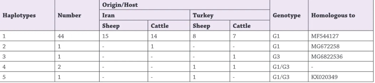
Moreover, two samples had not any homologous to the related sequences in the GenBank, and one sample had similarity to the Echinococcus granulosus from Armenia (KX020349). Therefore, these three samples from Turkey were considered as genotypes G1/G3 strain. All isolates of sheep and cattle from both countries clustered in one group within 5 different haplotypes.
Table 1. The specific primers for amplification of cox1 and
nad1 fragments
Sequences Primers
Genome
5’-TTT TTT GGG CAT CCT GAG GTT TAT-3’ JB3 (F)
cox1
5’-TAA AGA AAG AAC ATA ATG AAA ATG-3’ JB4.5 (R) 5’-AGATTCGTAAGGGGCCTAATA-3’ JB11 (F) nad1 5’-ACCACTAACTAATTCACTTTC-3’ JB12 (R)
Figure 1. Geographic regions of Turkey (left) and Iran (right)
Türkiye Parazitol Derg 2019;43(3):123-9
126
Forty-four samples from both countries were homologous to the
E. granulosus sensu stricto G1 from Turkey (MF544127) and one
sample from Iran was homologous to the E. granulosus G1 from Argentina (MG672258) described earlier. The third haplotype in our study was the only sample from Turkey (MH542404) which had similarity with the E. granulosus sensu stricto G3 from Turkey (MG682536) described earlier. Three of our samples from Turkey (MH542399, MH542406, and MH542395) were placed in two separate haplotypes compared to the rest of the samples (Table 3 and Figure 3, 4). The nad1 sequences were not available for these three isolates and if available, they could be useful in the phylogenetic analyses.
Phylogenetic analysis of the sequences of two cox1 and nad1 genes and alignment of the sequences with available related sequences in the GenBank revealed that the intra-species genetic variation were 0.0-0.6% and 0.0-1.4% for cox1 and nad1, respectively, while the polymorphism variation between the isolates or in other words, the isolates sharing the same haplotype was 0.0 (Figure 5, 6).
DISCUSSION
The Middle East countries have long been considered as important foci of both human and animal CE. The metacestodes
of E. granulosus has been reported in almost all countries of the
region, but its prevalence is higher in Iran, Turkey, and Iraq in comparison with the rest of the countries in the region (28).
Barazesh et al. Genotyping of Echinococcus granulosus
Figure 2. Electrophoresis of PCR products, using JB3 and JB4,
5 primers for cox1 and JB11 and JB12 for nad1, on 1.5% agarose gel. Lane 1: Molecular weight marker; Lane 2: Positive control for cox1, DNA extracted from sheep isolate of Iran; Lane 3: Positive control for nad1, DNA extracted from sheep isolate of Iran; Lane 4: Negative control; Lanes 5, 6: Sheep isolates of Iran and Turkey in the current study, targeting the cox1 gene; Lanes 7, 8: Sheep isolates of Iran and Turkey in the current study, targeting the nad1 gene
PCR: Polymerase chain reaction
Table 2. Host origin of Echinococcus granulosus isolates from
Turkey and Iran livestock and accession numbers deposited in GenBank, using cox1 and nad1 genomes
Sample
no. Code Host Origin Accession no. (cox1) Accession no. (nad1)
1 1SI Sheep Iran MH542362 MH557949
2 2SI Sheep Iran MH542363 MH557965
3 3SI Sheep Iran MH542364 MH557950
4 4SI Sheep Iran MH542365 MH557951
5 5SI Sheep Iran MH542366 MH557966
6 6SI Sheep Iran MH542367 MH557967
7 7SI Sheep Iran MH542368 MH557952
8 8SI Sheep Iran MH542369 MH557968
9 9SI Sheep Iran MH542370 MH557969
10 10SI Sheep Iran MH542371 MH557953
11 11SI Sheep Iran MH542372 MH557954
12 12SI Sheep Iran MH542373 MH557970
13 13SI Sheep Iran MH542374 MH557971
14 14SI Sheep Iran MH542375 MH557955
15 15SI Sheep Iran MH542376
-16 16CI Cattle Iran MH542377 MH557972
17 17CI Cattle Iran MH542378 MH557956
18 18CI Cattle Iran MH542379 MH557973
19 19CI Cattle Iran MH542380 MH557957
20 20CI Cattle Iran MH542381 MH557958
21 21CI Cattle Iran MH542382
-22 22CI Cattle Iran MH542383 MH557959
23 23CI Cattle Iran MH542384 MH557960
24 24CI Cattle Iran MH542385
-25 25CI Cattle Iran MH542386 MH557961
26 26CI Cattle Iran MH542387
-27 27CI Cattle Iran MH542388
-28 28CI Cattle Iran MH542389 MH557962
29 29CI Cattle Iran MH542390 MH557963
30 30CI Cattle Iran MH542391 MH557964
31 1ST Sheep Turkey MH542392 -32 2ST Sheep Turkey MH542393 -33 3ST Sheep Turkey MH542394 -34 4ST Sheep Turkey MH542395 ---35 5ST Sheep Turkey MH542396 ---36 6ST Sheep Turkey MH542397 ---37 7ST Sheep Turkey MH542398 ---38 8ST Sheep Turkey MH542399 ---39 9ST Sheep Turkey MH542400 ---40 10ST Sheep Turkey MH542401 ---41 11CT Cattle Turkey MH542402 ---42 12CT Cattle Turkey MH542403 ---43 14CT Cattle Turkey MH542404 ---44 15CT Cattle Turkey MH542405 ---45 16CT Cattle Turkey MH542406 ---46 17CT Cattle Turkey MH542407 ---47 18CT Cattle Turkey MH542408 ---48 19CT Cattle Turkey MH542409 ---49 20CT Cattle Turkey MH542410 ---Table 2. Continued
Various mitochondrial and nuclei genomes have been used for molecular evaluation and to determine the genotype of E.
granulosus. Regarding phylogenetic taxonomy of E. granulosus among closely related species, mtDNA has been reported more efficient than nuclear genomes due to the rapid sequence evolution and large datasets derived from mitochondrial genomes (29).The mitochondrial genes; including cox1, nad1, and atp6, as well as the fragment of the 12S rRNA gene have been used to identify the genotypes in different isolates. Findings of Rostami Nejad et al. (30) study on genetic diversity of E. granulosus in different hosts, revealed G1 and G6 genotypes in cattle, camels,
sheep, buffalo and goats in different geographic areas of Iran. Likewise, the ınternal transcribed spacer (ITS1) gene region has also been utilized for genotypic analysis of this parasite (31). In two separate studies, Ahmadi and Dalimi (32) and Harandi et al. (33) used ITS1 region gene to genotype the E. granulosus isolates. They found a similarity between strains in sheep and camel with cattle and humans.
However,cox1 and nad1 mitochondria genes, have been considered as the main and the best options for molecular characterization of CE. For distinction of intra- and interspecific variants, the gene
cox1 gene, can be used as a significant evolutionary marker (34). Table 3. Echinococcus granulosus haplotypes and genotypes detected in this study using cox1 and nad1 sequences
Haplotypes Number
Origin/Host
Genotype Homologous to
Iran Turkey
Sheep Cattle Sheep Cattle
1 44 15 14 8 7 G1 MF544127
2 1 - 1 - - G1 MG672258
3 1 - - - 1 G3 MG6822536
4 2 - - 1 1 G1/G3
-5 1 - - 1 - G1/G3 KX020349
Figure 3. Phylogenetic tree of representative sequences of
Echinococcus granulosus from Iran and Turkey and reference
sequences of other genotypes, using the maximum likelihood method based on cox1 gene. Taenia solium (AB086256) was used as the out-group sequence data
Intra-species genetic: 0-0.6%
Figure 4. Phylogenetic tree of representative sequences of
Echinococcus granulosus from Iran and reference sequences of
other genotypes, using the maximum likelihood method based on nad1 gene. Taenia solium (AB086256) was used as the out-group sequence data
Türkiye Parazitol Derg 2019;43(3):123-9
128
Barazesh et al. Genotyping of Echinococcus granulosusMahami-Oskouei et al. (35) used cox1 and nad1 genes to investigate the novel single-nucleotide polymorphism and reported that the
G1 genotype with 27 haplotypes was the main strain in human, sheep, goat, cattle and dog isolates. Their study showed that cross transmission of sheep-dog strain is circulating among potential intermediate/definitive hosts with heterogeneity traits of
Echinococcus in Iran and Turkey.
In the present study, we selected both cox1 and nad1 genomic fragments as the target and the resulting PCR product showed successful replication of the target genes. Recently, it has been reported that the differentiation between G1 and G3 genotypes for some cases is not possible and the identified genotypes has been reported as G1/G3 strain (36). In a recent study, carried out by Kinkar et al. (8), nad5 fragment has been introduced for proper differentiation of E. granulosus sensu stricto genotypes G1 and G3. Findings of the current study demonstrated the G1 strain as the dominant strain of E. granulosus in the livestock of the two studied
regions; Azerbaijan from Iran and Van from Turkey. Only one case of G3 strain and 3 cases of G1/G3 strains were found in this study. In general, E. granulosus sensu stricto (G1-G3) are the
predominant strains in CE cases throughout the world (7,8). Findings of the current study are in accordance with other reports from Iran, Turkey, and also the Middle East countries. In some studies, conducted in different geographical areas of Iran, the G1 strain of E. granulosus was reported as the dominance genotype
in intermediate hosts including cattle, sheep, human and camels (37,38). In one study conducted in Golestan province, northern Iran, G1 and G3 strains have been reported in 78.3% and 15% of CE cases respectively (39).The G1 strain also reported from the wild boar in Iran (14). This further emphasizes that the dominant strain of E. granulosus in Iran, not only in livestock but also in
wild animals is the G1-G3 strains. In a study by Simsek et al. (20) on cattle and sheep isolates of E. granulosus metacestodes from
eastern areas of Turkey, all of the 54 examined samples were found as G1-G3 strains.A study on the genetic characteristics of human and animals isolates of E. granulosus in the province of
Edirne from Turkey, DNA sequencing of the cox1 and nad1 genes was performed and authors indicated that the sheep strain G1 was the most common genotype of E. granulosus affecting humans,
sheep and cattle in the studied area. Moreover, 8 haplotypes of
Echinococcus species were identified in the region (17). In another
similar study for the molecular analysis of E. granulosus isolates
from different regions of Turkey, the cox1 gene was used for identification and molecular analysis of CE cases where all of the human hydatid cysts were belonged to the G1 (40). In 2008, Vural et al. (41) reported G1 strain of E. granulosus in 107 out of 112
samples whereas only 5 cases were determined as the G3 strain. The interesting point was that the parasites of the G3 genotype were identified only in the isolates derived from animals in the eastern regions of the country (41). This finding is fully consistent with our findings; where one of our samples derived from Van city (the eastern region in Turkey) was determined as the G3 strain and the rest of the isolates were identified as the G1 strain. It seems obvious that two regions evaluated in the present study have very close similarity in genetic features of E. granulosus as
there were no differences in terms of genotypes and also the diversity of isolates of the parasite in these two areas. Moreover, the isolates of both sheep and cattle from both countries were placed in the same cluster. It should be noted that only cox1 gene was used for genotype analysis of Turkish isolates in this study and this should be considered as a limitation of the current study.
CONCLUSION
Findings of the current study revealed that the sheep strain G1 is the dominant strain of E. granulosus in livestock isolates in Turkey
and Iran. The inter and intra heterogeneity of the isolates in the two countries were 0.0-0.6% and 0.0-1.4% for cox1 and nad1 genomes, respectively.
Findings of the study can be used for adopting the common policies and bilateral cooperation for prevention and also controlling the disease in these two countries. Further studies are needed to determine the dominant genotypes of E. granulosus in
human cases in these two regions. * Ethics
Ethics Committee Approval: The study was approved by the Research Ethics Committee of Shiraz University of Medical Sciences (SUMS, Iran).
Informed Consent: Patient consent was not obtained. Peer-review: Internally peer-reviewed.
* Authorship Contributions
Concept: A.B., B.S., G.S., Design: A.B., B.S., G.S., A.A., Data Collection or Processing: A.B., G.S., A.A., A.E., M.H., F.M., S.E., Analysis or Interpretation: A.B., B.S., F.M., Literature Search: A.B., B.S., F.M., Writing: A.B., B.S.
Conflict of Interest: No conflict of interest was declared by the authors.
Financial Disclosure: This study financially supported by the Vice-chancellor of Research of Shiraz University of Medical Sciences (Grant No. 95-01-106-13401).
REFERENCES
1. Deplazes P, Rinaldi L, Alvarez Rojas CA, Torgerson PR, Harandi MF,
Romig T, et al. Global Distribution of Alveolar and Cystic Echinococcosis. Adv Parasitol 2017;95:315-493.
2. Romig T, Deplazes P, Jenkins D, Giraudoux P, Massolo A, Craig PS, et al. Ecology and Life Cycle Patterns of Echinococcus Species. Adv Parasitol 2017;95:213-314.
3. Budke CM, Deplazes P, Torgerson PR. Global socioeconomic impact of cystic echinococcosis. Emerg Infect Dis 2006;12:296-303.
4. Thompson RC, McManus DP. Towards a taxonomic revision of the genus Echinococcus. Trends Parasitol 2002;18:452-7.
5. Thompson RC. Biology and systematics of Echinococcus. Adv Parasitol 2017;95:65-109.
6. Hüttner M, Nakao M, Wassermann T, Siefert L, Boomker JDF, Dinkel A, et al. Genetic characterization and phylogenetic position of Echinococcus felidis Ortlepp, 1937 (Cestoda: Taeniidae) from the African lion. Int J Parasitol 2008;38:861-8.
7. Rojas CAA, Romig T, Lightowlers MW. Echinococcus granulosus sensu
lato genotypes infecting humans–review of current knowledge. Int J Parasitol 2014;44:9-18.
8. Kinkar L, Laurimäe T, Acosta-Jamett G, Andresiuk V, Balkaya I, Casulli A, et al. Distinguishing Echinococcus granulosus sensu stricto genotypes G1 and G3 with confidence: A practical guide. Infect Genet Evol 2018;64:178-84.
9. Sarkari B, Sfedan AF, Moshfe A, Khabisi SA, Savardashtaki A, Hosseini F, et al. Clinical and molecular evaluation of a case of giant primary splenic hydatid cyst: A case report. Iranian J Parasitol 2016;11:585-90.
10. Sarkari B, Hosseini F, Khabisi SA, Sedaghat F. Seroprevalence of cystic echinococcosis in blood donors in Fars province, southern Iran. Parasite Epidemiol Control 2017;2:8-12.
11. Sarkari B, Sadjjadi SM, Beheshtian MM, Aghaee M, Sedaghat F. Human cystic Echinococcosis in Yasuj district in Southwest of Iran: an epidemiological study of seroprevalence and surgical cases over a ten‐year period. Zoonoses Public Health 2010;57:146-50.
12. Harandi MF, Budke CM, Rostami S. The monetary burden of cystic
echinococcosis in Iran. PLOS Negl Trop Dis 2012;6:e1915.
13. Rokni MB. Echinococcosis/hydatidosis in Iran. Iranian J Parasitol
2009;4:1-16.
14. Sarkari B, Mansouri M, Khabisi SA, Mowlavi G. Molecular characterization and seroprevalence of Echinococcus granulosus in wild boars (Sus scrofa) in south-western Iran. Ann Parasitol 2015;61:269-73.
15. Mansouri M, Sarkari B, Mowlavi GR. Helminth Parasites of Wild Boars, Sus scrofa, in Bushehr Province, Southwestern Iran. Iran J Parasitol 2016;11:377-82.
16. Utuk AE, Simsek S, Koroglu E, McManus DP. Molecular genetic
characterization of different isolates of Echinococcus granulosus in east and southeast regions of Turkey. Acta Trop 2008;107:192-4.
17. Eryıldız C, Sakru N. Molecular characterization of human and animal isolates of Echinococcus granulosus in the Thrace Region, Turkey. Balkan Med J 2012;29:261-7.
18. Altintas N. Past to present: echinococcosis in Turkey. Acta Trop
2003;85:105-12.
19. Esatgil MU, Tüzer E. Prevalence of hydatidosis in slaughtered animals in Thrace, Turkey. Turkiye Parazitol Derg 2007;31:41-5.
20. Simsek S, Balkaya I, Ciftci AT, Utuk AE. Molecular discrimination of sheep and cattle isolates of Echinococcus granulosus by SSCP and conventional PCR in Turkey. Vet Parasitol 2011;178:367-9.
21. Mor N, Allahverdi TD, Anuk T. The situation of cystic echinococcoses in Kars State Hospital for the last five years. Turkiye Parazitol Derg 2015;39:108-11.
22. Sánchez E, Cáceres O, Náquira C, Garcia D, Patiño G, Silvia H, et al. Molecular characterization of Echinococcus granulosusfrom Peru by sequencing of the mitochondrial cytochrome C oxidase subunit 1 gene. Mem Inst Oswaldo Cruz 2010;105:806-10.
23. Ozdal N, Gul A, Ilhan F, Deger S. Prevalence of Paramphistomum infection in cattle and sheep in Van Province, Turkey. Helminthologia 2010;47:20-4.
24. Daskiran I, Cedden F. Norduz goat of east anatolia. Journal of Animal and Veterinary Advances. 2004.
25. Barazesh A, Sarkari B, Ebrahimi S, Hami M. DNA extraction from hydatid cyst protoscolices: Comparison of five different methods. Vet World 2018;11:231.
26. Bowles J, Blair D, McManus DP. Genetic variants within the genus
Echinococcus identified by mitochondrial DNA sequencing. Mol Biochem Parasitol 1992;54:165-73.
27. Bowles J, McManus DP. NADH dehydrogenase 1 gene sequences
compared for species and strains of the genus Echinococcus. Int J Parasitol 1993;23:969-72.
28. Sadjjadi SM. Present situation of echinococcosis in the Middle East and Arabic North Africa. Parasitol Int 2006;55:S197-202.
29. Nakao M, McManus DP, Schantz PM, Craig PS, Ito A. A molecular
phylogeny of the genus Echinococcus inferred from complete mitochondrial genomes. Parasitology 2006;134:713-22.
30. RostamiNejad M, Taghipour N, Nochi Z, NazemalhosseiniMojarad E,
Mohebbi SR, Harandi MF, et al. Molecular identification of animal isolates of Echinococcus granulosus from Iran using four mitochondrial genes. J Helminthol 2012;86:485-92.
31. Nikmanesh B, Mirhendi H, Ghalavand Z, Alebouyeh M, Sharbatkhori M, Kia E, et al. Genotyping of Echinococcus granulosus isolates from human clinical samples based on sequencing of mitochondrial genes in Iran, Tehran. Iranian J Parasitol 2014;9:20-7.
32. Ahmadi N, Dalimi A. Characterization of Echinococcus granulosus isolates from human, sheep and camel in Iran. Infect Genet Evol 2006;6:85-90.
33. Harandi MF, Hobbs RP, Adams PJ, Mobedi I, Morgan-Ryan UM,
Thompson RCA. Molecular and morphological characterization of Echinococcus granulosusof human and animal origin in Iran. Parasitology 2002;125:367-73.
34. Nakao M, Lavikainen A, Yanagida T, Ito A. Phylogenetic systematics of the genus Echinococcus (Cestoda: Taeniidae). International Journal for Parasitology 2013;43:1017-29.
35. Mahami-Oskouei M, Kaseb-Yazdanparast A, Spotin A, Shahbazi
A, Adibpour M, Ahmadpour E, et al. Gene flow for Echinococcus granulosus metapopulations determined by mitochondrial sequences: a reliable approach for reflecting epidemiological drift of parasite among neighboring countries. Exp Parasitol 2016;171:77-83.
36. Umhang G, Richomme C, Boucher JM, Hormaz V, Boué F. Prevalence
survey and first molecular characterization of Echinococcus granulosus in France. Parasitol Res 2013;112:1809-12.
37. Nejad MR, Taghipour N, Nochi Z, Mojarad EN, Mohebbi SR, Harandi
MF, et al. Molecular identification of animal isolates of Echinococcus granulosus from Iran using four mitochondrial genes. J Helminthol 2012;86:485-92.
38. Harandi MF, Hobbs RP, Adams PJ, Mobedi I, Morgan-Ryan UM,
Thompson RCA. Molecular and morphological characterization of Echinococcus granulosus of human and animal origin in Iran. Parasitology. 2002;125:367-73.
39. Sharbatkhori M, Tanzifi A, Rostami S, Rostami M, Harandi MF.
Echinococcus granulosus sensu lato genotypes in domestic livestock and humans in Golestan province, Iran. Rev Inst Med Trop Sao Paulo 2016;58:38.
40. Ergin S, Saribas S, Yuksel P, Zengin K, Midilli K, Adas G, et al. Genotypic characterisation of Echinococcus granulosus isolated from human in Turkey. African J Microbiol Res 2010;4:551-5.
41. Vural G, Baca AU, Gauci CG, Bagci O, Gicik Y, Lightowlers MW. Variability in the Echinococcus granulosus cytochrome C oxidase 1 mitochondrial gene sequence from livestock in Turkey and a re-appraisal of the G1–3 genotype cluster. Vet Parasitol 2008;154:347-50.
View publication stats View publication stats