Cukurova Medical Journal
Cukurova Med J 2019;44(1):267-269ÇUKUROVA ÜNİVERSİTESİ TIP FAKÜLTESİ DOI: 10.17826/cumj.430740
Yazışma Adresi/Address for Correspondence: Dr. Mehmet Öztürk, Selcuk University Medical Faculty, Department of Radiology, Division of Pediatric Radiology, Konya, Turkey.
E-mail: [email protected], [email protected]
Geliş tarihi/Received: 05.06.2018 Kabul tarihi/Accepted: 13.06.2018 Çevrimiçi yayın/Published online: 05.07.2018
EDİTÖRE MEKTUP / LETTER TO THE EDITOR
Unilateral hypertrophy of the C5 vertebra spinous process in an
adolescent
Adölesan olguda C5 vertebra spinöz proçesinin tek taraflı hipertrofisi
İbrahim Başar
1, Mehmet Öztürk
2, Semih Sağlık
31Siirt State Hospital, Department of Nneurosurgery, 3Department of Radiology, Siirt, Turkey
2Selcuk University Medical Faculty, Department of Radiology, Division of Pediatric Radiology, Konya, Turkey.
Cukurova Medical Journal 2019;44(1):267-269 To the Editor,
Congenital anomalies of the cervical vertebrae are rarely observed situation1. These anomalies include
persistent apophysis of the transverse process, vertebral platyspondylia, fusion of vertebral bodies at various levels, vertebral hypoplasia and unilateral spinous process hypertrophy2. They may accompany
congenital abnormalities like Klippel-Feil syndrome, Morquio syndrome, diastrophic dwarfism, spondyloepiphyseal dysplasia, and osteogenesis imperfecta3, 4. Congenital abnormalities of the
transverse and spinous process are generally coincidentally identified on radiologic imaging but may cause clinical symptoms like pain or esthetic problems1. This report discusses the clinical and
radiologic findings of a male adolescent case with unilateral hypertrophy of the C5 vertebra spinous process.
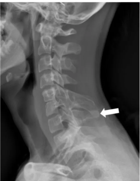
A 16-year old male patient attended the neurosurgery clinic due to noticeable painless swelling in the posterior cervical region. The patient had no history of trauma or previous operation for any reason. Physical examination identified a palpable, painless, hard and fixed mass in the posterior cervical region. Neurological examination did not identify any neurological deficit. Firstly, AP and lateral radiographs were obtained for the cervical vertebra with the aim of assessing the relationship of the lesion to the bone structures. The spinous process of C5 vertebra was assessed as
hypertrophic (Figure 1).
Figure 1. C5 vertebral spinous process hypertrophy on lateral AP radiograph (white arrow).
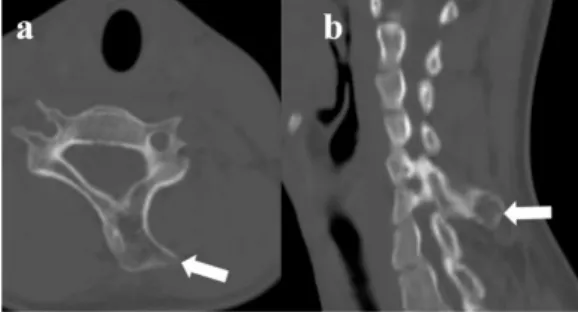
Cervical computed tomography (CT) identified left-side asymmetric hypertrophy of the C5 vertebral spinous process along with stenosis of the C5-C6 disk interval (Figure 2 a, b). For possible spinal and soft-tissue pathologies, magnetic resonance imaging (MRI) of the cervical, thoracic and lumbar vertebrae was performed. On MRI, the left half of the C5
Başar et al. Cukurova Medical Journal
268
vertebral spinous process was identified to have hypertrophy with similar intensity to bone structures in axial and sagittal planes and on all sequences (Figure 3 a, b).
Figure 2 a, b. Axial (a) and sagittal (b) CT images: hypertrophy of C5 vertebral spinous process on the left (white arrow).
Bilateral neural foramen widths were normal, with no additional spinal cord or paravertebral soft tissue pathology identified. The patient had no active complaint and did not accept surgical treatment so monitoring was considered appropriate.
Figure 3 a, b. Axial (a) and sagittal (b) T2-weighted MRI: hypertrophy of C5 vertebral spinous process on the left (white arrow).
The spine is a complicated and vital structure, serving as structural support for the body and as a channel for neural structures ensuring communication with the brain5. Vertebral arc
development begins in the 3-6th week intrauterine and continues until the second decade with the completion of fusion of secondary ossification centers3, 6. Dysfunction in the embryologic period
may cause one or more congenital anomalies. The cause of unilateral hypertrophy of the lamina or spinous process is not fully known. Additionally, abnormal extension of chondrification and other normal development and ossification of the vertebral column may explain present anomalies. Mostly it is asymptomatic and coincidentally
identified.
Developmental anomalies of the cervical vertebrae may accompany congenital syndromes like Klippel-Feil syndrome, Down syndrome, fibrodysplasia ossificans progressive or Rubinstein-Taybi syndrome7,8. In addition to developmental
abnormalities, acquired diseases like ankylosing spondylitis, psoriatic spondylitis, diffuse idiopathic skeletal hyperostosis, ossification of the posterior longitudinal ligament, sternoclavicular hyperostosis, juvenile chronic arthritis and acromegaly, and diseases causing hyperostosis or ossification of the cervical spine should be considered for differential diagnosis9. Hypertrophy of the spinous process of
the cervical vertebrae may be a part of a syndrome but may occur as an isolated abnormality. If it occurs as an isolated congenital anomaly, it rarely causes clinical symptoms. Clinical findings may occur in the form of cervical movement limitation, cervical pain, neurological deficits and esthetic abnormalities. In the absence of clinical findings, it may be coincidentally identified on radiologic assessments performed for different reasons like trauma10. In our case, there was no active complaint
other than esthetic anomaly.
The first choice for treatment of spinous process hypertrophy of the cervical vertebrae should be conservative methods like analgesics, muscle relaxants and physical therapy methods. If conservative methods fail or there are severe symptoms, the choice should be surgical resection10.
As our case did not have an active complaint, he did not accept surgical intervention, and the decision was made to monitor him. Additionally, as our case was an adolescent and the vertebral development process was continuing, close follow-up was important.
Definite diagnosis of congenital anomalies of the cervical vertebrae are placed with radiologic methods. The first and most commonly used method is direct radiography, but this may be insufficient for complicated bone anomalies or in the presence of trauma. If the diagnosis is uncertain the second method that should be used is CT. The use of CT and MRI together is the most useful choice to assess spinal anomalies, allowing detailed investigation. Though the majority of congenital spinal deformities do not have clinical symptoms, they have high incidence rates with intraspinal pathologies of 30-35%11. Additionally, in the
Cilt/Volume 44 Yıl/Year 2019 Unilateral hypertrophy of the C5 vertebra spinous process
269
pathologies like fracture, pseudoarthrosis and hematoma. MRI is superior to identify additional trauma or inflammation markers of bone marrow edema and traumatic spinal lesions. Cross-sectional imaging methods can assess paravertebral soft tissue planes, intervertebral disk intervals and bone marrow variations in detail10. In our case, CT and
MRI methods were applied together, with no additional anomalies or mass lesions identified. This data is very beneficial to plan the treatment and follow-up process for our case.
In conclusion, in adolescent cases attending with preliminary diagnosis of mass on the cervical vertebrae congenital anomalies like unilateral spinous process hypertrophy should be included in differential diagnosis. Along with clinical assessment, direct radiography, CT and MRI are very beneficial radiologic methods for diagnosis and monitoring.
Yazar Katkıları: Çalışma konsepti/Tasarımı: SS, MÖ; Veri toplama:
SS; Veri analizi ve yorumlama: MÖ; Yazı taslağı: İB; İçeriğin eleştirel incelenmesi: SS, MÖ; Son onay ve sorumluluk: İB, MÖ, SS; Teknik ve malzeme desteği: İB; Süpervizyon: MÖ; Fon sağlama (mevcut ise): yok.
Bilgilendirilmiş Onam: Katılımcılardan yazılı onam alınmıştır. Hakem Değerlendirmesi: Dış bağımsız.
Çıkar Çatışması: Yazarlar çıkar çatışması beyan etmemişlerdir. Finansal Destek: Yazarlar finansal destek beyan etmemişlerdir. Author Contributions: Concept/Design :SS, MÖ; Data acquisition: SS; Data analysis and interpretation: MÖ; Drafting manuscript: İB; Critical revision of manuscript: SS, MÖ; Final approval and accountability: İB, MÖ, SS; Technical or material support: İB; Supervision: MÖ; Securing funding (if available): n/a.
Informed Consent: Written consent was obtained from the
participants.
Peer-review: Externally peer-reviewed.
Conflict of Interest: Authors declared no conflict of interest. Financial Disclosure: Authors declared no financial support
REFERENCES
1. Hegde KK, Rashmi PS, Poosarla R, Vijayanath V. Unilateral hyperplasia of lamina and spinous process
of C6 vertebrae. International Journal of Pharmacy and Biological Sciences 2012; 2: 344-8.
2. Esposito G, de Bonis P, Tamburrini G, Massimi L, Byvaltsev V, Di Rocco CD et al. Unilateral hyperplasia of the left posterior arch and associated vertebral schisis at C6 level. Skeletal Radiol. 2009;38: 1191-5.
3. Rameez Farooqi R, Mehmood M, Kotwal AH
.
Hyperplasia of lamina and spinous process of C5 vertebrae and associated hemivertebra at C4 level. J Orthop Case Rep. 2017;7:79-81.4. Beals RK, Robbins JR, Rolfe B. Anomalies associated with vertebral malformations. Spine. 1993;18:1329-32.
5. Reddy OJ, Gafoor JA, Suresh B, Lakshmi KC. A case report on giant C5 spinous process with partial block vertebrae at C2, C3, and C4 levels. Journal of Dr. NTR University of Health Sciences. 2017;6:129-32.
6. Heyer CM, Nicolas V, Peters SA. Unilateral hyperplasia of a cervical spinous process as rare congenital variant of the spine. Clin Imaging. 2007;31:434-6.
7. Schaffer AA, Kaplan FS, Tracy MR, O’Brien ML, Dormans JP, Shore EM et al. Developmental anomalies of the cervical spine in patients with fibrodysplasia ossificans progressiva are distinctly different from those in patients with Klippel–Feil syndrome: clues from the BMP signaling pathway. Spine. 2005;30:1379-85.
8. Mete M, Ünsal ÜÜ, Duransoy YK, Umur AŞ, Selçuki M. A giant hypertrophy of C5 spinous process in Klippel-Feil syndrome. Journal of Neurological Sciences. 2015;32:575-9.
9. Resnick D. Hyperostosis and ossification in the cervical spine. Arthritis Rheum. 1984;27:564-9. 10. Kazanci B, Tehli O, Adilay U, Guclu B. Unilateral
hyperplasia of lamina and spinous process of C6 vertebra: case report. Surg Radiol Anat. 2012;34: 875-8.
11. Basu PS, Elsebaie H, Noordeen MH. Congenital spinal deformity: a comprehensive assessment at presentation. Spine. 2002;27:2255-9.