Abstract
In this preliminary case presentation, use of the minimally invasive cystoscopic pneumatic lithotripsy technique in the treatment of complete urethral obstruction in a dog has been described. The animal of the case comprised a 5-year old Chihuahua presenting with difficulty during urination and inability to urinate for the previous 2 days. Post-renal azotemia and hematuria was determined. Urethral stones causing complete urethral obstruction were visualized via cystoscopy, fragmented with a pneumatic lithotripter and the stone fragments were removed using the voiding urohydropulsion method. In conclusion, the pneumatic lithotripsy method was successfully used in the treatment of complete urethral obstruction.
Keywords: Pneumatic lithotripsy, Urethroscopy, Urethral stone, Obstruction, Dog
Bir Köpekte Tam Üretral Obstrüksiyonun Pnömatik Litotripsi
İle Tedavisi: İlk Rapor
Özet
Bu olgu sunumunda bir köpekte tam üretral obstrüksiyonun tedavisinde minimal invaziv sistoskopik pnömatik litotripsi tekniğinin kullanımı anlatıldı. Olgunun hayvan materyalini idrar yaparken zorlanma ve son iki gündür idrar yapamama şikâyetleri ile getirilen, postrenal azotemia ve hematuria tespit edilen Chihuahua ırkı, 5 yaşlı bir köpek oluşturdu. Tam üretral obstrüksiyona neden olan üretral taşlar sistoskopi ile görüntülendi, pnömatik litotriptör kullanılarak parçalandı ve taş fragmanları voiding urohydropulsiyon yöntemiyle dışarıya alındı. Sonuç olarak, pnömatik litotripsi yöntemi tam üretral obstrüksiyonun tedavisinde başarılı bir şekilde kullanıldı. Anahtar sözcükler: Pnömatik litotripsi, Üretroskopi, Üretral taş, Obstrüksiyon, Köpek
Treatment of Complete Urethral Obstruction by using Pneumatic
Lithotripsy in a Dog: A Preliminary Report
[1,2]Mehmet MADEN
1
Merve İDER
1Kurtuluş PARLAK
2Ahmet ÖZTÜRK
3 [1][2] 1 2 3
Presented at 11th Veterinary Internal Medicine Congress (Samsun, Turkey, 21-24 May 2015) as an oral presentation
Presented at 32nd World Veterinary Congress (Istanbul on September 13-17, 2015) as an poster presentation
Selcuk University, Faculty of Veterinary Medicine, Department of Internal Medicine, TR-42079 Konya - TURKEY Selcuk University, Faculty of Veterinary Medicine, Department of Surgery, TR-42079 Konya - TURKEY
Necmettin Erbakan University, Faculty of Medicine, Department of Urology, TR-42080 Konya - TURKEY
INTRODUCTION
Lithotripsy derives from the Greek words “lith” stone and “tripsis” to break and is a procedure literally meaning “breaking stones”. It was first used to fragment kidney stones in humans at Munich University on 7th February 1980. This technique was named “Extracorporeal Shock Wave Lithotrpisy” (ESWL). ESWL was followed by the development of intracorporeal techniques administered directly onto the stone using endoscopic methods. Fragmenting of bladder stones became easier with the development of lithotripters applied directly onto the surface of the stone guided by a cystoscope. The
electrohydrolic, ultrasonic, pneumatic and Holmium-YAG laser lithotripsy techniques are widely used in this field [1].
Pneumatic lithotripsy (PL) was developed in the 1990s. The method utilizes a rigid energy probe placed directly onto the stone and the stone is then broken into fragments by a drill-like effect due to the energy of the compressed air. The PL method provides a practical and low cost treatment approach in urethral stones due to its reusable probe [2]. Pneumatic lithotripsy is used extensively in human medicine [3-5]. In veterinary medicine, it was first demonstrated in fragmenting stones placed in the ureters of dogs and pigs in an experimental study [6].
İletişim (Correspondence)
+90 332 2233596
[email protected]KafKas Universitesi veteriner faKUltesi Dergisi
JoUrnal Home-Page: http://vetdergi.kafkas.edu.tr
online sUbmission: http://vetdergikafkas.org
Case Report
Kafkas Univ Vet Fak Derg 22 (2): 305-308, 2016
DOI: 10.9775/kvfd.2015.14298
306
Treatment of Complete ...
In this case report, the use of a pneumatic lithotripter under the guidance of a rigid cystoscope in the treatment of total urethral obstruction in a 5-year old Chihuahua dog has been described.
CASE HISTORY
The case was a 5-year old Chihuahua, brought to the Small Animal Hospital of Veterinary Faculty, Selçuk University. The case presented difficulty in urination (dysuria, stranguria), inappetence, lethargy and inability to urinate for the previous 2 days. Physical examination of the dog revealed normal body temperature, heart rate and respiratory rate. Distension and pain in the caudal abdomen along with a full urinary bladder were determined upon abdominal palpation. The dog frequently exhibited urination position but was unable to urinate. A full bladder was observed on direct radiographs with no other abnormalities detected. On ultrasound examination, structures presenting acoustic shadows were identified in the bladder and proximally to the os penis. The urethral catheterization attempt after decompressive cystocentesis was unsuccessful. Therefore, it was decided to perform cystoscopy in the dog. The patient’s haematological, venous blood gases, serum biochemistry and urine analysis data are shown in Table 1. Haematology and venous blood gas data was within the normal range, however, post-renal azotaemia and haematuria was determined. Prior to cystoscopy, the patient was anaesthetized with xylazine (Rompun® Bayer, 2 mg/kg, IM) and ketamine (Ketasol® Interhas, 10 mg/kg, IM). On rigid cystoscopic examination, 2 yellow and rough-surfaced stones proximal to the os penis were seen to have completely obstructed the urethra (Fig. 2). The stones were fragmented using a pneumatic
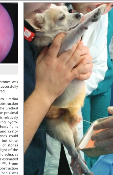
lithotripter (Fig. 1) and the urethra was unblocked. The bladder was then entered and stones in the bladder were fragmented. The stone fragments were removed via urination using the voiding urohydropulsion method (Fig. 3) [7-9]. The voiding urohydropulsion method was repeated 3 times.
DISCUSSION
In this preliminary case report, use of minimally invasive cystoscopic pneumatic lithotripsy method in the treatment
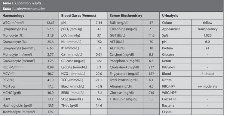
Table 1. Laboratory results Tablo 1. Laboratuar sonuçlar
Haematology Blood Gases (Venous) Serum Biochemistry Urinalysis
WBC (m/mm3) 12.67 pH 7.34 BUN (mg/dl) 57 Colour Yellow Lymphocyte (%) 52.5 pCO2 (mmHg) 37 Creatinine (mg/dl) 2.3 Appearence Transparancy Monocyte (%) 21.9 pO2 (mmHg) 37 GGT (IU/L) 11.0 SpG 1.020 Granulocyte (%) 25.6 Na+ (mmol/L) 152 ALT (IU/L) 70 pH 6.0 Lymphocyte (m/mm3) 6.65 K+ (mmol/L) 3.3 ALP (IU/L) 18 Protein +1 Monocyte (m/mm3) 2.77 Ca++ (mmol/L) 0.61 Calcium (mg/dl) 8.8 Glucose -Granulocyte (m/mm3) 3.25 Glucose (mg/dl) 122 Phosphorus (mg/dl) 6.8 Keton -RBC (M/mm3) 8.99 Lactate (mmol/L) 3.3 Cholesterol (mg/dl) 237 Bilirubin -MCV (fl) 46.7 HCO3- (mmol/L) 20.0 Triglyceride (mg/dl) 127 Blood -/+ intact PCV (%) 41.9 TCO2 (mmol/L) 21.1 Total Protein (g/dl) 6.1 Nitrite -MCH pg 17.2 BEecf (mmol/L) - 5.8 Albumin (g/dl) 4.0 RBC/HPF ++ moderate MCHC (g/dl) 36.9 BE(B) (mmol/L) - 5.2 Glucose (mg/dl) 215 WBC/HPF -RDW 12.1 SO2c (mmol/L) 66 T. Bilirubin (mg/dl) 1.8 Casts/HPF -Haemoglobin (g/dl) 15.5 THbc (g/dl) 14.6 Bacteria -Trombocyte (m/mm3) 159 Crystal
-Fig 1. Pneumatic lithotripter and rigid endoscope (A), lithotripter unit (B) Şekil 1. Pnömatik litotriptör ve rigid endoskop (A), litotriptör ünitesi (B)
307 MADEN, İDER PARLAK, ÖZTÜRK
of complete urethral obstruction caused by stones was described in a dog. The obstruction was successfully resolved and no complications were encountered.
The movement of stones from bladder into urethra is among the most frequent causes of urinary obstruction in dogs. Small breeds are more affected [10]. The urethral obstruction is most commonly reported in the proximal part of os penis, where the urethral diameter is relatively narrow. Urethral stones can be removed using hydro-pulsion, lithotripsy and basket retrieval methods [9], as well as the surgical options of cystotomy and cysto-lithotomy [11-14]. In this case report, no stones could be visualized on radiographic examination but ultra-sonographic examination revealed presence of stones in the bladder and acoustic shadows. In the light of the radioluscent stones present in the bladder and urethra, as well as the acidic nature of the urine pH, it was estimated that this could be either cysteine or urate [15]. Stone analysis could not be performed. The urethral obstruction determined to be located proximally to os penis was cleared using cystoscope-guided pneumatic lithotripsy [3-5] and voiding urohydropulsion [7-9].
Electrohydrolic (ELH) and Ho : YAG (holmium : yttrium, aluminium, garnett) laser lithotripsy methods are used to fragment stones in the urinary system [10,16-20]. ELH is recommended as a minimally invasive method in fragmenting urethral stones in males and bladder stones in females [18]. Endoscopic laser techniques remain limited in dogs weighing less than 6-8 kg and, in particular, due to the restriction of distensibility in the ventral groove of os penis [10,19]. This method has been reported to have advantageous in avoiding urethral mucosa damage [21], however, studies have also reported complications such as haematuria [19,20], superficial mucosal damage [21,22] and secondary stenosis resulting from thermal damage [6] caused by electrohydrolic/ultrasonic probes. High cost of the equipment and the need for technical experience is also among limiting factors [12].
Major perioperative complications in the laser litho-tripsy are death, urethra or bladder perforation and urethral obstruction; while minor perioperative complications are haematuria, leukocyturia and infection of the urinary system [20]. In studies where Ho : YAG laser lithotripsy was used, haematuria was seen in 18 of 25 dogs during the procedure and in 9 dogs after the procedure [19]. In another study, immediately after lithotripsy, focal lesions, erosions, hemorrhage and ulceration were seen in 4 of 19 dogs. These lesions were formed mostly due to the effect of the stone and tissue damage originating from laser had only developed in 1 case [21]. In another laser lithotripsy procedure performed under the guidance of a cystoscope, complication rates were found to be 17.9% and 13.3% in females and males, respectively [10].
Fig 2. Endoscopic appearance of urethral stones Şekil 2. Üretral taşların endoskopik görünümü
Fig 3. The application of voiding urohydropulsion Şekil 3. Voiding urohydropulsion uygulaması
308
Treatment of Complete ...
Pneumatic lithotripsy is recommended as a useful, effective and low cost method in the removal of ureter stones in human medicine [3-5,23]. In this case report, urethral stones causing complete urethral obstruction and stones in the bladder were visualized via cystoscopy, fragmented with a pneumatic lithotripter and stone fragment were removed using the voiding urohydropulsion method (Fig. 3). The pneumatic lithotripsy technique was successfully performed under the guidance of a 3-mm rigid endoscope in a relatively small dog of 3.4 kg/BW and this technique was tolerated well by the dog. Compared to other litho-tripsy methods [10,17-19], no complication other than minimal mucosal bleeding observed during fragmenting the urethral stone was seen after the procedure. Protective antibiotics (Baytril® Bayer, 5 mg/kg/daily, PO) were prescribed following the procedure.
In conclusion, the pneumatic lithotripsy method was successfully used in the treatment of complete urethral obstruction. As the pneumatic lithotripsy method was a useful, low-cost, practical and minimally invasive technique in the fragmenting of urethral and bladder stones, authors suggest that based on its use in human medicine, the pneumatic lithotripsy method coupled with a flexible cystoscope may be of help in treating ureter and bladder stone. However further clinical studies are needed for this purpose.
C
onfliCtofinterestAuthors disclose no conflict of interest.
REFERENCES
1. Lulich JP, Adams LG, Osborne CA: Lithotripsy. In, Elliott J, Gregory
F. Grauer GF (Eds): BSAVA Manual of Canine and Feline Nephrology and Urology. 2nd ed., 198-203, BSAVA, 2007.
2. Miller J, Stoller ML: Intracorporeal lithotripsy: Electrohydraulic,
pneumatic, and ultrasonic. In, Manoj Monga M (Ed): Ureteroscopy - Indications, Instrumentation & Technique. 149-160, Springer Science Business Media, New York, 2013.
3. Hofbauer J, Hobarth K, Marberger M: Electrohydraulic versus
pneumatic disintegration in the treatment of ureteral stones: A randomized, prospective trial. J Urol, 153, 623-625, 1995. DOI: 10.1016/ S0022-5347(01)67667-5
4. Atar M, Bodakci MN, Sancaktutar AA, Penbegul N, Soylemez H, Bozkurt Y, Hatipoglu NK, Cakmakci S: Comparison of pneumatic and
laser lithotripsy in the treatment of pediatric ureteral stones. J Pediatr
Urol, 9, 308-312, 2013. DOI: 10.1016/j.jpurol.2012.03.004
5. Degirmenci T, Gunlusoy B, Kozacioglu Z, Arslan M, Koras O, Arslan B, Minareci S: Comparison of Ho:YAG laser and pneumatic lithotripsy
in the treatment of impacted ureteral stones: An analysis of risk factors.
Kaohsiung J Med Sci, 30, 153-158, 2014. DOI: 10.1016/j.kjms.2013.08.007
6. Grasso M, Loisides P, Beaghler M, Bagley D: Treatment of urinary
calculi in a porcine and canine model using the browne pneumatic
impactor. Urology, 44, 937-941, 1994. DOI: 10.1016/S0090-4295(94)80190-8
7. Lulich JP, Osborne CA: Management of urocystoliths by voiding
urohydropropulsion. Vet Clin North Am: Small Anim Pract, 26, 629-637, 1996. DOI: 10.1016/S0195-5616(96)50088-4
8. Lulich JP, Osborne CA, Sanderson SL, Ulrich LK, Koehler LA, Bird KA, Swanson LL: Voiding urohydropropulsion lessons from 5 years of
experience. Vet Clin North Am: Small Anim Pract, 29, 283-291, 1999. DOI: 10.1016/S0195-5616(99)50016-8
9. Osborne CA, Lulich JP, Polzin DJ: Canine retrograde urohydroplusion.
Lessons from 25 years of experience. Vet Clin North Am: Small Anim Pract, 29, 267-281, 1999. DOI: 10.1016/S0195-5616(99)50015-6
10. Adams LG, Berent AC, Moore GE, Bagley DH: Use of laser lithotripsy
for fragmentation of uroliths in dogs: 73 cases (2005-2006). J Am Vet Med
Assoc, 232, 1680-1687, 2008. DOI: 10.2460/javma.232.11.1680
11. Grant DC, Harper TAM, Were SR: Frequency of incomplete urolith
removal, complications, and diagnostic imaging following cystotomy for removal of uroliths from the lower urinary tract in dogs: 128 cases (1994-2006). J Am Vet Med Assoc, 236, 763-766, 2010. DOI: 10.2460/ javma.236.7.763
12. Libermann SV, Doran IC, Bille CR, Bomassi EG, Rattez EP: Extraction
of urethral calculi by transabdominal cystoscopy and urethroscopy in nine dogs. J Small Anim Pract, 52, 190-194, 2011. DOI: 10.1111/j.1748-5827.2011.01045.x
13. Runge JJ, Berent AC, Mayhew PD, Weisse C: Transvesicular
percutaneous cystolithotomy for the retrieval of cystic and urethral calculi in dogs and cats: 27 cases (2006-2008). J Am Vet Med Assoc, 239, 344-349, 2011. DOI: 10.2460/javma.239.3.344
14. Jattennavar PS, Kalmath GP: Urethral obstruction by urinary calculi
in a pomeranian dog. Indian J Anim Res, 46(1), 100-102, 2012.
15. Lulich JP: Accurate and efficient management of canine urolithiasis:
Diagnosis to prevention. 1st Seminar of Veterinary Urology, 18-19
December, Ankara, 2014.
16. Fried NM: New technologies in endourology potential applications
of the Erbium:YAG laser in endourology. J Endourol, 15 (9): 889-894, 2001.
17. Adams LG, Williams Jr JC, McAteer JA, Hatt EK, Lingeman JE, Osborne CA: In vitro evaluation of canine and feline calcium oxalate
urolith fragility via shock wave lithotripsy. Am J Vet Res, 66, 1651-1654, 2005. DOI: 10.2460/ajvr.2005.66.1651
18. Defarges A, Dunn M: Use of electrohydraulic lithotripsy in 28 dogs
with bladder and urethral calculi. J Vet Intern Med, 22, 1267-1273, 2008. DOI: 10.1111/j.1939-1676.2008.0193.x
19. Grant DC, Were SR, Gevedon ML: Holmium:YAG laser lithotripsy
for urolithiasis in dogs. J Vet Intern Med, 22, 534-539, 2008. DOI: 10.1111/j.1939-1676.2008.0083.x
20. Lulich JP, Osborne CA, Albasan H, Monga M, Bevan M: Efficacy
and safety of laser lithotripsy in fragmentation of urocystoliths and urethroliths for removal in dogs. J Am Vet Med Assoc, 234, 1279-1285, 2009. DOI: 10.2460/javma.234.10.1279
21. Davidson EB, Ritchey JW, Higbee RD, Lucroy MD, Bartels KE: Laser
lithotripsy for treatment of canine uroliths. Vet Surg, 33, 56-61, 2004. DOI: 10.1111/j.1532-950x.2004.04002.x
22. Mustafa M, Pancaroglu K: Urine cytology to evaluate urinary
urothelial damage of shock-wave lithotripsy. Urol Res, 39, 223-227, 2011. DOI: 10.1007/s00240-010-0339-5
23. Farahat YA, Elbahnasy AEM, Elashry OM: A randomized prospective
controlled study for assessment of different ureteral occlusion devices in prevention of stone migration during pneumatic lithotripsy. Urology, 77, 30-35, 2011. DOI: 10.1016/j.urology.2010.05.063