East J Med 24(3): 386-389, 2019 DOI: 10.5505/ejm.2019.58855
*Corresponding Author: Fettah Eren, University of Health Sciences, Konya Training and Research Hospital, Clinic of Neurology, Konya, Turkey
E-mail: [email protected], Phone: +90 (505) 860 41 46
Published in: The case was presented as e-poster at 32st National Congress of EEG-EMG in Bodrum, Turkey at May 2016 Received: 13.01.2019, Accepted: 08.08.2019
CASE REPORT
Pulmonary Adenocarcinoma Presented With Lambert-
Eaton Myasthenic Syndrome
Fettah Eren1*, Recep Aygul2, Serefnur Ozturk2
1University of Health Sciences, Konya Training And Research Hospital, Neurology Clinic, Konya, Turkey 2Selcuk University Faculty of Medicine, Department of Neurology, Konya, Turkey
Introduction
Paraneoplastic neurological diseases are
autoimmune disorders, caused by the indirect effect of the tumor (1). Lambert- Eaton myasthenic syndrome (LEMS) is a paraneoplastic disease caused by autoantibodies against presynaptic voltage-gated calcium channels in neuromuscular junction. The most common symptoms are lower extremity proximal weakness and fatigue. Also, autonomic symptoms of LEMS include dry mouth, constipation, and erectile dysfunction. Small cell lung cancer and Merkel cell carcinoma are the most common tumors associated with LEMS (2,3). However, LEMS
disease associated with pulmonary
adenocarcinoma is quite rare.
In literature, total seven cases with pulmonary adenocarcinoma-associated LEMS disease were reported until now (4-10). Therefore, it is aimed to evaluate the clinical, radiological and electrophysiological features of this rare case.
Case Report
A 65-year-old male patient admitted to hospital with the complaint of difficulty in walking and climbing stairs for 6 months. In addition, muscle cramps were found in the proximal left lower extremity for the last year. He had hypertension in his medical history. He had not consumed alcohol or smoking cigarettes. In neurologic examination, he had minimal muscle weakness at the proximal of lower extremities. He could walk short distance without support, but could stand up with support. Deep tendon reflexes were hypoactive in the
lower and upper extremities. Babinski
reflex (plantar reflex) was negative. He had no sensory deficit.
Carotid Doppler ultrasonography (USG) showed 3x1.3 millimeter (mm) diameter hyperechogenic plate in the left carotid bulb. There was no stenosis. The right carotid and vertebral arteries were normal. Brain computed tomography (CT) and diffusion-weighted magnetic resonance imaging (MRI) were normal.
The laboratory findings were as follows; White blood cells (WBC): 6.39 K/uL (3.5-10.5), hemoglobin: 12.8 ABSTRACT
Paraneoplastic neurological syndrome is an autoimmune disease caused by distant effect of tumor. Lambert - Eaton myasthenic syndrome (LEMS) is a paraneoplastic disease that is associated with voltage-dependent calcium channel antibodies. These autoantibodies affect especially presynaptic neuromuscular area. A 65 -year-old male patient admitted to hospital with complaint of dizziness, walking difficulty, and difficulty in getting up stairs. Neur ological examination showed mild muscle weakness proximal of lower extremities. Deep tendon reflexes were hypoactive. Sensory nerve conduction was normal in the electroneuromyography. All motor nerve compound muscle action potential (CMAP) amplitudes were decreased. After 10 seconds of exercise, amplitude increased more than 100%. More than 10% decrement was observed at low frequencies in ulnar repetitive nerve stimulation. More than 200% increment was observed with 50 Hz tetanic stimulation. Lung adenocarcinoma was diagnosed with thorax computerized to mography and bronchoscopic biopsy. In the electrophysiological analyses of LEMS, significant amplitude increment is diagnostic with high frequency tetanic stimulation. Increment following short-term exercise is an easier alternative method to tetanic stimulation.
Eren et al / Adenocarcinoma and neuromuscular disease
East J Med Volume:24, Number:3, July-September/2019 387
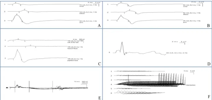
Fig. 1. Electroneuromyography: A) Ulnar motor nerve conduction, B) Median motor nerve conduction, C) Peroneal motor nerve conduction, D) Median sensory nerve conduction, E) Tibial F reflex, F) Ulnar nerve repetitive stimulation
g/dL (13.5-17.5), thrombocytes: 224 K/uL (150-450), B12: 312 pg/mL (211-911), folic acid: 10.8 ng/mL (9-24), glucose: 96 mg/dL (70-110), creatinine: 1.13 mg/dL (0,7-1,2), urea: 31.6 mg/dL (18-55) and HbA1c: 6 (4-6). Thyroid stimulating hormone, free T3 and T4, thyroglobulin, thyroid antibodies, liver function tests, creatinine phophokinase, electrolytes and arterial blood gas test resulted within normal limits. Procalcitonin, C-reactive protein (CRP) and erythrocyte sedimentation rate was normal. Electrocardiography (ECG) and echocardiography were normal.
The laboratory findings were as follows; Sensory nerve conduction was normal. The compound muscle action potential (CMAP) amplitudes of all motor nerves were low. After 10 seconds of exercise, more than 100% amplitude increment was observed. Repetitive nerve stimulation recorded with ulnar nerve stimulation showed more than 10% decrement at low frequencies. More than 200% increment was observed with 50 Hz tetanic stimulation (Figure 1). In conclusion, electrophysiological findings like LEMS that is a presynaptic pathology were detected. Serum acetylcholine receptor (AchR) antibody was negative.
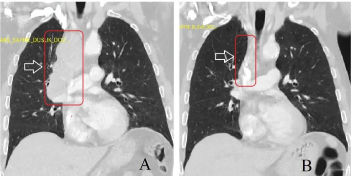
Since LEMS could be a paraneoplastic disease, thorax CT was performed for primary disease. In right paratracheal area, 3.5x5.5 centimeter (cm) diameter lesion like lymphadenopathy was observed. There were also lymphadenopathies in the right hilar region. A 5.5 cm diameter tumor that is on the upper, middle and lower lobes of right lung, surrounding the
pulmonary artery was detected. Upper part of tumor was compressing to vena cava superior (Figure 2A). 1 cm diameter nodular lesion was detected in both adrenal glands with abdominal CT scan. No metastase was detected in scintigraphy. There were no symptoms such as cough, chest pain and weight loss. Also, there were not chest radiographs in other hospitals for at least months.
The specimen that was taken by bronchoscopic biopsy was centrifuged and stained with
Papanikolaou, May Grünwald-Giemsa and
Hematoxylin Eosin. Bronchial epithelial cells, squamous epithelial cells, alveolar macrophages and lymphocytes were observed. There were irregular groups with restricted cytoplasm, pleomorphic, hyperchromatic nucleus, and no nucleoli selectable. Adenocarcinoma was revealed with cytological analysis.
After the chemotherapy treatment, initial complaints of patient were minimally improved. His neurological examination did not differ. In thorax CT scan, the lymph nodes in right hilar region were regressed. The largest was 12x25 mm in the subcarinal area. Tumor was regressed and diameter was 22x25 mm. Also, compressing to vena cava superior was regressed (Figure 2B).
Discussion
LEMS was initially considered to be a paraneoplastic disease associated with lung cancer (11). However, approximately half of the patients did not have lung cancer. These patients were called idiopathic LEMS.
Eren et al / Adenocarcinoma and neuromuscular disease
East J Med Volume:24, Number:3, July-September/2019 388
Fig. 2. Thoracic computed tomography: A) Before treatment B) After treatment Paraneoplastic LEMS was detected especially in
elderly patients with smoking (12). But in our case, lung malignancy was detected despite not smoking. It is important to distinguish LEMS from myasthenia graves (MG). Both diseases may present with similar symptoms. MG especially presents with ocular and bulbar symptoms and LEMS with muscular pain and weakness in lower extremities. In addition, LEMS has autonomic symptoms such as dry mouth, erectile dysfunction, postural hypotension and bowel motility disorder (2). Because, LEMS is a disorder of reduced acetylcholine release from the presynaptic nerve terminals due to antibodies to voltage-gated calcium channels (11). Electrophysiological analysis may provide important information for differential diagnosis. In LEMS; CMAP amplitudes of motor nerves may be low. Conduction of sensory nerves are normal. In repetitive stimulation, decrement is observed at low frequencies. After 3-10 seconds of exercise or 50 Hz stimulation, facilitation is monitored. Approximately 10% decrement is monitored with MG patients, When these clinical symptoms and electrophysiological analysis were evaluated, LEMS was considered in our patient. Diabetes mellitus (DM), pernicious anemia, hypothyroidism and celiac disease are other diseases that may be associated with LEMS. Therefore, biochemical and hormonal analysis is important in these patients (14). The type and stage of cancer is important for treatment. Other associated diseases may also affect prognosis. Each patient's treatment should be personalized. Treatment of cancer also enables successful treatment of this disease. Some patients can be treated with immunotherapies, pridositigmine and 3,4-diaminopyridine (15).
Immunotherapy, pridostigmine and
3,4-diaminopyridine were not given to our patient because her symptoms were not severe. His symptoms decreased with the treatment of adenocarcinoma.
LEMS is especially associated with small cell lung cancer (2). It is rarely associated with lung adenocarcinoma. In literature, there are seven case reports of pulmonary adenocarcinoma-associated LEMS disease. The mean age of cases is 61.4 ± 15.27. Tumor is on the left in only one case. It is located in upper lobe for 3 cases, in middle lobe for 1 case and in lower lobe for 3 cases. Four cases were treated with chemotherapy, 1 case with immunotherapy and 1 case with palliative care. Four of these cases died within two years (4-9). In our case, right lung adenocarcinoma was detected like literature. In the third month of treatment, his symptoms partially decreased with chemotherapy treatment. Lung adenocarcinoma regressed.
In conclusion, LEMS is a rare paraneoplastic syndrome. LEMS-associated malignancy can be detected before, at the same time or after the disease. Therefore, patients with LEMS should be screened for cancer at regular intervals, even if no tumor is detected initially. Typical symptoms and electrophysiological analysis are important for diagnosis of LEMS. Also, it should be noted that
LEMS may be associated with lung
adenocarcinoma.
Conflict of Interest: No conflict of interest was
declared by the authors
Financial Disclosure: The authors declared that
Eren et al / Adenocarcinoma and neuromuscular disease
East J Med Volume:24, Number:3, July-September/2019 389
Informed Consent: The informed consent on
sharing patient’s details and images with scientific purposes was obtained from patient.
References
1. Kleopa KA, Teener JW, Scherer SS, et al. Chronic multiple paraneoplastic syndromes. Muscle Nerve 2000; 23: 1767-1772.
2. Titulaer MJ, Wirtz PW, Kuks JB, et al. The Lambert- Eaton myasthenic syndrome 1988– 2008: a clinical picture in 97 patients. J Neuroimmunol 2008; 201: 153-158.
3. Nakao YK, Motomura M, Fukudome T, et al.
Seronegative Lambert- Eaton myasthenic
syndrome: study of 110 Japanese patients. Neurol 2002; 59: 1773–1775.
4. Ramos-Yeo YL, Reyes CV. Myasthenic syndrome (Eaton- Lambert syndrome) associated with pulmonary adenocarcinoma. J Surg Oncol 1987; 34: 239-242.
5. Sumitomo M, Uyama T, Kimura S, Hashioka K, Nobuhara K, Monden Y. A case of Lambert-Eaton myasthenic syndrome associated with adenocarcinoma of the lung. Haigan 1989; 29: 167-172.
6. Okudera K, Ebina A, Imai S, et al. A case with adenocarcinoma of the lung accompanied Lambert- Eaton myasthenic syndrome. Nikkyo 1996; 55: 902-907.
7. Milanez FM, Pereira CA, Trindade PH,
Milinavicius R, Coletta EN. Lung
adenocarcinoma, dermatomyositis, and Lambert- Eaton myasthenic syndrome: a rare combination. J Bras Pneumol 2008; 34: 333-336.
8. Arai H, Inui K, Hashimoto K, et al. Lung adenocarcinoma with Lambert-Eaton myasthenic syndrome indicated by voltage-gated calcium channel: a case report. J Med Case Rep 2012; 6: 281.
9. Bukhari S, Soomro R, Fawwad S, Alvarez C, Wallach S. Adenocarcinoma of lung presenting as Lambert-Eaton myasthenic syndrome. J Investig Med High Impact Case Rep 2017; 5: 1-4.
10. Tang Y, Wang K, Chen Z, et al. Ophthalmoplegia associated with lung adenocarcinoma in a patient with the Lambert–Eaton myasthenic syndrome: A case report. Medicine 2017; 96: e6484.
11. Sanders DB. Lambert-Eaton myasthenic
syndrome: clinical diagnosis, immune-mediated mechanisms, and update on therapies. Ann Neurol 1995; 37: 63-73.
12. Titulaer MJ, Maddison P, Sont JK, et al. Clinical Dutch- English Lambert- Eaton myasthenic syndrome (LEMS) tumor association prediction score accurately predicts small-cell lung cancer in the LEMS. J Clin Oncol 2011; 29: 902-908. 13. Oh SJ, Kim DE, Kuruoglu R, Brooks J, Claussen
G. Electrophysiological and clinical correlations in the Lambert–Eaton myasthenic syndrome. Muscle Nerve 1996; 19: 903-906.
14. O’Neill JH, Murray NM, Newsom-Davis J. The Lambert- Eaton myasthenic syndrome. A review of 50 cases. Brain 1988; 111: 577-596.
15. Chalk CH, Murray NM, Newsom-Davis J, O’Neill JH, Spiro SG. Response of the Lambert-Eaton myasthenic syndrome to treatment of associated small-cell lung carcinoma. Neurol 1990; 40: 1552-1556.