CASE
REPORT
–
OPEN
ACCESS
InternationalJournalofSurgeryCaseReports5(2014)858–860ContentslistsavailableatScienceDirect
International
Journal
of
Surgery
Case
Reports
jo u r n al ho me p a g e :w w w . c a s e r e p o r t s . c o m
Nerve
sheath
myxoma
of
the
dorsal
paravertebral
space
Melih
Malkoc
a,∗,
Tugrul
Ormeci
b,
Mert
Keskinbora
a,
Adem
Yılmaz
c,
Ozgur
Korkmaz
a,
Canan
Besleyici
Tanik
daDepartmentofOrthopedicsandTraumatology,IstanbulMedipolUniversity,SchoolofMedicine,Turkey bDepartmentofRadiology,IstanbulMedipolUniversity,SchoolofMedicine,Turkey
cDepartmentofNeurosurgery,SisliResearchandEducationHospitalIstanbul,Turkey dDepartmentofPathology,SisliResearchandEducationHospitalIstanbul,Turkey
a
r
t
i
c
l
e
i
n
f
o
Articlehistory: Received24April2014
Receivedinrevisedform31July2014 Accepted6October2014
Availableonline16October2014
Keywords: Nervesheathtumor Myxoma
Neurothekeoma Paravertebralspace
a
b
s
t
r
a
c
t
INTRODUCTION:Nervesheathmyxomas(NSM)arerarebenignsofttissuetumors.Thedorsal
paraverte-bralplacedNMSdiagnosiscanbedifficult.
PRESENTATIONOFCASE:Thisarticlepresentsclinical,radiologicalfindingsandtreatmentoftheNSMof
thedorsalparavertebralspaceina32-year-oldmanpresentedwitharightshoulderandbackpainfor4
years.
DISCUSSION:NSMisarareandbenigntumorandthatmostoftenoccursintheskinofthehead,neck
orupperlimbsofyoungerpatients.Rarelocationssuchasintracranial,spinalcanal,trunk,lowerlimb
andoralcavitywerealsoreported.TheappropriatetreatmentofNSMissurgicalexcision.Diagnosisis
difficultinanuncommonpresentation.
CONCLUSION:AlthoughthemostpresentedcaseofNMSare dermaltumors,itmay alsobe found
extremelyrarelocations.Weconcludethat,thedefinitivetreatmentofNSMissurgicalexcisionwith
safemarginsevenwhenitispossible.
©2014TheAuthors.PublishedbyElsevierLtd.onbehalfofSurgicalAssociatesLtd.Thisisanopen
accessarticleundertheCCBY-NC-SAlicense(http://creativecommons.org/licenses/by-nc-sa/3.0/).
1. Introduction
Nervesheathmyxomas(NSM)arerarebenignsofttissuetumors thathave apredilictiontohead–neckregionand upperlimbs.1
Although,themajorityofNSMarecommonlydermal,other loca-tionslike mucosal,intracranial and intraspinal have also been described.Wereporta caseofdorsalNSM withmisdiagnoseof nearly4years.Tothebestofourknowledge,itwasthefirstcase ofNSMinvolvingthedorsalparavertebralspace,reportedinthe Englishliteraturetodate.
2. Presentationofcase
A32-year-oldotherwise healthymanpresentedwitharight shoulderandbackpainfor4years.Recently,painstartstocontinue atrestandpatientdescribingdifficultyinfallingasleepandhad difficultytoperformhisdailyactivitiesandworkingschedule.
PositivityofJobetest,dropping sign,Hawkinstest,Yergason testandSpeedtestobservedinthephysicalexaminationofright shoulder.Scapularinstabilitywasdetermined.Thepatientdefines
∗ Correspondingauthorat:DepartmentofOrthopedicandTraumatology,Istanbul MedipolUniversity,SchoolofMedicine,TemAvrupaOtoyoluGoztepeCıkısi,No.1 Bagcilar,34214Istanbul,Turkey.Tel.:+902124607777;fax:+902124607070.
E-mailaddress:[email protected](M.Malkoc).
slighttendernesswithdeeppalpationofspinalprocessbetweenT2 andT5,butnomassidentified.Therewasnomotorandsensorial deficiencydeterminedontheshoulderandarmmusculature.Many differentdiagnosticandtreatmentmodalitieswasperformed ini-tially,likeacupuncture,electromyographyorshoulderandcervical magneticresonanceimaging(MRI).Noneofthesemodalitiescould achievetoshowanabnormalityorreducethepain.AthoracalMRI wasperformedinourhospital.
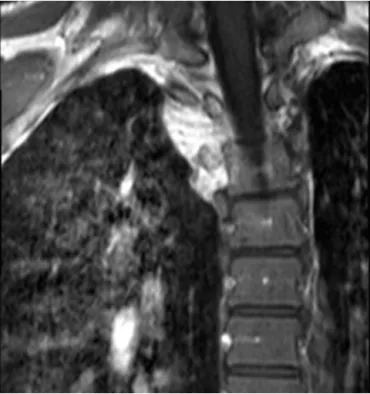
Magneticresonanceimagingshowedanill-defined,solidtumor locatedintherightparavertebralspacebetweenT2andT4 verte-brae. The lesion extends into thorax with an extrapulmonary fashionatthelevelofT2–3whereitdevelopsdestructiononthe adjacentribandtransverseprocess.Thelesionhadslight heterge-neoushighintensitywhencomparedwithmuscletissueonT1-and brightsignalintensityechoonT2-weightedimages.Enhancement identifiedinthelesionafterintravenouscontrastadministration. Thelesionmeasured15mm×17mmintheaxialplane(Fig.1)and 20mminthecoronalplane(Fig.2).
Inthedifferentialdiagnosisofnervesheathmyxoma, aggres-siveangiomyxoma,myxoidneurofibroma,lowgradefibromyxoid sarcoma,myxoidliposarcoma,lowgrademyxofibrosarcoma, cel-lularmyxoma,juxta-articularmyxoma,nodularfasciitismustbe inmind.Forthisreasonpatientundergoneaclosedbiopsywith administration of local anesthesia and sedation. From a lancet tipsmallincision,aJamshidineedleinsertedbytheguidanceof ComputerizedTomography.Threetissuesamplesweretakenfrom
http://dx.doi.org/10.1016/j.ijscr.2014.10.003
2210-2612/© 2014The Authors.Publishedby Elsevier Ltd.onbehalf of SurgicalAssociates Ltd.This is an openaccessarticle under theCC BY-NC-SA license (http://creativecommons.org/licenses/by-nc-sa/3.0/).
CASE
REPORT
–
OPEN
ACCESS
M.Malkocetal./InternationalJournalofSurgeryCaseReports5(2014)858–860 859
Fig.1. InaxialTSET2MRimage;thereisslightlyheterogeneoushyperintenselesion
isseenatthelevelofT2–T3.
differentpartsofthelesion.Pathologicalinterpretationrevealeda myxomatouslesionwithoutanysignsofmalignancy(Figs.3and4). Completeexcisionofthelesionwasperformedbya longitu-dinalmidlineincisionover thelesionside. Bybluntdissections of therightparavertebral muscles, pseudocapsuleof thelesion wasreached. Although,thecloserelationof themasswiththe nearbyvertebralbodiesandtheribs,noinvasiontothebonytissue wasobserved.Theadjacentpleuralmembranewasintact. Intra-operativefrozensectionwasconfirmedthebenignnatureofthe lesion.Twopiecesof grayish-pinktumortissuewasexcisedby carefulbluntdissections.Accordingtopathologyreporttherewas nomitotic activityand nolipoblasts. The excisedmaterialwas hypocellular,composedofblandcellsandscantbloodvessels.The tumorwasimmunohistochemicallycharacterizedasaNSMgiven itspositiveimmunoreactivityforS-100protein,CD10and epithe-lialmembraneantigen,withnoimmunoreactivitytocytokeratin.
Fig.2.PostcontrastcoronalSPIRTSET1imageshowscontrastenhancedlesion locatedbetweenT2andT4vertebraewithextrapulmonaryfashion.
Fig.3. microscopyofnervesheathmyxoma,redarrowindicatethetumoraltissue, whitearrowindicatestheperipheralnerve.
The patient’s symptoms disappeared immediately after the operation. The post-operative coursewas uneventful. One year afterhisoperation,clinicalfollow-upandMRIshowednoevidence ofarecurrentlesionat3rd,6th,9thandinthelastfollowup1year aftersurgery.Patientreturnedtohisdailyactivitieswithoutpain andrestriction.
3. Discussion
NSMisararetumorthatmostoftenoccursintheskinofthe head,neckorupperlimbsofyoungerpatients.Likeourdorsally paravertebrallocatedcase,somerarelocationssuchasintracranial, spinalcanal,trunk,lowerlimbandoralcavitywerealsoreported. Thereportedagesofthepatientsrangebetween15monthsand 84years.2Thepreviouslyreportedcaseshadapeakincidencein
thefourthdecadeoflife,butareextremelyrareininfancy.3Ithas
aremarkablepredilectionforthefemalegender.4
Thesebenign lesionswerefirstdescribed in1969byHarkin andReed.Although,thetumorwaslaterre-namedbyGallagerand Helwigasneurothekeoma,recentgenestudiesstronglysupports that, nerve sheath myxomas and neurothekeomas are distinct neoplasms. Schwann-cell differentiation in the ultrastructural observationofNSM cells, suggestsanoriginfromnerve sheath
Fig.4.microscopyofnervesheathmyxoma,redarrowindicatesthetumoraltissue, whitearrowindicatesperipheralnerve.
CASE
REPORT
–
OPEN
ACCESS
860 M.Malkocetal./InternationalJournalofSurgeryCaseReports5(2014)858–860
precursorcells.However,therearestillsometheoriesproposinga proliferationofotherperineuralcells,whichbringscontroversies tothecelloforigin.5TherearesomesimilarfeaturesofNSMwith
otherneuraltissuetumors,likeschwannomaandneurofibroma. That seems the reason why various names such as pacinian neurofibroma, cutaneouis lobular neurofibroma and perineural neurofibromahave beenusedforthislesion.Theseoverlapping featuresareleadingtodifficultiesindiagnosis.
Threevariantsof NSM weredescribed basedon histopatho-logicalevaluation.These subtypesincludethemyxoid type,the cellulartypeandthemixedtype.Theclassicalmyxoidtypeis char-acterizedbylowcellularityandlargeamountsofmyxoidmatrix. ThelesionusuallyborderedanddiffuselypositiveforS-100.Asin ourcase,thestrongpositivityoftumorcellsforS-100,whichis awell-establishedmarkerforSchwanncellsandmyelinsheath, favorsthe Schwann cell origin.The cellulartypes of NSM typ-ically are notwell circumscribed, and themixed type includes variedcellularitywithinfocalmyxoidregions.6,7Thedifferential
diagnosesinclude schwannoma (alsothemaligntype), neurofi-broma,neurilemmoma,leiomyoma,intramuscularmyxoma and low-gradesarcoma.
TheappropriatetreatmentofNSMissurgicalexcision.Inour case,thecloserelationofthetumoralmasswithpleuraand adja-centvertebra-ribjunctionmakesthetotalexcisiondifficulttoget withsafe margins. Afteran intra-operative frozen section that revealedthebenignnatureofthelesion,amarginalexcisionwas performed.Anintra-operativeconsultationfromthoracicsurgery wasalsotaken,inordernottopenetratethepleuralmembrane. Althoughamarginal excisionwasperformedinourpatient,no recurrenceobservedinthepatientsfirstyearfollow-up.Usually, NSMsupposed to havea low recurrence rateafterlocal recur-rence,butsomeauthorsreporteda recurrencerateinabout7% ofpatients.8,9
4. Conclusion
Althoughthemajorityofreportedcasesaredermaltumors,NSM mayalsobelocatedinthe extremelyrare locations.Weaimed toincrease theawareness aboutthis disease,in orderto avoid delays in diagnosis.We concludethat, thedefinitivetreatment of NSM is surgical excision withsafe marginseven when it is possible. Conflictofinterest None. Funding None. Ethicalapproval
Writteninformedconsentwasobtainedfromthepatientfor publicationofthiscasereportandaccompanyingimages.
Authorcontributions
MelihMalkoccontributedtostudydesign.TugrulOrmeci con-tributedtodatacollecting.MertKeskinboracontributedtowriting. AdemYılmazcontributedtodatacollectingandanalysis.Ozgur Korkmazcontributedtofigurepreparation.CananBesleyiciTanik contributedtopathologicspecimenanalysis.
References
1.VijM,JaiswalS,AgrawalV,JaiswalA,BehariS.Nervesheathmyxoma (neu-rothekeoma)ofcerebellopontineangle:casereportofararetumorwithbrief reviewofliterature.TurkNeurosurg2013;23(1):113–6.
2.PapadopoulosEJ,CohenPR,HebertAA.Neurothekeoma: reportofacasein aninfantandreviewoftheliterature.JAmAcad Dermatol2004;50(January (1)):129–34.
3.FetschJF,LaskinWB,MiettinenM.Nervesheathmyxoma:aclinicopathologicand immunohistochemicalanalysisof57morphologicallydistinctive,S-100 protein-andGFAP-positive,myxoidperipheralnervesheathtumorswitha predilec-tionfor theextremities andahigh localrecurrencerate.AmJSurgPathol 2005;29(December(12)):1615–24.
4.O’RourkeH,MeyersSP,KatzmanPJ.Neurothekeomaintheupperextremity: magneticresonanceimagingandcomputedtomographyfindings.JComputAssist Tomogr2005;29(November–December(6)):847–50.
5.GallagerRL,HelwigEB.Neurothekeoma–abenigncutaneoustumorofneural origin.AmJClinPathol1980;74(December(6)):759–64.
6.LeeD,SuhYL,HanJ,KimES.Spinalnervesheathmyxoma(neurothekeoma).Pathol Int2006;56(March(3)):144–9.
7.AkhtarK,ZaheerS,RayPS,SherwaniRK.Myxoidneurothekeoma:araresofttissue tumorofhandinamaletoddler.NigerJSurg2013Jan;19(1):32–4.
8.HornickJL,FletcherCD.Cellularneurothekeoma:detailedcharacterizationina seriesof133cases.AmJSurgPathol2007;31:329–40.
9.WollinaU,HanselG,SchönlebeJ,HaroskeG.Myxoidneurothekeoma–apainful caseinalesscommonlocation.DermatolOnlineJ2009;15(April(4)):3.
OpenAccess
ThisarticleispublishedOpenAccessatsciencedirect.com.ItisdistributedundertheIJSCRSupplementaltermsandconditions,which permitsunrestrictednoncommercialuse,distribution,andreproductioninanymedium,providedtheoriginalauthorsandsourceare credited.
View publication stats View publication stats