REPRODUCTIVE PHYSIOLOGY AND DISEASE
Expression of CCM2 and CCM3 during mouse gonadogenesis
Aylin Yaba1&N. Ece Gungor Ordueri2&Gamze Tanriover2&Pinar Sahin2&Necdet Demir2&Ciler Celik-Ozenci2
Received: 22 May 2015 / Accepted: 20 August 2015 / Published online: 19 September 2015 # Springer Science+Business Media New York 2015
Abstract
Purpose Three cerebral cavernous malformation (CCM) pro-teins, CCM1, CCM2, and CCM3, regulate cell-cell adhesion, cell shape and polarity, and most likely cell adhesion to extra-cellular matrix. Recently, CCM2 and CCM3 are known to be expressed in control and varicocele-induced rat testes, but little is known about these proteins during gonadogenesis. This led us to study the CCM proteins during the mouse gonadogenesis. Methods Neonatal (PND 0), postnatal, and adult mice testes and ovaries were obtained from mice. CCM2 and CCM3 expression were analyzed during mouse testicular and ovarian development by immunohistochemistry and quantitative real-time PCR. Results The results showed that in both sexes, Ccm2 and Ccm3 mRNA and protein were first detectable after gonadogenesis when the gonads were well differentiated and remained present until the adult stage. In the testis, CCM2 and CCM3 expression were restricted to the nuclei of Sertoli cells, suggesting a conserved role in testicular differentiation. In the ovary, the CCM2 and CCM3 proteins were localized in the cytoplasm of oocytes, suggesting an unexpected role during oogenesis. Quantitative real-time PCR (qRT-PCR) results showed that expression of Ccm2 and Ccm3 genes could play
a role in the regulation of mouse gonadogenesis translational activation upon testicular and ovarian development.
Conclusions The localization of CCM2 and CCM3 proteins show their different functions for CCM2 and CCM3 which may have important roles in testicular and ovarian differenti-ation. In conclusion, CCM2 and CCM3 may be involved in establishing the differential expression pattern in developing mouse testis and ovary.
Keywords CCM2 . CCM3 . Mouse . Gonadogenesis
Introduction
Adult fertility requires appropriate and coordinated instruction of somatic and germ cell activity during lineage specification, development, and maturation. Germ cell development con-sists of three fundamental periods: i) the primordial germ cells (PGCs), the first cells of the germline lineage in the embryo; ii) spermatogonial stem cells (SSCs)/oogonial stem cells (OSCs); and iii) spermatozoon/oocyte, respectively. The first two periods cover the fetal and neonatal periods leading to the formation of the SSCs/OSCs and include two main types of germ cells, PGCs and gonocytes.
In the male, PGC differentiates into gonocytes which form type A spermatogonia. [1]. Gonad differentiation in the mouse involves the coordinated behaviors of multiple cell types over the fetal and neonatal developmental periods. In male mouse, testis development starts around E 10.5 days due to expression within somatic pre-Sertoli cells [2–4]. Testicular cords are formed by these somatic cells, and factors they secrete also direct the gonocytes into the male differentiation pathway [5]. Gonocytes proliferate until approximately E 13.5 and then enter mitotic arrest until birth [6,7]. Following birth, the gon-ocytes migrate from the seminiferous cord center to the Capsule Expressions of CCM2 and CCM3 during mouse gonadal
differentiation may be involved in establishing the regulation of gonad development.
* Ciler Celik-Ozenci [email protected]
1
Department of Histology and Embryology,İstanbul Medipol University School of Medicine,İstanbul, Turkey
2 Department of Histology and Embryology, Akdeniz University
periphery and resume proliferation, forming the spermatogo-nial cell population [8, 9]. Thus, postnatal development of testicular cell populations in mice at day 5 contains only sper-matogonia and somatic cells. The first wave of spermatogen-esis begins around postnatal day 5 (PND 5). At PND 9, preleptotene spermatocytes appear and zygotene and tene spermatocytes by PND 15–PND 18. By PND 20, pachy-tene spermatocytes are present, and by PND 35 (adult), the full complement of germ cells begins to be present.
Similarly, adult female reproductive capacity is thought to be determined during ovarian development. Follicle formation in mice begins as perinatal when germ cell cysts are invaded by pregranulosa cells to establish individual primordial folli-cles [10]. Once formed, the pool of primordial follicles serves as a source of developing follicles and oocytes [11–14]. Fol-licular organization in the ovary of the mouse begins after birth, and during the infantile period small primary follicles with two layers of typical granulosa cells are developed [15]. This transition from primordial to growing follicles is poorly defined at the molecular level. One hallmark of primordial follicle activation is growth of the oocyte, which, although still arrested in the first meiotic prophase, is transcriptionally active [16]. Follicular activation is characterized by rapid oo-cyte growth and proliferation of a few flattened pregranulosa cells into cuboidal granulosa cells [17] and is believed to be a continuous process that starts just after follicle formation, long before the onset of puberty [11]. This process is followed by further follicular development, ovulation, and luteinization during each repetitive menstrual/estrous cycle [11,18,19].
Cerebral cavernous malformations (CCM) are vascular le-sions histologically characterized by hyperpermeable blood ves-sels in the brain without intervening brain parenchyma [20,21]. Ccm involves the homozygous inactivating mutations of one of three genes, Ccm1, Ccm2, or Ccm3. CCM1, CCM2, and CCM3 proteins form a complex to organize the signaling networks controlling endothelial cell physiology including actin dynamics, tube formation, and adherens junctions [22]. Ccm genes, which are very well conserved among different organisms [23], have been mapped and identified in the recent years which are impor-tant for understanding of the mechanisms of this disorder. In addition to the identification of CCM genes, Ccm1, Ccm2, and Ccm3, a number of recent biochemical in vitro studies and in vivo CCM animal model studies have helped to unravel the functional roles of these proteins [23,24]. It has been shown that KRIT1 (CCM1), OSM (CCM2), and PDCD10 (CCM3) pro-teins form a complex to organize the signaling networks [25]. Mice lacking either Ccm1 or Ccm2 die in mid-gestation with vascular defects at the same developmental stage, and with a similar phenotype [26–29]. The complete loss of Ccm1 or Ccm2 results in vascular defects with a failure to connect the developing heart to the developing aorta; as a result, circulation is not established as expected at E 8.5 [28], and developmental arrest and death ensue. Studies using mice with tissue-specific
deletions of Ccm2 have shown that there is an absolute require-ment for CCM2 in the endothelium during developrequire-ment while the neuronal expression of CCM2 was not required for develop-ment [28,29]. Recently, a plethora of new data has emerged on the signaling pathways in which they are involved. They regu-late diverse aspects of endothelial cell morphogenesis and blood vessel stability such as cell-cell junctions, cell shape and polarity, or cell adhesion to extracellular matrix [24].
We have previously shown the expression patterns of two brain proteins, CCM2 and CCM3, in control and varicocele-induced rat testes, suggesting a role during normal and path-ological spermatogenesis [30]. Our findings were the first demonstration that these proteins may have a role during gonadogenesis. Here, we show that mouse Ccm2 and Ccm3 expressions exist in male and female gonad during embryo-genesis. In the present study, we aimed to identify the Ccm2 and Ccm3 genes involved in male- and female-specific gonad-al development. To gain a better understanding of these mol-ecules in reproductive biology, we characterized the expres-sion patterns of the CCM2 and CCM3 proteins in mouse ova-ry and testis at different stages of fetal and postnatal develop-ment. The results of the immunohistochemistry were partly supported by a qRT-PCR analysis. This is the first study to examine the mRNA and protein expression patterns of CCM2 and CCM3 throughout gonadal development in the male mouse, from E 13.5 until PND 20, and in the female mouse, from PND 1 until adult. These diverse expressions of CCM2 and CCM3 during gonadal differentiation clue a role of this factor in regulation of mouse gonadal development.
Materials and methods
Tissue collection and processing
Mouse embryos from timed matings of BALB/C female mice were obtained from pregnant dams between E 13.5 and E 17.5 in half-day increments, with noon on the day of vaginal plug-ging designated as 0.5 dpc (day post coitus). Embryos were sexed by morphology. Neonatal (PND 0) and postnatal mice testes at 3, 5, 9, 15, 20, and adult were examined. All tissues were fixed in Bouin’s fixative overnight at 4 °C and embedded in paraffin. Ovaries, obtained from postnatal days 1, 3, 5, and 9 and adult mice, were fixed in 10 % formalin overnight and embedded in paraffin. Experiments were performed on three mice of each sex and age. Sections of 5μm were placed on superfrost plus slides (Menzel-Glaser, Braunschweig, Germa-ny). Testis and ovary tissue samples for mRNA preparation (PND 1, PND 3, PND 5, PND 9, and adult) were snap frozen immediately after collection and stored at−80 °C until use. All animal handling and experimental procedures were approved by the Animal Care and Usage Committee of Akdeniz University.
Immunohistochemistry
Immunohistochemistry with anti-CCM2 and anti-CCM3 an-tibodies was performed essentially as previously described [30]. Antigen retrieval was performed in 50 mM glycine (pH 3.5; 90 °C maintained for 8 min), and primary antibodies were applied at 1/250 dilution for overnight incubation at 4 °C in 0.1 % bovine serum albumin/TBS. Control sections were in-cubated with normal rabbit IgG serum (Vector Laboratories, Burlingame, CA) at the same concentration. Subsequent steps were performed at room temperature, with TBS washes be-tween incubations. Primary antibody binding was detected using a biotinylated goat anti-rabbit antibody (Vector Labora-tories). Secondary antibody was applied to the sections for 30 min and diluted at 1:400 in 0.1 % bovine serum albumin/ TBS. The slides were then incubated with Vectastain Elite ABC kit (Vector Laboratories, Burlingame, CA) according to the manufacturer’s instructions. Antibody binding was de-tected as a brown precipitate after development with diamino-benzidine chromogene and Mayer’s hematoxylin counter-stain. The sections were mounted under glass cover slips in Permount (Fisher Chemicals, Zurich, Switzerland). For the fetal data, immunohistochemistry was performed with each antibody on gonads from three distinct embryos from different mothers. Two sections from each of these gonads were ana-lyzed for CCM2 and CCM3 protein localization. In the post-natal testis and ovary, at least two sections were analyzed for CCM2 and CCM3 protein localization.
Total RNA isolation and reverse transcription
Total RNA was obtained from neonatal and adult ovary and testis tissues of BALB/C mice by using TRIzol (GIBCO/ BRL) according to the manufacturer’s instructions. RNA sam-ples were quantified spectrophotometrically. Reverse-transcription (RT) reactions with oligo d(T) primers and the Omniscript kit (Qiagen) were performed 2μg of total RNA from each tissue according to the manufacturer’s instructions. Quantitative real-time PCR
Gene expression studies by quantitative real-time PCR (qRT-PCR) were carried out on an iCycler (Bio-Rad, CA, USA). Tissue cDNA was assayed in duplicate, and each experiment was repeated three times. qRT-PCR for Ccm2 and Ccm3 was performed in a 25-μl final reaction that contained 12.5 μl
SYBR Green supermix (Thermo Scientific), 0.4 mM of each primer, and 1μl of template. q RT-PCR was carried out using the following parameters: one cycle of 94 °C for 5 min (de-naturation), followed by 35 cycles (amplification) at 94 °C for 30 s, 59 °C for 1 min, and 72 °C for 45 s. Melt curve analyses were run with each series to confirm the specificity of the amplified products. In order to confirm the amplification spe-cific for mRNA, no reverse transcriptase controls were also performed for each sample. The standard curves were pre-pared for Ccm2, Ccm3, and Beta-actin. All expression data were normalized by dividing the amount of target gene by the amount of beta-actin used as control. All primers are shown in Table1.
Identification of follicles
Follicles were classified separately for the neonatal 1-, 3-, and 5-day-old ovaries and for the older (9- and 28-day) ovaries. In the newborn mice, follicle classification was performed as described [31]: oocytes that were not surrounded by somatic cells were classified as naked oocytes; oocytes with a single full layer of flattened granulosa cells were termed primordial. In the older animals, follicle classification was done according to previously described terms [32]. A primary follicle has one layer of cuboidal granulosa cell [33]; a secondary follicle is comprised of an oocyte surrounded by two layers of granulosa cell; a preantral follicle was distinguished by the presence of more than two layers of granulosa cells, with the follicle hav-ing no antrum; while the antral follicles were the larger ones that had a fluid-filled cavity known as the antrum.
Statistical analysis
qRT-PCR results from each group were compared by one-way ANOVA. Statistical calculations were performed using Sigma Stat for Windows, version 3.0 (Jandel Scientific Corp., San Rafael, CA). P<0.05 was considered statistically significant.
Results
CCM2 and CCM3 protein localization during testis morphogenesis
To determine the cellular localization of CCM2 and CCM3 proteins in developing male gonad, immunohistochemistry Table 1 Primers
Gene Forward primer Reverse primer
Ccm2 TCT GCT CAG TCT GTC TGC CTA CAA ATA TTG CTC GGT CCA GAA Ccm3 CCC AGA TGA GAT CAA TGA CAGA ATG AAT TAG TCG GT TGG CAC TT Β-actin GAT GAC GAT ATC GCT GCG CTG GTA CGA CCA GAG GCA TAC AGG
was performed on gonad sections from several fetal, neonatal, and postnatal ages. Data from these analyses were summa-rized in Table2.
Localization of CCM2 protein in prenatal (E 13.5, E 14.5, E 15.5, E 17.5), neonatal (PND 0), postnatal (PND 3, PND 5, PND 9, PND 15, PND 20), and adult mouse testis In the fetal testis E 13.5 and E 14.5, CCM2 protein was pre-dominantly localized to the interstitial cells (yellow arrows) not in gonocytes (black arrows) and Sertoli cells (red arrows) (Fig.1a, b). Cells within the seminiferous cords of the fetal testis generally showed increased CCM2 expression at E 15.5 (Fig.1c). We also observed moderate nuclear CCM expres-sion in some gonocytes, and interstitial cells around the cords also showed CCM2 expression (Fig.1c). By E 17.5, while gonocytes and interstitial cells present CCM2 expression, Sertoli cells did not show any expression (Fig.1d).
At birth, PND 0, at the end of the mitotic silence, the CCM2 expression was unable to be detected in the gonocyte; however, some of the interstitial cells expressed nuclear and cytoplasmic CCM2 protein (Fig.1e). At PND 3, some gono-cytes had migrated to the basal side of the tubule showed cytoplasmic localization of the CCM2 protein and additional-ly some interstitial cells showed nuclear and cytoplasmic ex-pressions (Fig.1f). At PND 5, we observed cytoplasmic CCM2 expression in interstitial cells and in some gonocytes, which were located at the center of the seminiferous cords but not in the spermatogonial cells at the basal side (Fig.1g). Cells within the seminiferous cords of the fetal testis generally showed CCM2 expression between E 13.5 and PND 5 (Fig. 1a–g). The CCM2 protein is evident in the nuclei of pachytene spermatocytes at PND 9 (Fig.1h). Round sperma-tids also showed nuclear CCM2 staining in the different
seminiferous tubules, and interstitial cells present nuclear/ cytoplasmic CCM2 staining in the different areas. At PND 15, while the CCM2 protein was evident in the nuclei of preleptotene and pachytene spermatocytes (Fig. 1i), round spermatids had cytoplasmic CCM2 expression at PND 20 (Fig.1j). In the adult mouse testis, the nuclear and cytoplasmic CCM2 expression was detected in elongated spermatocytes at stages II–III, IX, and X of the adult testis (Fig.1k).
Localization of CCM3 protein in prenatal (E 13.5, E 14.5, E 15.5, E 17.5), neonatal (PND 0), postnatal (PND 0, PND 3, PND 5, PND 9, PND 15, PND 20), and adult mouse testis In the cords, CCM3 appeared to be localized to some gono-cytes cytoplasm and to some of the gonogono-cytes, spermatogonia, and spermatocytes from birth through PND 15.
At E 13.5, gonocytes in the lumen of the seminiferous cord, Sertoli cells, and interstitial cells around the cords showed cytoplasmic CCM3 expression (Fig. 2a). At E 14.5, while we detected increased perinuclear cytoplasmic CCM3 expres-sion in gonocytes, Sertoli cells and interstitial cells showed cytoplasmic expression (Fig.2b). At E 15.5, some gonocytes showed cytoplasmic CCM3 expression while others exhibit nuclear localization (Fig.2c). At E 17.5, some gonocytes in mitotic silence and Sertoli cells continue to express cytoplas-mic CCM3 expression but thereafter its expression start to decrease in Sertoli cells (Fig.2d). Cells within the interstitial area showed cytoplasmic CCM3 expression between E 13.5 and E 17.5 (Fig.2a–d). At birth, PND 0, while some gono-cytes at the end of the mitotic silence present very weak CCM3 expression, some of the interstitial cells showed de-creased CCM3 expression (Fig.2e). In addition, we did not observe any Sertoli cell staining with CCM3 after birth. In the meantime, we detected nuclear CCM3 expression in the
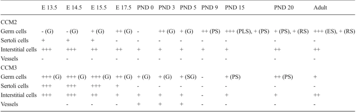
Table 2 Summary of CCM2 and CCM3 expression patterns in developing male gonad
E 13.5 E 14.5 E 15.5 E 17.5 PND 0 PND 3 PND 5 PND 9 PND 15 PND 20 Adult CCM2
Germ cells - (G) - (G) + (G) ++ (G) - ++ (G) + (G) ++ (PS) +++ (PLS), + (PS) + (PS), + (RS) +++ (ES), + (RS)
Sertoli cells + + + - - - -Interstitial cells +++ +++ ++ ++ + + + + + ++ ++ Vessels - - - -CCM3 Germ cells +++ (G) +++ (G) +++ (G) ++ (G) + (G) + (G) + (SG) - + (PS) ++ (PS) + Sertoli cells +++ +++ +++ + - - - -Interstitial cells +++ +++ ++ + + + + - + + ++ Vessels - - - + + + - - -
-+ low expression, -+-+ moderate expression, -+-+-+ high expression, - not detected, G gonocyte, SG spermatogonium, PLS preleptotene spermatocyte, PS pachytene spermatocyte, RS round spermatid, ES elongated spermatid
endothelial cells of the vessels (Figures2e). At PND 3, while interstitial cells showed cytoplasmic CCM3 immunoreactivi-ty, endothelial cells present nuclear CCM3 expression
(Fig.2f). At PND 5, we detected CCM3 expression in the spermatogonial cells’ cytoplasm and CCM3 expres-sion in the endothelial cells continues (Fig. 2g). At Fig. 1 Immunohistochemical analysis on fetal, neonatal, postnatal, and
adult mouse testis. Transverse sections showing the presence of CCM2 protein expression during embryonic period (E), neonatal (PND 0), postnatal period (PND 3, 5, 9, 15, 20), and adult mouse testis. a, b E 13.5 mouse testis, CCM2 expression was not detected in the cytoplasm of gonocytes (black arrow) but in Sertoli cells (red arrow) and interstitial cells (yellow arrow). c E 15.5 mouse testis, gonocytes did not show any cytoplasmic or nuclear CCM2 expression (black arrow), Sertoli cells (red arrow) and interstitial cells (yellow arrow) showed cytoplasmic expression. d E 17.5 mouse testis, cytoplasmic CCM2 expression was detected in gonocytes (black arrow) and interstitial cells (yellow arrow), not in Sertoli cells (red arrow). e At PND 0, there is no CCM2 expression in gonocytes (black arrow) but interstitial cells showed cytoplasmic staining (yellow arrow). f At PND 3, CCM2 expression was seen in the cytoplasm of gonocytes (black arrow) and interstitial cells (yellow
arrow). g At PND 5, CCM2 staining was detected in the cytoplasm of gonocytes (black arrow) and interstitial cells (yellow arrow). h At PND 9, pachytene spermatocytes (black arrow) showed nuclear CCM2 expression, interstitial cells presented cytoplasmic and nuclear CCM2 expression. i At PND 15, CCM2 expression was detected in the nuclei of preleptoten (black arrow) and cytoplasm of pachytene spermatocytes (blue arrow) and cytoplasm and nuclei of interstitial cells (yellow arrow). j At PND 20, cytoplasmic CCM2 expression was seen in pachytene (blue arrow) and round spermatid (black arrow) and interstitial cells (yellow arrow). k In adult mouse testis, CCM2 expression was detected in elongated spermatids in stage II–III, X–XII tubules (black arrow), round spermatids in stage IX tubules (black arrowhead). Interstitial cells showed nuclear and cytoplasmic CCM2 staining (yellow arrow). Scale bar 80 μm. Data shown in panel are representative findings of three mice. There is no staining in negative control slides (insert, j)
PND 9, CCM3 immunoreactivity was not detected in any cells (Fig. 2h). At PND 15, pachytene spermato-cytes present cytoplasmic expression but most of the interstitial cells did not (Fig. 2i). At PND 20, we de-tected increased CCM3 expression in pachytene
spermatocytes. In addition, we once again observed CCM3 protein expression in interstitial cells at PND 20 (Fig. 2j). In the adult mouse testis, CCM3 expres-sion was shown to be very weak cytoplasmic in pachy-tene spermatocytes in VII–VIII stage: distinctive, round Fig. 2 Immunohistochemical analysis on fetal, neonatal, postnatal, and
adult mouse testis. Transverse sections showing the presence of CCM3 protein expression during embryonic period (E), neonatal (PND 0), postnatal period (PND 3, 5, 9, 15, 20) and adult mouse testis. a, b E 13.5 mouse testis, CCM3 expression was detected in the cytoplasm of gonocytes (black arrow), Sertoli cells (red arrow), and interstitial cells (yellow arrow). c E 15.5 mouse testis, gonocytes showed nuclear and cytoplasmic CCM3 expression (black arrow), Sertoli cells (red arrow) and interstitial cells (yellow arrow) showed cytoplasmic expression. d E 17.5 mouse testis, cytoplasmic CCM3 expression was detected in gonocytes (black arrow) and interstitial cells (yellow arrow) in same intensity with E 15.5, but we detected decreased CCM3 expression intensity in Sertoli cells (red arrow). e At PND 0, CCM3 showed weak staining in gonocyte cytoplasm (black arrow), cytoplasmic staining in some of interstitial cells. Endothelial cells of vessels showed nuclear CCM3 expression (blue arrow). f At PND 3, we detected cytoplasmic
CCM3 staining in the cytoplasm of some of the interstitial cells (yellow arrow) and nuclei of endothelial cells (blue arrow). g At PND 5, CCM3 expression was presented spermatogonia (black arrow) and endothelial cells (blue arrow). h PND 9, there is no CCM3 expression. i At PND 15 days, pachytene spermatocytes (black arrow) and interstitial cells (yellow arrow) showed CCM3 expression. j At PND 20 days, we detected increased CCM3 expression in the cytoplasm of pachytene spermatocytes (black arrow), and interstitial cells (yellow arrow) showed same CCM3 staining pattern with PND 15 days testis. k Adult mouse testis, pachytene spermatocytes (black arrow), and round spermatids (black arrowhead) showed detectable staining in stage VII– VIII tubules, elongated spermatids presented cytoplasmic CCM3 expression in stage XI–XII tubules (black arrow, left corner). CCM3 expression was ongoing in interstitial cells (yellow arrow). Scale bar 80μm. Data shown in panels are representative findings of three mice. There is no staining in negative control slides (insert, e)
spermatid, and elongated spermatids in XI–XII stage (Fig. 2k). A similar pattern was observed in the inter-stitial cells with PND 20.
Localization of CCM2 and CCM3 proteins during mouse ovarian development
Ovaries stained with antibody to CCM2 and CCM3 showed similar histology at postnatal day 1–9 (Fig. 3a–h). Mouse ovarian germ cell cysts undergo programmed breakdown to form primordial follicles between the day of birth and PND 5; virtually all of the oocytes expressed CCM2 (Fig.3a, c, e) and CCM3 proteins (Fig.3b, d, f).
At PND 5, when the primordial follicles had already been formed, CCM2 expression was initiated in the oocyte nuclei of primordial follicles and growing transient follicles (Fig.3e). Expression of CCM2 was also observed in pregranulosa cells in primordial follicles (Fig.3e). In growing primary follicles and secondary follicles, CCM2 was found to be expressed in both oocyte nuclei and in granulosa cells at PND 5–9 (Fig.3e, g). In further-developed follicles from 12 weeks old, where oocytes are grown, however, CCM2 was no longer found to be expressed in oocyte nuclei (Fig.3i.) but was still expressed in granulosa cells (Fig.3i).
CCM3 immunostaining was observed within granulosa cells, the squamous granulosa cells of the neonatal primordial follicles, and differentiating granulosa cells in different stages of growing follicles (Fig.3b, d, f and j). Throughout the PND 1, 3, 5, 9, and adult ovary, granulosa cells of primordial, pri-mary, secondary, and preantral follicles expressed CCM3 pro-tein; in these developmental days, they were also detected in the oocyte cytoplasm of all follicular stages (Fig.3b, d, f, and j). In the primordial and primary follicles of PND 3 and 5 mouse ovaries, cytoplasmic CCM3 immunoreactivity was shown in oocytes. On granulosa cells of the PND 1–9 mouse ovary, CCM3 was expressed regardless of the follicular devel-opmental stage.
CCM2 and CCM3 expression in oocytes of primary folli-cles and further-developed follifolli-cles were dramatically in-creased through PND 1 to 9. In adult (12 weeks old), the corpora lutea (CL) could be identified, indicating that ovula-tion had taken place.
In the neonatal ovary, both CCM2 and CCM3 were detect-ed in the oocyte cytoplasm in primordial follicle (Fig.3a–d). At PND 5, CCM2 was strongly expressed in the oocyte nuclei within primary follicles (Fig.3e). At PND 9, both proteins localized to the nucleolus of the primary oocyte (Fig.3g, h). In the adult, CCM2 was essentially present in the cytoplasm of granulosa cells and in granulosa lutein cells in the corpus luteum whereas (Fig.3i) CCM3 expression was detected strongly in granulosa lutein cells in the corpus luteum (Fig.3j). This implicates the important roles of CCM2 and CCM3 during folliculogenesis.
Expression ofCcm2 and Ccm3 mRNA during testis and ovary morphogenesis
The expression of Ccm2 and Ccm3 mRNA were analyzed by qRT-PCR during neonatal and adult testis and ovary (Fig.4). qRT-PCR analysis revealed that Ccm2 and Ccm3 mRNA were expressed at higher levels in the developing testis than in the developing ovary (Fig.4) (P<0.05).
Level of Ccm2 expression was very high in day 1 ovary and then gradually decreased by day 7; during day 7, its level increased. We detected decreased CCM2 expression during day 9 and then its’ expression level increased in adult testis (Fig.4a). Ccm2 showed a similar expression pattern through day 1 to day 1, and its expression increased in adult ovary (Fig.4b) (P<0.05).
Ccm3 gene expression increased gradually from day 1 to day 7 testis. Then, its level decreased on day 9. In adult testis, we detected increased Ccm3 level when compared with day 9 (Fig.4c). Level of Ccm3 decreased from day 1 to day 9 during neonatal ovarian development; however, we detected in-creased Ccm3 gene expression in the adult ovary (P<0.05).
Discussion
Three Ccm genes have been mapped and identified in the recent years which are important for understanding of the mechanisms of Ccms, a commonly seen vascular malforma-tion in the populamalforma-tion with a prevalence of 1 in 200–250 indi-viduals [24]. Moreover, a number of recent biochemical in vitro studies and in vivo CCM animal model studies have helped to highlight that the CCM proteins are connected to the plasma membrane and regulate cell-cell adhesion, cell shape and polarity, and most likely cell adhesion to extracellular matrix.
CCM1, CCM2, and CCM3 proteins are found in a complex with various proteins in cells [25]. The fact that none of the three CCM proteins have definable catalytic functions charac-terized by domain architecture within protein databases strongly suggests that the function of KRIT1, OSM, and PDCD10 is to organize protein complexes in cells. Interaction of CCM1 and CCM2 proteins has been demonstrated by two independent groups utilizing similar but not identical experi-ments [34,35]. CCM1 interacts with CCM2 and may shuttle it through the nuclei via its nuclear localization signal and nu-clear export signals, thereby regulating its cellular function [35]. The CCM2 protein is cytoplasmic at steady state, but shuttles between the nuclei and cytoplasm, despite lacking either a nuclear localization signal or a nuclear export signal in its sequence. It has been suggested that the subcellular compartmentalization of these proteins that results from this interaction may be important in the regulation of cellular sig-naling involved in the pathogenesis of CCMs.
In this study, we suggested that CCM2 and CCM3 proteins may have different physiological roles during mouse testicular and ovarian development according to different staining pat-terns of these proteins. CCM3 expression was detected in gonocytes which are known in the proliferative stage at E 13.5 and E 14.5. Sertoli cells and interstitial cells showed
same staining pattern in these days. Therefore, we suggested that CCM3 proteins may have different functions in germ cells as opposed to somatic cells. We observed CCM2 protein ex-pression in gonocyte cytoplasm E 15.5 and E 17.5 until PND 0, afterwards CCM3 expression was seen. At PND 3, CCM2 expression was more intense than the CCM3 expression in Fig. 3 Immunohistochemical
analysis of CCM2 and CCM3 on developing (d1: PND 1; d3: PND 3; d5: PND 5; d9: PND 9) and adult mouse ovary. Transverse sections showing the presence of CCM2 and CCM3 protein expressions in developing follicles. Arrows nuclei of secondary oocyte, Arrowheads primordial oocytes. Scale bar 80μm. Data shown in panels are representative findings of three mice. There is no staining in negative control slides (insert, j)
gonocytes. AT PND 5 days, we established a baseline expres-sion of CCM2 and CCM3 in differentiated gonocytes and noticed that the CCM3 expression had decreased in these cells. These findings might suggest that CCM2 protein plays a more effective role in cellular differentiation than CCM2 protein. At PND 9, CCM2 localization in the nuclei has been shown in different germ cells while there is no CCM3 expres-sion. According to this, CCM2 protein may have nuclear and/ or cytoplasmic functions depending on its localization in the cell. Thereafter, CCM2 and CCM3 proteins were detected in different maturation stages in the meiotic process in spermato-cytes and thereafter, suggesting a potential role of these pro-teins in the process. In the female, both CCM2 and CCM3 proteins generally localize in the cytoplasm of oocyte and granulosa cells during postnatal ovarian development; how-ever, CCM2 expression level is more intense than CCM3 expression level. Expression of CCM2 was particularly dense in the nuclei of the oocyte as distinct from CCM3 during PND 5, PND 9, and adult mouse ovary. Therefore, CCM2 may have a role during meiotic division of the oocyte. This data suggests that CCM2 and CCM3 proteins could participate in the regu-lation of oocyte maturation and follicular development.
Our findings also showed that although CCM2 is mostly cytoplasmic in general, it is also located in the nuclei of both male and female germ cells during particular stages of gonad development. In the male, CCM2 is present in the nuclei of preleptotene spermatocytes which initiate the first wave of meiosis at PND 9 and PND 15 in the mouse. In the female
gonad, CCM2 is strongly expressed in the nuclei of primary follicles with cubic granulosa cells around them which initiate the first wave of folliculogenesis around PND 5 in the mouse. While our data supports the literature with new findings, it also suggests a new role for CCM2, such as the control of meiotic divisions in germ cells of both sexes which indeed needs further investigation.
In this study, we have analyzed the developmental expres-sion of Ccm2 and Ccm3 that have demonstrated a heteroge-neous level of mRNA expression in the testis and ovary. We have shown that Ccm2 and Ccm3 gene expressions detected by qRT-PCR analysis is upregulated in adult mouse testis and ovary, correlating with CCM2 and CCM3 protein expression. Therefore, we suggested that Ccm2 and Ccm3 genes could play a role in the regulation of mouse gonadogenesis transla-tional activation upon testicular and ovarian maturation through day 1 to adult.
As suggested above, during gonadogenesis, CCM2 and CCM3 may be required in each of their functional roles, and the literature indicates that in tissues this is the case; regulation of the subcellular localization of CCM2 and CCM3 are both essential and complex. Further functional studies will provide an insight into whether CCM2 and CCM3 participate in de-velopmental signal mechanisms during male and female post-natal gonadal development. When we evaluated the expres-sional pattern of CCM2 and CCM3 proteins’ cellular locali-zations, we found these two proteins have physiological roles taken together or independently. CCM3 localization in round Fig. 4 The expression ofCcm2
and Ccm3 mRNA were analyzed by qRT-PCR during neonatal and adult testis and ovary. (a, b) Ccm2 mRNA expression on mouse testis and ovaries (respectively). (c, d) Ccm3 mRNA expression on mouse testis and ovaries (respectively). qRT-PCR analysis revealed that Ccm2 and Ccm3 mRNA were expressed at higher levels in developing testis than developing ovary (P<0.05). The relative levels of Ccm2 and Ccm3 mRNAs when compared with levels of the housekeeping gene, Beta-actin from day 1 to 9 and adult testis and ovaries. Y axis shows relative mRNA expressions
spermatids and then CCM2 expression in elongated sperma-tids may present a function during acrosomal rearrangement and cellular differentiation during spermatogenesis, sugges-tive of the fact that these two proteins are thought to have a consecutive role.
In the study, we systematically examined the stage and cell-specific expression of CCM2 and CCM3 during mouse gonadogenesis. Unlike the male testis that has an unlimited supply of renewable germ cells that differentiate into millions of spermatozoa, the mammalian ovary is endowed with a quo-ta of non-renewable primordial follicles conquo-taining oocytes at birth. Nevertheless, it is accepted that, in both sexes, the es-tablishment of the populations of both the germ cells and their essential supporting cells during fetal and neonatal develop-ment particularly determines adult reproductive function.
Although extensive data has already been presented about the molecular mechanisms underlying gonad development, the topic still needs further investigation. This study provides the first assessment of the expression of CCM2 and CCM3 during gonad development in the normal male and female mice. It provides baseline data about the possible roles of these proteins in normal testicular and ovarian development and would assist future studies in genetically modified or experi-mentally manipulated mice.
In brief, CCM2 and CCM3 reflected sex-nonspecific and at the same time cell-specific patterning of the gonad. Our time course of CCM2 and CCM3 component expression during gonadogenesis correlates with new developmental mecha-nisms for the testis and ovary. It is inviting to suggest CCM2 and CCM3 proteins play a role in organizing cells of the testis and ovary during postnatal gonadal development.
References
1. Lonnie D. R. ER. Histological and histopathological evaluation of the testis: Cache River Pr; 1 edition 1990.
2. Morrish BC, Sinclair AH. Vertebrate sex determination: many means to an end. Reproduction. 2002;124:447–57.
3. Koopman P, Munsterberg A, Capel B, Vivian N, Lovell-Badge R. Expression of a candidate sex-determining gene during mouse testis differentiation. Nature. 1990;348:450–2.
4. Wilhelm D, Koopman P. The makings of maleness: towards an integrated view of male sexual development. Nat Rev Genet. 2006;7:620–31.
5. Itman C, Miyamoto Y, Young J, Jans DA, Loveland KL. Nucleocytoplasmic transport as a driver of mammalian gametogen-esis. Semin Cell Dev Biol. 2009;20:607–19.
6. Bendel-Stenzel M, Anderson R, Heasman J, Wylie C. The origin and migration of primordial germ cells in the mouse. Semin Cell Dev Biol. 1998;9:393–400.
7. Western PS, Miles DC, van den Bergen JA, Burton M, Sinclair AH. Dynamic regulation of mitotic arrest in fetal male germ cells. Stem Cells. 2008;26:339–47.
8. de Rooij DG, Russell LD. All you wanted to know about spermato-gonia but were afraid to ask. J Androl. 2000;21:776–98. 9. Nagano R, Tabata S, Nakanishi Y, Ohsako S, Kurohmaru M, Hayashi
Y. Reproliferation and relocation of mouse male germ cells (gonocytes) during prespermatogenesis. Anat Rec. 2000;258:210–20. 10. Pepling ME, Spradling AC. Mouse ovarian germ cell cysts undergo programmed breakdown to form primordial follicles. Dev Biol. 2001;234:339–51.
11. McGee EA, Hsueh AJ. Initial and cyclic recruitment of ovarian follicles. Endocr Rev. 2000;21:200–14.
12. Vanderhyden B. Molecular basis of ovarian development and func-tion. Front Biosci. 2002;7:d2006–22.
13. Albertini DF. Regulation of meiotic maturation in the mammalian oocyte: interplay between exogenous cues and the microtubule cy-toskeleton. Bioessays. 1992;14:97–103.
14. Buccione R, Schroeder AC, Eppig JJ. Interactions between somatic cells and germ cells throughout mammalian oogenesis. Biol Reprod. 1990;43:543–7.
15. Ben-Or S. Morphological and functional development of the ovary of the mouse. I. Morphology and histochemistry of the developing ovary in normal conditions and after FSH treatment. J Embryol Exp Morphol. 1963;11:1–11.
16. Bachvarova R. Gene expression during oogenesis and oocyte de-velopment in mammals. Dev Biol (N Y 1985). 1985;1:453–524. 17. Cheng A, Le T, Palacios M, Bookbinder LH, Wassarman PM,
Suzuki F, et al. Sperm-egg recognition in the mouse: characteriza-tion of sp56, a sperm protein having specific affinity for ZP3. J Cell Biol. 1994;125:867–78.
18. Knobil E, Neill’s JD, et al. The physiology of reproduction. 2nd ed. New York: Raven; 1994. p. 79–122.
19. Matzuk MM, Burns KH, Viveiros MM, Eppig JJ. Intercellular com-munication in the mammalian ovary: oocytes carry the conversa-tion. Science. 2002;296:2178–80.
20. Riant F, Bergametti F, Ayrignac X, Boulday G, Tournier-Lasserve E. Recent insights into cerebral cavernous malformations: the mo-lecular genetics of CCM. FEBS J. 2010;277:1070–5.
21. Nussbaum ES. Vascular malformations of the brain. Minn Med. 2013;96:40–3.
22. Richardson BT, Dibble CF, Borikova AL, Johnson GL. Cerebral cavernous malformation is a vascular disease associated with acti-vated RhoA signaling. Biol Chem. 2013;394:35–42.
23. Chan AC, Li DY, Berg MJ, Whitehead KJ. Recent insights into cerebral cavernous malformations: animal models of CCM and the human phenotype. FEBS J. 2010;277:1076–83.
24. Faurobert E, Albiges-Rizo C. Recent insights into cerebral cavern-ous malformations: a complex jigsaw puzzle under construction. FEBS J. 2010;277:1084–96.
25. Hilder TL, Malone MH, Bencharit S, Colicelli J, Haystead TA, Johnson GL, et al. Proteomic identification of the cerebral cavern-ous malformation signaling complex. J Proteome Res. 2007;6: 4343–55.
26. Kleaveland B, Zheng X, Liu JJ, Blum Y, Tung JJ, Zou Z, et al. Regulation of cardiovascular development and integrity by the heart of glass-cerebral cavernous malformation protein pathway. Nat Med. 2009;15:169–76.
27. Whitehead KJ, Plummer NW, Adams JA, Marchuk DA, Li DY. Ccm1 is required for arterial morphogenesis: implications for the etiology of human cavernous malformations. Development. 2004;131:1437–48.
28. Whitehead KJ, Chan AC, Navankasattusas S, Koh W, London NR, Ling J, et al. The cerebral cavernous malformation signaling path-way promotes vascular integrity via Rho GTPases. Nat Med. 2009;15:177–84.
29. Boulday G, Blecon A, Petit N, Chareyre F, Garcia LA, Niwa-Kawakita M, et al. Tissue-specific conditional CCM2 knockout mice establish the essential role of endothelial CCM2 in
angiogenesis: implications for human cerebral cavernous malformations. Dis Model Mech. 2009;2:168–77.
30. Tanriover G, Sati L, Tekcan M, Demir N, Gunel M, Celik-Ozenci C. Presence of the brain proteins cerebral cavernous malformation-2 and cerebral cavernous malformation-3 in rat testes and their potential role in experimental varicocele. Fertil Steril. 2010;93:2716–22.
31. Rajah R, Glaser EM, Hirshfield AN. The changing architecture of the neonatal rat ovary during histogenesis. Dev Dyn. 1992;194: 177–92.
32. Gougeon A. In: Filicori M, Flamigni C, editors. The ovary: regula-tion, dysfunction and treatment. Amsterdam: Elsevier Science B.V; 1996. p. 3–12.
33. Gougeon A, Busso D. Morphologic and functional determinants of primordial and primary follicles in the monkey ovary. Mol Cell Endocrinol. 2000;163:33–42.
34. Zawistowski JS, Stalheim L, Uhlik MT, Abell AN, Ancrile BB, Johnson GL, et al. CCM1 and CCM2 protein interactions in cell signaling: implications for cerebral cavernous malformations path-ogenesis. Hum Mol Genet. 2005;14:2521–31.
35. Zhang J, Rigamonti D, Dietz HC, Clatterbuck RE. Interaction between krit1 and malcavernin: implications for the pathogen-esis of cerebral cavernous malformations. Neurosurgery. 2007;60:353–9.